Advertisement

Main menu

  • Home
  • Content
    • Current Issue
    • Accepted Manuscripts
    • Article Preview
    • Past Issue Archive
    • Video Articles
    • AJNR Case Collection
    • Case of the Week Archive
    • Case of the Month Archive
    • Classic Case Archive
  • Special Collections
    • AJNR Awards
    • Low-Field MRI
    • Alzheimer Disease
    • ASNR Foundation Special Collection
    • Photon-Counting CT
    • View All
  • Multimedia
    • AJNR Podcasts
    • AJNR SCANtastic
    • Trainee Corner
    • MRI Safety Corner
    • Imaging Protocols
  • For Authors
    • Submit a Manuscript
    • Submit a Video Article
    • Submit an eLetter to the Editor/Response
    • Manuscript Submission Guidelines
    • Statistical Tips
    • Fast Publishing of Accepted Manuscripts
    • Graphical Abstract Preparation
    • Imaging Protocol Submission
    • Author Policies
  • About Us
    • About AJNR
    • Editorial Board
    • Editorial Board Alumni
  • More
    • Become a Reviewer/Academy of Reviewers
    • Subscribers
    • Permissions
    • Alerts
    • Feedback
    • Advertisers
    • ASNR Home

User menu

  • Alerts
  • Log in

Search

  • Advanced search
American Journal of Neuroradiology

American Journal of Neuroradiology

ASHNR American Society of Functional Neuroradiology ASHNR American Society of Pediatric Neuroradiology ASSR
  • Alerts
  • Log in

Advanced Search

  • Home
  • Content
    • Current Issue
    • Accepted Manuscripts
    • Article Preview
    • Past Issue Archive
    • Video Articles
    • AJNR Case Collection
    • Case of the Week Archive
    • Case of the Month Archive
    • Classic Case Archive
  • Special Collections
    • AJNR Awards
    • Low-Field MRI
    • Alzheimer Disease
    • ASNR Foundation Special Collection
    • Photon-Counting CT
    • View All
  • Multimedia
    • AJNR Podcasts
    • AJNR SCANtastic
    • Trainee Corner
    • MRI Safety Corner
    • Imaging Protocols
  • For Authors
    • Submit a Manuscript
    • Submit a Video Article
    • Submit an eLetter to the Editor/Response
    • Manuscript Submission Guidelines
    • Statistical Tips
    • Fast Publishing of Accepted Manuscripts
    • Graphical Abstract Preparation
    • Imaging Protocol Submission
    • Author Policies
  • About Us
    • About AJNR
    • Editorial Board
    • Editorial Board Alumni
  • More
    • Become a Reviewer/Academy of Reviewers
    • Subscribers
    • Permissions
    • Alerts
    • Feedback
    • Advertisers
    • ASNR Home
  • Follow AJNR on Twitter
  • Visit AJNR on Facebook
  • Follow AJNR on Instagram
  • Join AJNR on LinkedIn
  • RSS Feeds

AJNR Awards, New Junior Editors, and more. Read the latest AJNR updates

Research ArticleBRAIN

Accuracy of the Alberta Stroke Program Early CT Score during the First 3 Hours of Middle Cerebral Artery Stroke: Comparison of Noncontrast CT, CT Angiography Source Images, and CT Perfusion

K. Lin, O. Rapalino, M. Law, J.S. Babb, K.A. Siller and B.K. Pramanik
American Journal of Neuroradiology May 2008, 29 (5) 931-936; DOI: https://doi.org/10.3174/ajnr.A0975
K. Lin
  • Find this author on Google Scholar
  • Find this author on PubMed
  • Search for this author on this site
O. Rapalino
  • Find this author on Google Scholar
  • Find this author on PubMed
  • Search for this author on this site
M. Law
  • Find this author on Google Scholar
  • Find this author on PubMed
  • Search for this author on this site
J.S. Babb
  • Find this author on Google Scholar
  • Find this author on PubMed
  • Search for this author on this site
K.A. Siller
  • Find this author on Google Scholar
  • Find this author on PubMed
  • Search for this author on this site
B.K. Pramanik
  • Find this author on Google Scholar
  • Find this author on PubMed
  • Search for this author on this site
  • Article
  • Figures & Data
  • Info & Metrics
  • Responses
  • References
  • PDF
Loading

Article Figures & Data

Figures

  • Tables
  • Fig 1.
    • Download figure
    • Open in new tab
    • Download powerpoint
    Fig 1.

    A 62-year-old man imaged <3 hours after sudden onset of right hemiparesis. From left to right, baseline noncontrast CT (NCCT), CTA source images (CTA-SI), CT perfusion CBV color maps (CT-CBV), and follow-up DWI. Noncontrast CT, by using a window center of 35 HU and a width of 15 HU to visualize optimally the gray-white interface, fails to demonstrate a focal region of parenchymal hypoattenuation, yielding an ASPECTS of 10. CTA source images reveal a focal hypoattenuation at the caudate nucleus consistent with acute infarction, yielding an ASPECTS of 9. CT perfusion CBV map shows relative hypoperfusion in the caudate nucleus as well as at the anterior inferior frontal lobe (M1) and anterior superior frontal lobe (M4), yielding an ASPECTS of 7. Follow-up DWI at 37 hours post-CT imaging confirms acute infarctions in the caudate nucleus, M1, and M4, yielding a final ASPECTS of 7.
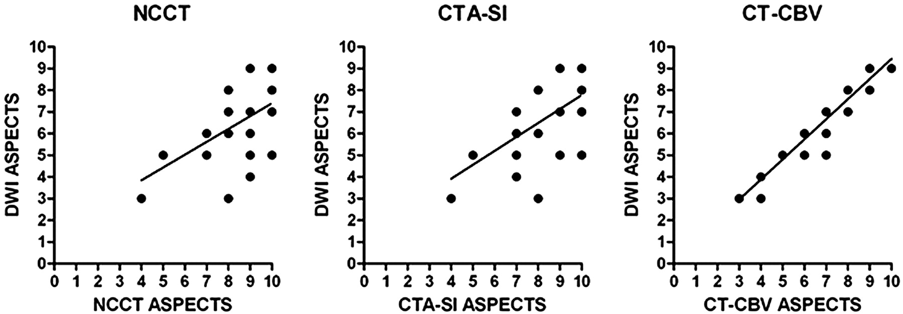
  • Fig 2.
    • Download figure
    • Open in new tab
    • Download powerpoint
    Fig 2.

    Plots of linear regression between noncontrast CT (NCCT) ASPECTS (r2 = 0.34, P = .0011), CTA source images (CTA-SI) ASPECTS (r2 = 0.42, P = .0002), and CT perfusion CBV (CT-CBV) ASPECTS (r2 = 0.91, P < .0001), with follow-up DWI ASPECTS.

Tables

  • Figures
    • View popup
    Table 1:

    Sensitivity, specificity, and accuracy of detecting ASPECTS regional infarctions <3 hours old (n = 280) with a follow-up DWI reference standard*

    ImagingTPFPFNTNSensitivitySpecificityAccuracy
    NCCT4405818044.0 [28.5–56.1]100 [97.3–100]80.0 [73.3–85.4]
    CTA-SI5714317957.0 [41.4–68.1]99.4 [96.3–99.9]84.3 [77.7–89.2]
    CT-CBV910918091.0†‡ [83.5–95.3]100 [97.3–100]96.8†‡ [94.1–98.3]
    • Note:—NCCT indicates noncontrast CT; CTA-SI, CT angiography source image; CT-CBV, CT perfusion cerebral blood volume maps; TP, true-positive; FP, false-positive; FN, false-negative; TN, true-negative.

    • * Sensitivity, specificity, and accuracy values are expressed in percentages with 95% CIs in brackets.

    • † Significantly different compared with NCCT.

    • ‡ Significantly different compared with CTA-SI.

    • View popup
    Table 2:

    Comparison of mean NCCT, CTA-SI, CT-CBV, and follow-up DWI ASPECTS (repeated measures ANOVA with the Bonferroni multiple comparisons test)*

    NCCTCTA-SICT-CBVDWI
    Mean Score (SD)8.4 (1.8)8.0 (1.8)6.8 (1.9)6.5 (1.8)
    NCCT[−0.23–1.2]‡[0.95–2.3]†[1.3–2.7]†
    CTA-SI[0.49–1.9]†[0.81–2.2]†
    CT-CBV[−0.37–1.0]‡
    • Note:—NCCT indicates noncontrast CT; CTA-SI, CT angiography source images; CT-CBV, CT perfusion cerebral blood volume maps; ANOVA, analysis of variance.

    • * 95% CIs of the difference in mean ASPECTSs are in brackets.

    • † Significant P value <.05.

    • ‡ Not significant.

PreviousNext
Back to top

In this issue

American Journal of Neuroradiology: 29 (5)
American Journal of Neuroradiology
Vol. 29, Issue 5
May 2008
  • Table of Contents
  • Index by author
Advertisement
Print
Download PDF
Email Article

Thank you for your interest in spreading the word on American Journal of Neuroradiology.

NOTE: We only request your email address so that the person you are recommending the page to knows that you wanted them to see it, and that it is not junk mail. We do not capture any email address.

Enter multiple addresses on separate lines or separate them with commas.
Accuracy of the Alberta Stroke Program Early CT Score during the First 3 Hours of Middle Cerebral Artery Stroke: Comparison of Noncontrast CT, CT Angiography Source Images, and CT Perfusion
(Your Name) has sent you a message from American Journal of Neuroradiology
(Your Name) thought you would like to see the American Journal of Neuroradiology web site.
CAPTCHA
This question is for testing whether or not you are a human visitor and to prevent automated spam submissions.
Cite this article
K. Lin, O. Rapalino, M. Law, J.S. Babb, K.A. Siller, B.K. Pramanik
Accuracy of the Alberta Stroke Program Early CT Score during the First 3 Hours of Middle Cerebral Artery Stroke: Comparison of Noncontrast CT, CT Angiography Source Images, and CT Perfusion
American Journal of Neuroradiology May 2008, 29 (5) 931-936; DOI: 10.3174/ajnr.A0975

Citation Manager Formats

  • BibTeX
  • Bookends
  • EasyBib
  • EndNote (tagged)
  • EndNote 8 (xml)
  • Medlars
  • Mendeley
  • Papers
  • RefWorks Tagged
  • Ref Manager
  • RIS
  • Zotero
0 Responses
Respond to this article
Share
Bookmark this article
Accuracy of the Alberta Stroke Program Early CT Score during the First 3 Hours of Middle Cerebral Artery Stroke: Comparison of Noncontrast CT, CT Angiography Source Images, and CT Perfusion
K. Lin, O. Rapalino, M. Law, J.S. Babb, K.A. Siller, B.K. Pramanik
American Journal of Neuroradiology May 2008, 29 (5) 931-936; DOI: 10.3174/ajnr.A0975
del.icio.us logo Twitter logo Facebook logo Mendeley logo
  • Tweet Widget
  • Facebook Like
  • Google Plus One
Purchase

Jump to section

  • Article
    • Abstract
    • Methods
    • Results
    • Discussion
    • Conclusion
    • References
  • Figures & Data
  • Info & Metrics
  • Responses
  • References
  • PDF

Related Articles

  • No related articles found.
  • PubMed
  • Google Scholar

Cited By...

  • CT perfusion and angiographic assessment of pial collateral reperfusion in acute ischemic stroke: the CAPRI study
  • Automated CT Perfusion Ischemic Core Volume and Noncontrast CT ASPECTS (Alberta Stroke Program Early CT Score): Correlation and Clinical Outcome Prediction in Large Vessel Stroke
  • Performance of CT ASPECTS and Collateral Score in Risk Stratification: Can Target Perfusion Profiles Be Predicted without Perfusion Imaging?
  • Dynamic Angiography and Perfusion Imaging Using Flat Detector CT in the Angiography Suite: A Pilot Study in Patients with Acute Middle Cerebral Artery Occlusions
  • Impact of the ASPECT scores and distribution on outcome among patients undergoing thrombectomy for acute ischemic stroke
  • Computed Tomographic Angiography and Cerebral Blood Volume Can Predict Final Infarct Volume and Outcome After Recanalization
  • Length of Occlusion Predicts Recanalization and Outcome After Intravenous Thrombolysis in Middle Cerebral Artery Stroke
  • Alberta Stroke Program Early CT Scale Evaluation of Multimodal Computed Tomography in Predicting Clinical Outcomes of Stroke Patients Treated With Aspiration Thrombectomy
  • Pre-intervention cerebral blood volume predicts outcomes in patients undergoing endovascular therapy for acute ischemic stroke
  • Computed Tomography Angiography in Hyperacute Ischemic Stroke: Prognostic Implications and Role in Decision-Making
  • Guidelines for the Early Management of Patients With Acute Ischemic Stroke: A Guideline for Healthcare Professionals From the American Heart Association/American Stroke Association
  • Location of the Clot and Outcome of Perfusion Defects in Acute Anterior Circulation Stroke Treated with Intravenous Thrombolysis
  • Interobserver Agreement of ASPECT Score Distribution for Noncontrast CT, CT Angiography, and CT Perfusion in Acute Stroke
  • Dramatically Reducing Imaging-to-Recanalization Time in Acute Ischemic Stroke: Making Choices
  • Stroke and CT Perfusion
  • CT Angiographic Source Images: Flow- or Volume-Weighted?
  • Evaluation of CT Perfusion in the Setting of Cerebral Ischemia: Patterns and Pitfalls
  • Optimal Brain Perfusion CT Coverage in Patients with Acute Middle Cerebral Artery Stroke
  • Identification of Infarct Core and Penumbra in Acute Stroke Using CT Perfusion Source Images
  • Advances in Interventional Neuroradiology
  • Crossref (87)
  • Google Scholar

This article has been cited by the following articles in journals that are participating in Crossref Cited-by Linking.

  • Guidelines for the Early Management of Patients With Acute Ischemic Stroke
    Edward C. Jauch, Jeffrey L. Saver, Harold P. Adams, Askiel Bruno, J.J. (Buddy) Connors, Bart M. Demaerschalk, Pooja Khatri, Paul W. McMullan, Adnan I. Qureshi, Kenneth Rosenfield, Phillip A. Scott, Debbie R. Summers, David Z. Wang, Max Wintermark, Howard Yonas
    Stroke 2013 44 3
  • Certainty of Stroke Diagnosis: Incremental Benefit with CT Perfusion over Noncontrast CT and CT Angiography
    Julia Hopyan, Anthony Ciarallo, Dar Dowlatshahi, Peter Howard, Verity John, Robert Yeung, Liying Zhang, Jisung Kim, Genevieve MacFarlane, Ting-Yim Lee, Richard I. Aviv
    Radiology 2010 255 1
  • CT Protocol for Acute Stroke: Tips and Tricks for General Radiologists
    Enrique Marco de Lucas, Elena Sánchez, Agustín Gutiérrez, Andrés González Mandly, Eva Ruiz, Alejandro Fernández Flórez, Javier Izquierdo, Javier Arnáiz, Tatiana Piedra, Natalia Valle, Itziar Bañales, Fernando Quintana
    RadioGraphics 2008 28 6
  • Evaluation of CT Perfusion in the Setting of Cerebral Ischemia: Patterns and Pitfalls
    Y.W. Lui, E.R. Tang, A.M. Allmendinger, V. Spektor
    American Journal of Neuroradiology 2010 31 9
  • Time-resolved assessment of collateral flow using 4D CT angiography in large-vessel occlusion stroke
    Andreas M. J. Frölich, Sarah Lena Wolff, Marios N. Psychogios, Ernst Klotz, Ramona Schramm, Katrin Wasser, Michael Knauth, Peter Schramm
    European Radiology 2014 24 2
  • Automated CT Perfusion Ischemic Core Volume and Noncontrast CT ASPECTS (Alberta Stroke Program Early CT Score)
    Diogo C. Haussen, Seena Dehkharghani, Srikant Rangaraju, Leticia C. Rebello, Mehdi Bouslama, Jonathan A. Grossberg, Aaron Anderson, Samir Belagaje, Michael Frankel, Raul G. Nogueira
    Stroke 2016 47 9
  • Interobserver Agreement of ASPECT Score Distribution for Noncontrast CT, CT Angiography, and CT Perfusion in Acute Stroke
    Olga Finlayson, Verity John, Robert Yeung, Dar Dowlatshahi, Peter Howard, Liying Zhang, Rick Swartz, Richard I. Aviv
    Stroke 2013 44 1
  • Length of Occlusion Predicts Recanalization and Outcome After Intravenous Thrombolysis in Middle Cerebral Artery Stroke
    Vladimir Rohan, Jan Baxa, Radek Tupy, Lenka Cerna, Petr Sevcik, Michal Friesl, Jiri Polivka, Jiri Polivka, Jiri Ferda
    Stroke 2014 45 7
  • CT Angiographic Source Images: Flow- or Volume-Weighted?
    M. Sharma, A.J. Fox, S. Symons, A. Jairath, R.I. Aviv
    American Journal of Neuroradiology 2011 32 2
  • CT Perfusion Imaging in Acute Stroke
    Angelos A. Konstas, Max Wintermark, Michael H. Lev
    Neuroimaging Clinics of North America 2011 21 2

More in this TOC Section

  • Multimodal CT Provides Improved Performance for Lacunar Infarct Detection
  • Optimal MRI Sequence for Identifying Occlusion Location in Acute Stroke: Which Value of Time-Resolved Contrast-Enhanced MRA?
  • Evaluating the Effects of White Matter Multiple Sclerosis Lesions on the Volume Estimation of 6 Brain Tissue Segmentation Methods
Show more BRAIN

Similar Articles

Advertisement

Indexed Content

  • Current Issue
  • Accepted Manuscripts
  • Article Preview
  • Past Issues
  • Editorials
  • Editor's Choice
  • Fellows' Journal Club
  • Letters to the Editor
  • Video Articles

Cases

  • Case Collection
  • Archive - Case of the Week
  • Archive - Case of the Month
  • Archive - Classic Case

More from AJNR

  • Trainee Corner
  • Imaging Protocols
  • MRI Safety Corner
  • Book Reviews

Multimedia

  • AJNR Podcasts
  • AJNR Scantastics

Resources

  • Turnaround Time
  • Submit a Manuscript
  • Submit a Video Article
  • Submit an eLetter to the Editor/Response
  • Manuscript Submission Guidelines
  • Statistical Tips
  • Fast Publishing of Accepted Manuscripts
  • Graphical Abstract Preparation
  • Imaging Protocol Submission
  • Evidence-Based Medicine Level Guide
  • Publishing Checklists
  • Author Policies
  • Become a Reviewer/Academy of Reviewers
  • News and Updates

About Us

  • About AJNR
  • Editorial Board
  • Editorial Board Alumni
  • Alerts
  • Permissions
  • Not an AJNR Subscriber? Join Now
  • Advertise with Us
  • Librarian Resources
  • Feedback
  • Terms and Conditions
  • AJNR Editorial Board Alumni

American Society of Neuroradiology

  • Not an ASNR Member? Join Now

© 2025 by the American Society of Neuroradiology All rights, including for text and data mining, AI training, and similar technologies, are reserved.
Print ISSN: 0195-6108 Online ISSN: 1936-959X

Powered by HighWire