
Article Figures & Data
Figures
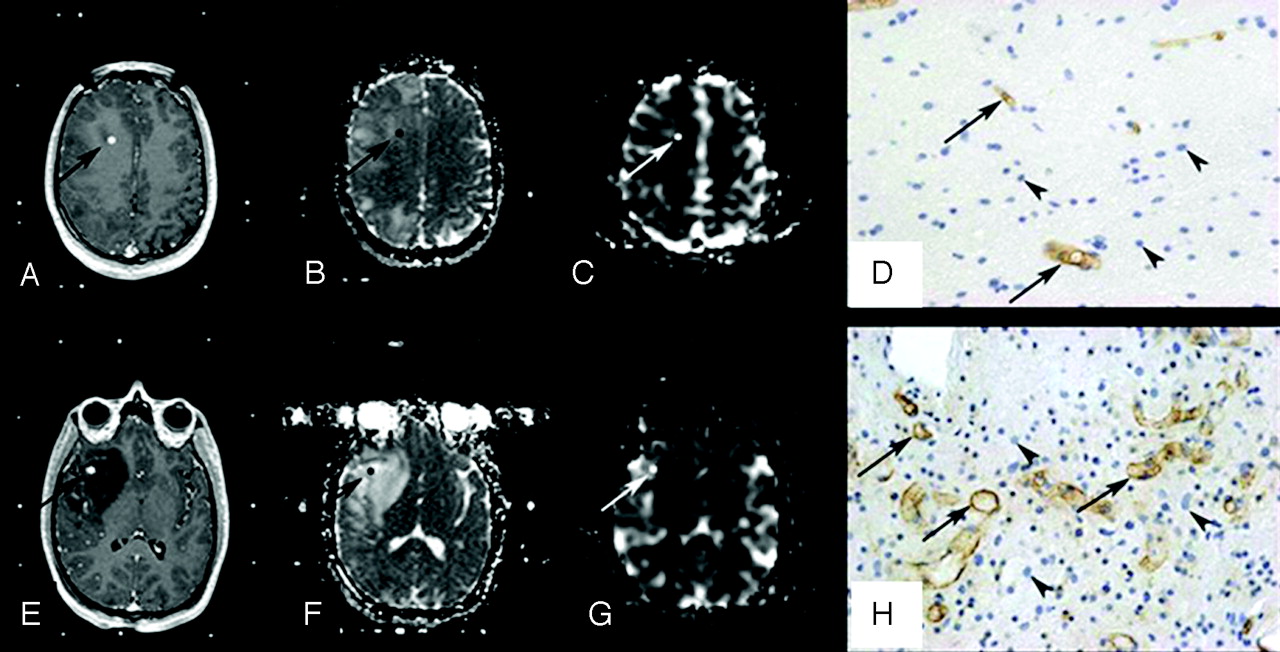
- Fig 1.
Histopathologic correlation of MR imaging data with stereotactic biopsy specimens in a 19-year-old patient with a right frontotemporal grade II astrocytoma. A–D, Top row shows axial 3D T1-weighted images (TR/TE, 20/4.6 ms) with contrast (A), coregistered ADC map (B), coregistered rCBV map (C), and corresponding histopathologic results for a “peritumoral tissue” sample (D). E–H, Bottom row shows axial 3D T1-weighted images (TR/TE, 20/4.6 ms) with contrast (E), coregistered ADC map (F), coregistered rCBV map (G), and corresponding histopathologic results for a “infiltrated tissue” sample (H). Regions of interest where ADC and rCBV have been measured are illustrated (arrows). The samples were immunohistochemically stained by using a monoclonal antibody against the CD34 antigen to assess microvessel density (arrows) and counterstained with hematoxylin to assess cell density (arrowheads) (original magnification, × 400).
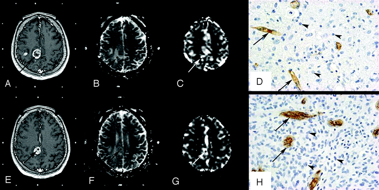
- Fig 2.
Histopathologic correlation of MR imaging data with stereotactic biopsy specimens in a 50-year-old patient with a right parietal grade IV astrocytoma. A–D, Top row shows axial 3D T1-weighted images (TR/TE, 20/4.6 ms) with contrast (A), coregistered ADC map (B), coregistered rCBV map (C), and corresponding histopathologic results for an “infiltrated tissue” sample (D). E–H, Bottom row shows axial 3D T1-weighted images (TR/TE, 20/4.6 ms) with contrast (E), coregistered ADC map (F), coregistered rCBV map (G), and corresponding histopathologic results for a “bulk tumor” sample (H). Regions of interest where ADC and rCBV have been measured are illustrated (arrows). The samples were immunohistochemically stained by using a monoclonal antibody against the CD34 antigen to assess microvessel density (arrows) and counterstained with hematoxylin to assess cell density (arrowheads) (original magnification, × 400).
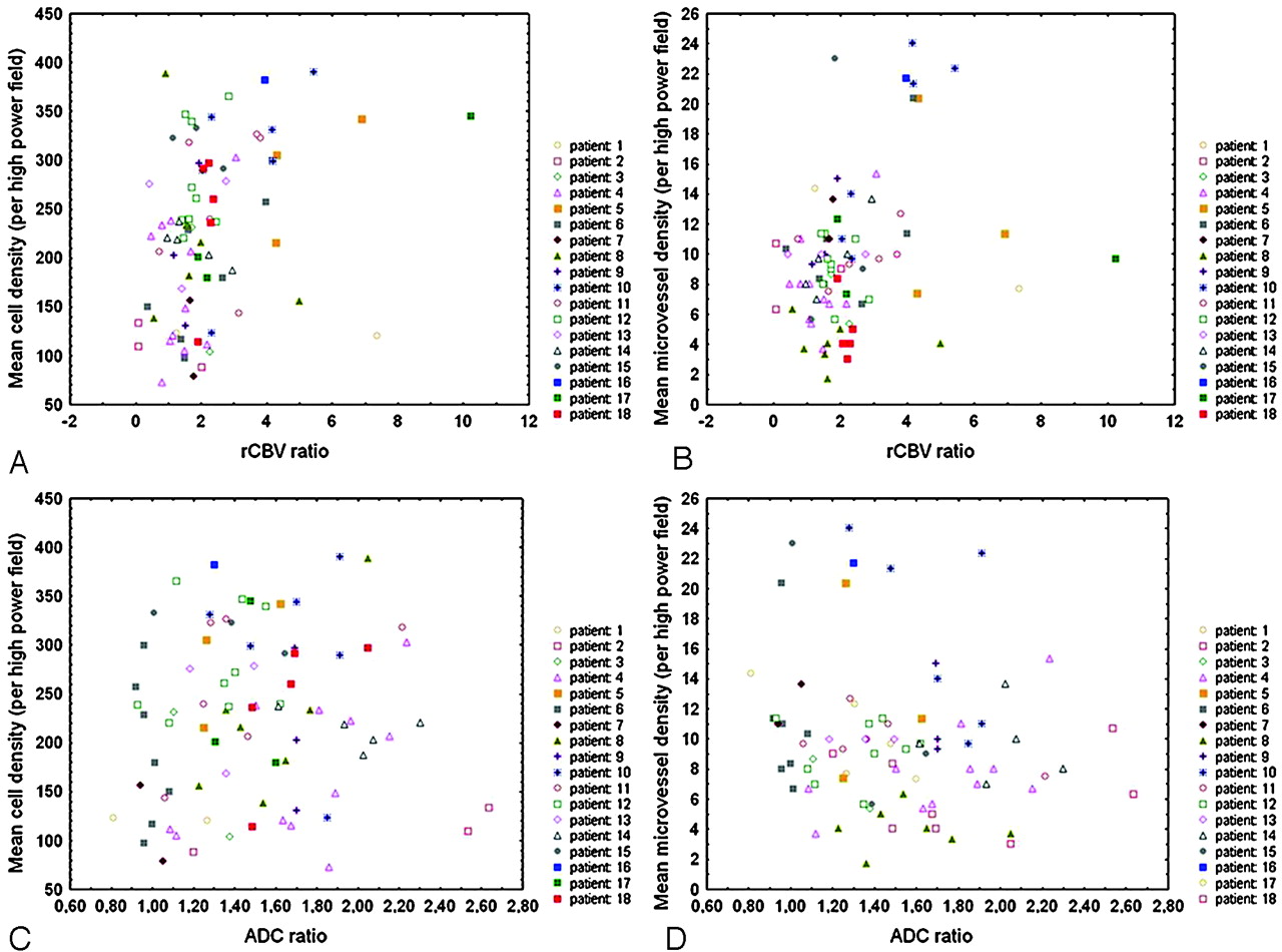
- Fig 3.
Scatterplots show a positive correlation between rCBV ratios and both cell density (n = 81, r = 0.37, P < .001) (A) and microvessel density (n = 81, r = 0.26, P < .05) (B) in the whole set of samples. There is no significant correlation between ADC ratios and either cell density (n = 81, r = 0.11, P = .34) (C) or microvessel density (n = 81, r = −0.20, P = .08) (D). Spearman rank correlation test was used, and P < .05 was considered to be statistically significant.
- Fig 4.
Scatterplots show higher correlation between rCBV ratios and both cell density (n = 33, r = 0.57, P < .001) (A) and microvessel density (n = 33, r = 0.46, P < .01) (B) in bulk tumor than in the whole set of samples (Fig 3). In bulk tumor, there is no significant correlation between ADC ratios and cell density (n = 33, r = −0.20, P = .26) (C), whereas an inverse correlation between ADC and microvessel density is found (n = 33, r = −0.36, P < .05) (D). Spearman rank correlation test was used, and P < .05 was considered to be statistically significant.


Tables
Patient No./Age (yr)/Sex Histologic Diagnosis, Grade Location PET No. of Biopsy Specimens 1/25/F LA, 2 L temporal MET 3 2/55/F LA, 2 R frontoparietal MET 4 3/39/F LA, 2 R frontal MET 2 4/19/M LA, 2 R frontotemporal MET 11 5/78/F LA, 2 R temporal MET 3 6/67/F LA, 2 L parietal MET 8 7/58/F LA, 2 R parietooccipital FDG 4 8/24/M LA, 2 R frontotemporal MET 7 9/47/M O, 2 L frontal MET 3 10/62/M O, 2 L frontotemporal FDG 6 11/34/F OA, 2 R cerebellum FDG 6 12/19/M AA, 3 L frontoparietal MET 9 13/33/M AA, 3 L temporoparietal MET 3 14/42/M AA, 3 R thalamus FDG 5 15/39/F O, 3 L temporoparietal MET 3 16/35/F O, 3 R thalamus FDG 3 17/50/M GB, 4 R parietal MET 3 18/58/M GB, 4 L temporal MET 5 Note:—LA indicates low-grade astrocytoma; O, oligodendroglioma; OA, mixed oligoastrocytoma; AA, anaplastic astrocytoma; GB, glioblastoma; L, left; R, right.
- Table 2:
Results of Spearman rank correlation test between rCBV and ADC ratios versus cell and microvessel density
Density All Samples (n = 18, N = 81) Peritumoral Tissue (n = 4, N = 9) Infiltrated Tissue (n = 17, N = 39) Bulk Tumor (n = 13, N = 33) r (P) r (P) r (P) r(P) ADC-cell 0.11 (.34) 0.02 (.97) −0.02 (.90) −0.20 (.26) ADC-microvessel −0.20 (.08) −0.62 (.08) −0.06 (.72) −0.36 (.04)* rCBV-cell 0.37 (.0006)* −0.07 (.86) 0.09 (.59) 0.57 (.0005)* rCBV-microvessel 0.26 (.02)* 0.03 (.93) −0.18 (.28) 0.46 (.008)* Note:—nindicates number of patients; N, number of biopsy samples.
* Statistically significant with P < .05.
In this issue


Jump to section
Related Articles
Cited By...
- Detection of local microvascular proliferation in IDH wild-type Glioblastoma using relative Cerebral Blood Volume
- Voxelwise and Patientwise Correlation of 18F-FDOPA PET, Relative Cerebral Blood Volume, and Apparent Diffusion Coefficient in Treatment-Naive Diffuse Gliomas with Different Molecular Subtypes
- Estimating Local Cellular Density in Glioma Using MR Imaging Data
- Non-Contrast-Enhancing Tumor: A New Frontier in Glioblastoma Research
- Glioma grade map: a machine-learning based imaging biomarker for tumor characterization
- Accurate Patient-Specific Machine Learning Models of Glioblastoma Invasion Using Transfer Learning
- A Multiparametric Model for Mapping Cellularity in Glioblastoma Using Radiographically Localized Biopsies
- Correlation of Tumor Immunohistochemistry with Dynamic Contrast-Enhanced and DSC-MRI Parameters in Patients with Gliomas
- Mitotic Activity in Glioblastoma Correlates with Estimated Extravascular Extracellular Space Derived from Dynamic Contrast-Enhanced MR Imaging
- A Prognostic Model Based on Preoperative MRI Predicts Overall Survival in Patients with Diffuse Gliomas
- Comparison of 18F-FET PET and Perfusion-Weighted MR Imaging: A PET/MR Imaging Hybrid Study in Patients with Brain Tumors
- Assessment of Angiographic Vascularity of Meningiomas with Dynamic Susceptibility Contrast-Enhanced Perfusion-Weighted Imaging and Diffusion Tensor Imaging
- Differentiation of Primary Central Nervous System Lymphomas and Glioblastomas: Comparisons of Diagnostic Performance of Dynamic Susceptibility Contrast-Enhanced Perfusion MR Imaging without and with Contrast-Leakage Correction
- Phase I Study of GRN1005 in Recurrent Malignant Glioma
- Persistent Diffusion-Restricted Lesions in Bevacizumab-Treated Malignant Gliomas Are Associated with Improved Survival Compared with Matched Controls
- The Added Value of Apparent Diffusion Coefficient to Cerebral Blood Volume in the Preoperative Grading of Diffuse Gliomas
- Imaging biomarkers of angiogenesis and the microvascular environment in cerebral tumours
- Correlation of MR Relative Cerebral Blood Volume Measurements with Cellular Density and Proliferation in High-Grade Gliomas: An Image-Guided Biopsy Study
- Distinguishing Recurrent Primary Brain Tumor from Radiation Injury: A Preliminary Study Using a Susceptibility-Weighted MR Imaging-Guided Apparent Diffusion Coefficient Analysis Strategy
- Switching on the Lights for Real-Time Multimodality Tumor Neuroimaging: The Integrated Positron-Emission Tomography/MR Imaging System
- Candidate Biomarkers of Extravascular Extracellular Space: A Direct Comparison of Apparent Diffusion Coefficient and Dynamic Contrast-Enhanced MR Imaging--Derived Measurement of the Volume of the Extravascular Extracellular Space in Glioblastoma Multiforme