
Article Figures & Data
Figures
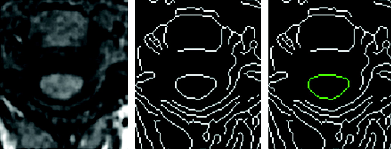
- Fig 1.
Cervical cord identification and quantification. Left, Original cervical 3D SPGR-T1WI. Center, Edge-detection image. Right, Cord edges identified by operator.
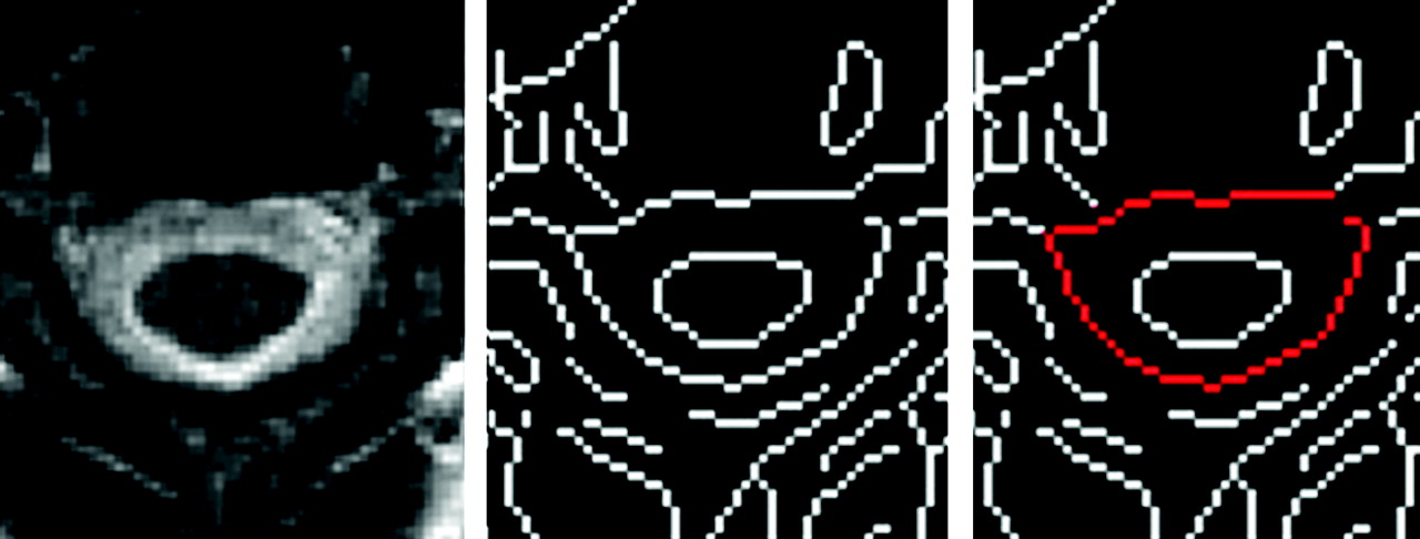
- Fig 2.
Cervical canal identification and quantification. Left, Original cervical T2WI. Center, Edge-detection image. Right, Spinal canal edge identified by operator.
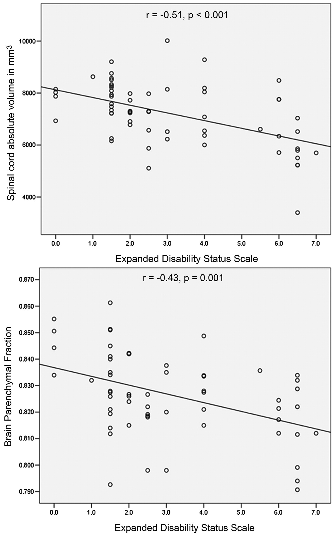
- Fig 3.
Correlation of cervical cord absolute volume and brain parenchyma fraction with disability, as measured by EDSS.


Tables
- Table 1:
Demographic and clinical characteristics of normal control subjects and multiple sclerosis patients, according to disease type
Variable NC (n= 19) MS (n = 65) CIS (n = 11) RR (n = 34) SP (n = 14) PP (n = 7) Female, n (%) 10 (52.6) 48 (72.7) 9 (81.8) 26 (76.5) 9 (64.3) 4 (57.1) Age in years, mean (SD) 30.4 (12) 41.2 (12.4) 36.9 (9.1) 42.4 (8.6) 47.2 (11.4) 59.9 (12) Disease duration in years, mean (SD) NA 11.8 (10.7) 1.1 (1.4) 11.1 (8.2) 17.1 (9.9) 19.6 (18.6) EDSS, mean (SD) NA 3.1 (2.1) 1.6 (0.3) 2.1 (1.3) 5.0 (1.8) 5.8 (1.3) Note:—NC indicates normal control subjects; MS, multiple sclerosis; RR, relapsing-remitting; SP, secondary-progressive; PP, primary-progressive; CIS, clinically isolated syndrome; NA, not applicable.
Cervical Atrophy Measure Scan-Rescan COV, Mean % (95% CI) Intrarater COV, Mean % (95% CI) Interrater COV, Mean % (95% CI) CCAV 1.29 (0.62–2.37) 0.25 (0.12–0.46) 1.26 (0.60–2.30) CCF 1.4 (0.67–2.60) 0.6 (0.29–1.10) 1.1 (0.53–2.00) CCAV/ICV 1.6 (0.77–2.90) 0.7 (0.34–1.30) 1.6 (0.77–2.90) BPF 0.1 (0.048–0.184) 0 0 MPD 0.84 (0.40–1.55) 0 0 Note:—COV indicates coefficient of variation; CCAV, cervical cord absolute volume in cubic millimeters; CCF, cervical cord fraction; CCAV/ICV, cervical cord absolute volume to intracranial volume; CI, confidence interval; BPF, brain parenchyma fraction; MPD, mean parenchyma diffusivity.
- Table 3:
Cervical cord atrophy and lesion MR imaging measures in normal control subjects and multiple sclerosis patients, according to disease type
Variable NC (n= 19), Mean (SD) MS (n= 66), Mean (SD) CIS (n= 11), Mean (SD) RR (n= 34), Mean (SD) SP (n= 14), Mean (SD) PP (n= 7), Mean (SD) CCAV, mm3 7691.7 (1136.2) 7063.2 (1206.8)** 7461.1 (599.5) 7281.6 (758.9) 5990.7 (1060.9)*** 6907.3 (1796.2)* CCF 0.343 (0.29) 0.318 (0.23)** 0.344 (0.04) 0.307 (0.06) 0.307 (0.06)* 0.294 (0.04)* CCAV/ICV 0.012 (0.002) 0.016 (0.02) 0.012 (0.002) 0.015 (0.005) 0.015 (0.016) 0.01 (0.005) Cervical T2-LV, mL NA 4.6 (5.8) 0.7 (0.02) 4.2 (3.8) 9.7 (8) 2.6 (3.2) Note:—NC indicates normal control subject; MS, multiple sclerosis; RR, relapsing-remitting; SP, secondary-progressive; PP, primary-progressive; CIS, clinically isolated syndrome; CCAV, cervical cord absolute volume; CCF, cervical cord fraction; CCAV/ICV, cervical cord absolute volume to intracranial volume; LV, lesion volume; NA, nonapplicable. A general linear model analysis was performed to test significant differences between normal control subjects and multiple sclerosis patients in which the age was entered as a covariate and, due to the multiple comparisons, a post hoc Bonferroni correction was applied directly in the SPSS analysis model.
P values are provided between normal control subject and multiple sclerosis patients
* P < .05;
** P < .01;
*** P < .001.
- Table 4:
Brain MR imaging measures in normal control subjects and multiple sclerosis patients, according to disease type
Variable NC (n= 19), Mean (SD) MS (n= 66), Mean (SD) CIS (n= 11), Mean (SD) RR (n= 34), Mean (SD) SP (n= 14), Mean (SD) PP (n= 7), Mean (SD) BPF 0.845 (0.006) 0.827 (0.02) 0.834 (0.01) 0.832 (0.01) 0.815 (0.02) 0.815 (0.009) MPD, × 10−6 mm2/s 1130.1 (65.8) 1204.3 (89.2) 1143.4 (59.1) 1222.8 (100.4) 1196.3 (71) 1222.9 (61) T2-LV, mL NA 11 (12.6) 5.1 (6.5) 12.1 (14.4) 18.3 (13.5) 6.9 (7.7) T1-LV, mL NA 1.7 (3.2) 0.5 (0.7) 1.2 (1.7) 3.2 (5.4) 1.2 (1.5) Note:—NC indicates normal control subjects; MS, multiple sclerosis; RR, relapsing-remitting; SP, secondary-progressive; PP, primary-progressive; CIS, clinically isolated syndrome; BPF, brain parenchyma fraction; MPD, mean parenchyma diffusivity; LV, lesion volume; NA, nonapplicable.
- Table 5:
Correlation analysis between cervical and brain MR imaging measures and clinical variables
Variable EDSS Disease Duration r P r P CCAV, mm3 −0.51* <0.0001* −0.15 0.246 CCF −0.31* 0.018* −0.14 0.260 CCAV/ICV −0.12 0.459 −0.12 0.438 Cervical T2-LV, mL 0.38* 0.027* 0.33* 0.04* BPF −0.43* 0.001* −0.36* 0.004* MPD 0.31* 0.04* 0.25 0.069 Brain T2-LV, mL 0.16 0.232 0.25* 0.047* Brain T1-LV, mL 0.36* 0.009* 0.26 0.055 Note:—CCAV indicates cervical cord absolute volume; CCF, cervical cord fraction; CCAV/ICV, cervical cord absolute volume to intracranial volume; BPF, brain parenchyma fraction; MPD, mean parenchyma diffusivity; LV, lesion volume.
* Values are significant (P < .05).


