
Article Figures & Data
Figures
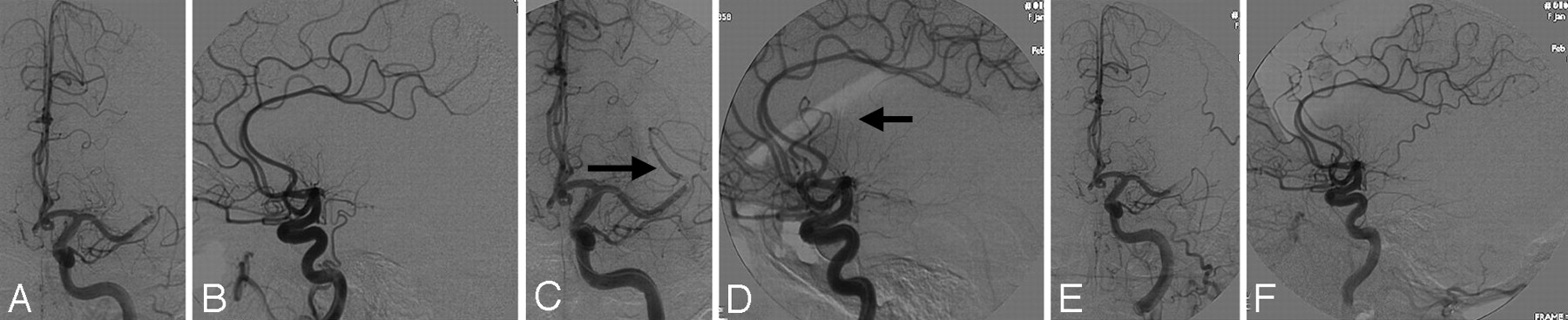
- Fig 1.
Arterial reocclusion. A 54-year-old woman, status post IV tissue plasminogen activator, has a left M1 occlusion seen in the anteroposterior (A) and lateral (B) planes (Qureshi grade 3A). After mechanical thrombolysis, there is improved flow in the superior division, frontal branches of M2 (arrows, C and D; Qureshi grade 2). However, the final angiogram shows reocclusion of the M1 stem similar to her initial angiographic appearance (E and F, Thrombolysis in Myocardial Infarction grade 0, Qureshi grade 3A).
- Fig 2.
Distal embolization. A 53-year-old woman has right carotid terminus occlusion seen in the anteroposterior (panel A) and lateral (panel B) projections (Qureshi grade 5). Status post IA reteplase and intravenous abciximab, there is flow through the anterior cerebral artery seen in the anteroposterior (C) and lateral (D) projections. The arrow (D) indicates an occlusion of the callosomarginal branch, whereas the pericallosal branch is filling well (Qureshi grade 3B).


Tables
Total (n = 91) Reocclusion (n = 16) No Reocclusion (n = 75) P Value Mean age (years) 67 ± 15 70 ± 14 66 ± 16 .58 Male patients (No., %) 48 (53) 8 (50) 40 (53) .81 Occlusion site: .92 MCA 48 (53%) 9 (56%) 39 (52%) ICA 29 (32%) 5 (31%) 24 (32%) ACA 2 (2%) 0 (0%) 2 (3%) BA 12 (13%) 2 (13%) 10 (13%) Study protocol* .18 Group 1 47 (52%) 9 (56%) 38 (51%) Group 2 12 (13%) 4 (25%) 8 (11%) Group 3 19 (21%) 3 (19%) 16 (21%) Group 4 13 (14%) 0 (0%) 13 (17%) Mean time to treatment (h) 4.36 ± 2.18 4.05 ± 1.27 4.42 ± 2.33 .06 Initial Qureshi grade 0–3B 53 (58%) 9 (56%) 44 (59%) .86 Initial Qureshi grade 4A-5 38 (42%) 7 (44%) 31 (41%) Final recanalization† <.01 None 19 (21%) 2 (13%) 17 (23%) Partial 41 (45%) 13 (82%) 28 (37%) Complete 29 (32%) 1 (6%) 28 (37%) Symptomatic ICH (No.) 10 3 7 .46 Median initial NIHSS score 19 (±8) 19 (±7) 19 (±8) .90 Median 24-hour NIHSS score 18 ± 13 22 (±11) 17 (±13) .18 Median 7-day NIHSS score 18 ± 16 25 (±16) 17 (±16) .06 Favorable mRS‡ at 1–3 months (No., %) 29 (32) 1 (6) 28 (37) .02 Favorable 1–3 mRS at 1–3 months among treatment responders§ (n = 70) 24/70 (34%) 1/16 (6%) 23/56 (41%) .01 Note:—MCA indicates middle cerebral artery; ICA, internal carotid artery; ACA, anterior cerebral artery; BA, basilar artery; h, hour; ICH, intracerebral hemorrhage; NIHSS, National Institutes of Health Stroke Scale; mRS, modified Rankin Scale.
* Group 1, IA reteplase and MTD; group 2, EKOS MicroLysUS North American trial; group 3, MTD following IV thrombolysis; group 4, IA reteplase and IV abciximab (FDA IND 9180).
† Defined as ≤2.
‡ Data were missing on 2 patients.
§ Patients who never demonstrated recanalization were excluded from the analysis.
Total (n = 91) Distal Embolization (n = 15) No Distal Embolization (n = 76) P Value Mean age (years) 67 ± 15 60 ± 13 68 ± 16 .16 Male/female 48 (53%) 6 (40%) 45 (59%) .28 Occlusion site .58 MCA 48 (53%) 6 (40%) 42 (55%) ICA 29 (32%) 6 (40%) 23 (30%) ACA 2 (3%) 0 (0%) 2 (3%) BA 12 (13%) 3 (20%) 9 (12%) Study protocol* .00 Group 1 47 (52%) 2 (13%) 45 (59%) Group 2 12 (13%) 6 (40%) 6 (8%) Group 3 19 (21%) 4 (27%) 15 (20%) Group 4 13 (14%) 3 (20%) 10 (13%) Mean time to treatment (hours) 4.36 ± 2.18 4.59 ± 3.13 4.31 ± 1.97 .14 Initial Qureshi grade 0–3B 53 (58%) 6 (40%) 47 (62%) .12 Initial Qureshi grade 4A-5 38 (42%) 9 (60%) 29 (38%) Final recanalization† .48 None 19 (21%) 2 (13%) 17 (22%) Partial 41 (45%) 8 (50%) 33 (43%) Complete 29 (32%) 3 (19%) 26 (34%) Symptomatic ICH (No., %) 10 3 (21)c 6 (8) .12 Median initial NIHSS score 19 (±8) 19 (±8) 19 (±8) .98 Mean 24-hour NIHSS score 18 ± 13 24 (±15) 17 (±12) .07 Mean 7-day NIHSS score 18 ± 16 21 (±17) 18 (±16) .46 Favorable 1- to 3-month mRS‡ (No., %) 29 (32%) 2 (13%) 27 (36%) .09 Favorable 1–3 mRS at 1–3 months among treatment responders§ 24/70 (34%) 23/24 (96%) 1/11 (9%) .05 Note:—MCA indicates middle cerebral artery; ICA, internal carotid artery; ACA, anterior cerebral artery; BA, basilar artery; ICH, intracerebral hemorrhage; NIHSS, National Institutes of Health Stroke Scale; mRS, modified Rankin Score.
* Group 1, IA reteplase and MDT; group 2, EKOS MicroLysUS North American trial; group 3, MDT following IV thrombolysis; group 4, IA reteplase and IV abciximab (FDA IND 9180).
† Defined as ≤ 2.
‡ Data were missing for 1 patient.
§ Patients who never demonstrated recanalization were excluded from the analysis.
Study No Recanalization Partial Recanalization Complete Recanalization Study 1 (IA reteplase and MDT), n = 47 6 (13%) 27 (57%) 14 (30%) Study 2 (EKOS), n = 12a 1 (10%)* 4 (40%)* 5 (50%)* Study 3 (IV thrombolysis and MDT) n = 19 6 (32%) 8 (42%) 5 (26%) Study 4 (IV abciximab and IA reteplase) n = 13 6 (46%) 2 (15%) 5 (39%) * Data were missing for 2 patients; percentages are of 10 patients.
Variable P Value Odds Ratio 95% Confidence Interval Lower Upper Age .04 4.4 0.91 1.00 Sex .72 0.1 0.26 2.55 Initial NIHSS score .03 4.7 0.82 0.99 Initial Qureshi grade* .79 0.1 0.24 2.99 Time to treatment .06 3.7 1.00 1.65 Symptomatic ICH .25 1.3 0.02 2.80 Recanalization complete partial none (reference) .26 1.3 0.59 7.29 .03 4.8 0.03 0.82 Reocclusion .05 3.9 0.01 0.98 Distal embolization .09 3.0 0.03 1.26 Note:—ICH indicates intracerebral hemorrhage; NHSS, National Institutes of Health Stroke Scale.
* Dichotomized as grades 0–3B and 4A-5.


