Advertisement

Main menu

  • Home
  • Content
    • Current Issue
    • Accepted Manuscripts
    • Article Preview
    • Past Issue Archive
    • Video Articles
    • AJNR Case Collection
    • Case of the Week Archive
    • Case of the Month Archive
    • Classic Case Archive
  • Special Collections
    • Advancing NeuroMRI with High-Relaxivity Contrast Agents
    • AJNR Awards
    • Low-Field MRI
    • Alzheimer Disease
    • ASNR Foundation Special Collection
    • Photon-Counting CT
    • View All
  • Multimedia
    • AJNR Podcasts
    • AJNR SCANtastic
    • Trainee Corner
    • MRI Safety Corner
    • Imaging Protocols
  • For Authors
    • Submit a Manuscript
    • Submit a Video Article
    • Submit an eLetter to the Editor/Response
    • Manuscript Submission Guidelines
    • Statistical Tips
    • Fast Publishing of Accepted Manuscripts
    • Graphical Abstract Preparation
    • Imaging Protocol Submission
    • Author Policies
  • About Us
    • About AJNR
    • Editorial Board
    • Editorial Board Alumni
  • More
    • Become a Reviewer/Academy of Reviewers
    • Subscribers
    • Permissions
    • Alerts
    • Feedback
    • Advertisers
    • ASNR Home

User menu

  • Alerts
  • Log in

Search

  • Advanced search
American Journal of Neuroradiology

American Journal of Neuroradiology

ASHNR American Society of Functional Neuroradiology ASHNR American Society of Pediatric Neuroradiology ASSR
  • Alerts
  • Log in

Advanced Search

  • Home
  • Content
    • Current Issue
    • Accepted Manuscripts
    • Article Preview
    • Past Issue Archive
    • Video Articles
    • AJNR Case Collection
    • Case of the Week Archive
    • Case of the Month Archive
    • Classic Case Archive
  • Special Collections
    • Advancing NeuroMRI with High-Relaxivity Contrast Agents
    • AJNR Awards
    • Low-Field MRI
    • Alzheimer Disease
    • ASNR Foundation Special Collection
    • Photon-Counting CT
    • View All
  • Multimedia
    • AJNR Podcasts
    • AJNR SCANtastic
    • Trainee Corner
    • MRI Safety Corner
    • Imaging Protocols
  • For Authors
    • Submit a Manuscript
    • Submit a Video Article
    • Submit an eLetter to the Editor/Response
    • Manuscript Submission Guidelines
    • Statistical Tips
    • Fast Publishing of Accepted Manuscripts
    • Graphical Abstract Preparation
    • Imaging Protocol Submission
    • Author Policies
  • About Us
    • About AJNR
    • Editorial Board
    • Editorial Board Alumni
  • More
    • Become a Reviewer/Academy of Reviewers
    • Subscribers
    • Permissions
    • Alerts
    • Feedback
    • Advertisers
    • ASNR Home
  • Follow AJNR on Twitter
  • Visit AJNR on Facebook
  • Follow AJNR on Instagram
  • Join AJNR on LinkedIn
  • RSS Feeds

AJNR Awards, New Junior Editors, and more. Read the latest AJNR updates


Improved Turnaround Times | Median time to first decision: 12 days

Research ArticleNeurointervention

Management and Clinical Outcome of Acute Basilar Artery Dissection

B.M. Kim, S.H. Suh, S.I. Park, Y.S. Shin, E.C. Chung, M.H. Lee, E.J. Kim, J.S. Koh, H.-s. Kang, H.G. Roh, Y.S. Won, P.-W. Chung, Y.-B. Kim and B.C. Suh
American Journal of Neuroradiology November 2008, 29 (10) 1937-1941; DOI: https://doi.org/10.3174/ajnr.A1243
B.M. Kim
  • Find this author on Google Scholar
  • Find this author on PubMed
  • Search for this author on this site
S.H. Suh
  • Find this author on Google Scholar
  • Find this author on PubMed
  • Search for this author on this site
S.I. Park
  • Find this author on Google Scholar
  • Find this author on PubMed
  • Search for this author on this site
Y.S. Shin
  • Find this author on Google Scholar
  • Find this author on PubMed
  • Search for this author on this site
E.C. Chung
  • Find this author on Google Scholar
  • Find this author on PubMed
  • Search for this author on this site
M.H. Lee
  • Find this author on Google Scholar
  • Find this author on PubMed
  • Search for this author on this site
E.J. Kim
  • Find this author on Google Scholar
  • Find this author on PubMed
  • Search for this author on this site
J.S. Koh
  • Find this author on Google Scholar
  • Find this author on PubMed
  • Search for this author on this site
H.-s. Kang
  • Find this author on Google Scholar
  • Find this author on PubMed
  • Search for this author on this site
H.G. Roh
  • Find this author on Google Scholar
  • Find this author on PubMed
  • Search for this author on this site
Y.S. Won
  • Find this author on Google Scholar
  • Find this author on PubMed
  • Search for this author on this site
P.-W. Chung
  • Find this author on Google Scholar
  • Find this author on PubMed
  • Search for this author on this site
Y.-B. Kim
  • Find this author on Google Scholar
  • Find this author on PubMed
  • Search for this author on this site
B.C. Suh
  • Find this author on Google Scholar
  • Find this author on PubMed
  • Search for this author on this site
  • Article
  • Figures & Data
  • Supplemental
  • Info & Metrics
  • Responses
  • References
  • PDF
Loading

Article Figures & Data

Figures

  • Fig 1.
    • Download figure
    • Open in new tab
    • Download powerpoint
    Fig 1.

    A 42-year-old man presenting with progressive deterioration of consciousness. CT angiogram (A) and vertebral angiogram (B) reveal near-occlusion of the basilar artery by an intramural hematoma (arrows). C, After emergency stent insertion, the basilar artery and left posterior cerebral artery are visualized, but the right posterior cerebral artery is not seen on vertebral angiogram. D, A 2-week (not seen) and 18-month follow-up vertebral angiogram show a patent basilar artery and its branches. The patient had a favorable outcome (mRS score, 0).

  • Fig 2.
    • Download figure
    • Open in new tab
    • Download powerpoint
    Fig 2.

    A 30-year-old man presenting with signs of stem compression 3 years after initial presentation of brain stem infarction. A, Initial MR angiogram reveals focal asymmetric dilation in the distal basilar artery. B, CT angiogram 3 years after initial MR imaging as a result of brain stem compression. A large asymmetric pseudoaneurysm is demonstrated in the same location. C, Vertebral angiogram in lateral projection after overlapping double-stent deployment shows flow redirection through the stent and the basilar artery with stasis of contrast media in the pseudoaneurysmal sac (arrows). D and E, A 4-month follow-up vertebral artery angiogram (D) and right internal carotid artery angiogram (E) reveal occlusion of the basilar artery and hypertrophied right posterior communicating artery (arrow), which supplies the distal basilar artery and its branches.
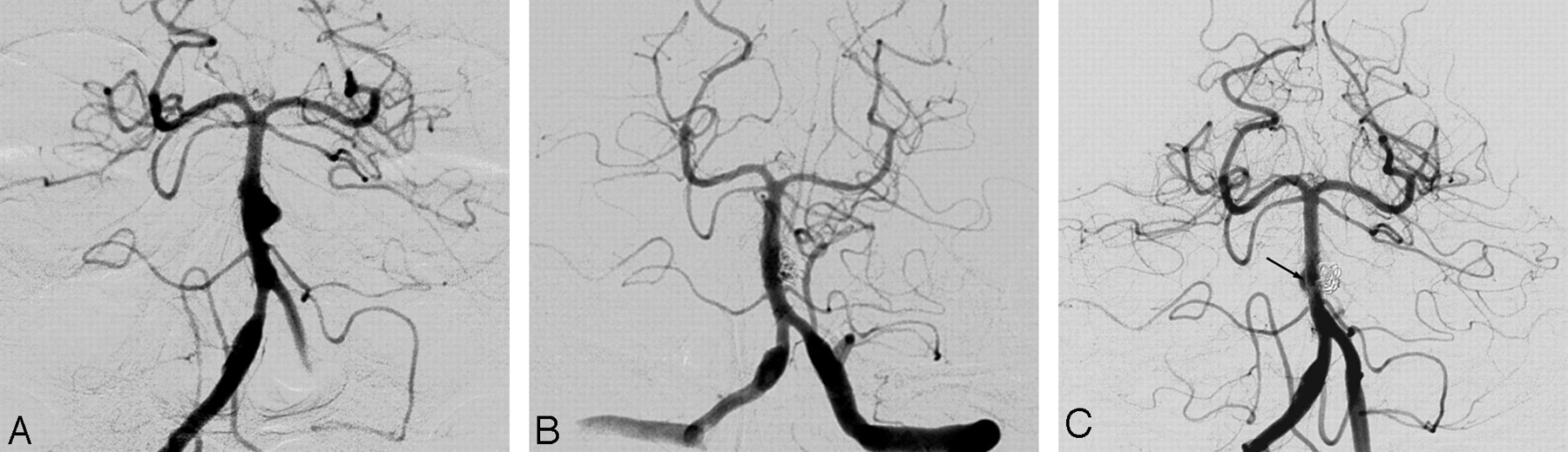
  • Fig 3.
    • Download figure
    • Open in new tab
    • Download powerpoint
    Fig 3.

    A 48-year-old woman presenting with a subarachnoid hemorrhage. A, Initial vertebral angiogram reveals focal asymmetric dilation of the basilar artery. B, Minimal contrast media filling remains in the coil-embolized pseudoaneurysmal sac outside the stents. C, A 9-month follow-up angiogram shows complete obliteration of the pseudoaneurysmal sac and minimal contrast media filling portion in the right side outside the stents. Note the gap (arrow) between the stents and contrast media filling portion.

PreviousNext
Back to top

In this issue

American Journal of Neuroradiology: 29 (10)
American Journal of Neuroradiology
Vol. 29, Issue 10
November 2008
  • Table of Contents
  • Index by author
Advertisement
Print
Download PDF
Email Article

Thank you for your interest in spreading the word on American Journal of Neuroradiology.

NOTE: We only request your email address so that the person you are recommending the page to knows that you wanted them to see it, and that it is not junk mail. We do not capture any email address.

Enter multiple addresses on separate lines or separate them with commas.
Management and Clinical Outcome of Acute Basilar Artery Dissection
(Your Name) has sent you a message from American Journal of Neuroradiology
(Your Name) thought you would like to see the American Journal of Neuroradiology web site.
CAPTCHA
This question is for testing whether or not you are a human visitor and to prevent automated spam submissions.
Cite this article
B.M. Kim, S.H. Suh, S.I. Park, Y.S. Shin, E.C. Chung, M.H. Lee, E.J. Kim, J.S. Koh, H.-s. Kang, H.G. Roh, Y.S. Won, P.-W. Chung, Y.-B. Kim, B.C. Suh
Management and Clinical Outcome of Acute Basilar Artery Dissection
American Journal of Neuroradiology Nov 2008, 29 (10) 1937-1941; DOI: 10.3174/ajnr.A1243

Citation Manager Formats

  • BibTeX
  • Bookends
  • EasyBib
  • EndNote (tagged)
  • EndNote 8 (xml)
  • Medlars
  • Mendeley
  • Papers
  • RefWorks Tagged
  • Ref Manager
  • RIS
  • Zotero
0 Responses
Respond to this article
Share
Bookmark this article
Management and Clinical Outcome of Acute Basilar Artery Dissection
B.M. Kim, S.H. Suh, S.I. Park, Y.S. Shin, E.C. Chung, M.H. Lee, E.J. Kim, J.S. Koh, H.-s. Kang, H.G. Roh, Y.S. Won, P.-W. Chung, Y.-B. Kim, B.C. Suh
American Journal of Neuroradiology Nov 2008, 29 (10) 1937-1941; DOI: 10.3174/ajnr.A1243
del.icio.us logo Twitter logo Facebook logo Mendeley logo
  • Tweet Widget
  • Facebook Like
  • Google Plus One
Purchase

Jump to section

  • Article
    • Abstract
    • Materials and Methods
    • Results
    • Discussion
    • Conclusion
    • Footnotes
    • References
  • Figures & Data
  • Supplemental
  • Info & Metrics
  • Responses
  • References
  • PDF

Related Articles

  • PubMed
  • Google Scholar

Cited By...

  • CTA Evaluation of Basilar Septations: An Entity Better Characterized as Aberrant Basilar Fenestrations
  • Stent alone treatment for dissections and dissecting aneurysms involving the basilar artery
  • Incidence and Risk Factors of Recurrence After Endovascular Treatment of Intracranial Vertebrobasilar Dissecting Aneurysms
  • Outcomes and prognostic factors of intracranial unruptured vertebrobasilar artery dissection
  • Reconstructive Endovascular Treatment of Intracranial Fusiform Aneurysms: A 1-Stage Procedure with Stent and Balloon
  • Endovascular Strategies for Vertebrobasilar Dissecting Aneurysms
  • Clinical and Angiographic Follow-Up of Stent-Only Therapy for Acute Intracranial Vertebrobasilar Dissecting Aneurysms
  • Crossref
  • Google Scholar

This article has not yet been cited by articles in journals that are participating in Crossref Cited-by Linking.

More in this TOC Section

  • Prophylactic Endovascular Treatments for CBS
  • Smoking & Aneurysm Outcomes: A Meta-Analysis
  • “Buddy-wire anchoring” technique for TVE
Show more NEUROINTERVENTION

Similar Articles

Advertisement

Indexed Content

  • Current Issue
  • Accepted Manuscripts
  • Article Preview
  • Past Issues
  • Editorials
  • Editor's Choice
  • Fellows' Journal Club
  • Letters to the Editor
  • Video Articles

Cases

  • Case Collection
  • Archive - Case of the Week
  • Archive - Case of the Month
  • Archive - Classic Case

More from AJNR

  • Trainee Corner
  • Imaging Protocols
  • MRI Safety Corner
  • Book Reviews

Multimedia

  • AJNR Podcasts
  • AJNR Scantastics

Resources

  • Turnaround Time
  • Submit a Manuscript
  • Submit a Video Article
  • Submit an eLetter to the Editor/Response
  • Manuscript Submission Guidelines
  • Statistical Tips
  • Fast Publishing of Accepted Manuscripts
  • Graphical Abstract Preparation
  • Imaging Protocol Submission
  • Evidence-Based Medicine Level Guide
  • Publishing Checklists
  • Author Policies
  • Become a Reviewer/Academy of Reviewers
  • News and Updates

About Us

  • About AJNR
  • Editorial Board
  • Editorial Board Alumni
  • Alerts
  • Permissions
  • Not an AJNR Subscriber? Join Now
  • Advertise with Us
  • Librarian Resources
  • Feedback
  • Terms and Conditions
  • AJNR Editorial Board Alumni

American Society of Neuroradiology

  • Not an ASNR Member? Join Now

© 2025 by the American Society of Neuroradiology All rights, including for text and data mining, AI training, and similar technologies, are reserved.
Print ISSN: 0195-6108 Online ISSN: 1936-959X

Powered by HighWire