
Abstract
SUMMARY: We report the imaging features of 4 cases of patients with papillary tumor of the pineal region, a tumor newly recognized in the 2007 World Health Organization “Classification of Tumors of the Nervous System.” In each case, the tumor was intrinsically hyperintense on T1-weighted images with a characteristic location in the posterior commissure or pineal region. The pathologic hallmarks of the tumor are discussed, including a possible explanation for the MR imaging characteristics in our cases.
Primary papillary tumors of the central nervous system and particularly the pineal region are rare. Papillary tumor of the pineal region (PTPR) is a recently described neoplasm that has been formally recognized in the 2007 World Health Organization (WHO) “Classification of Tumors of the Nervous System.”1 Histologic features and immunohistochemical staining distinguish this type of papillary tumor from other papillary-like tumors that occur in the region.2–7 It is postulated that the masses arise from the specialized ependymocytes of the subcommissural organ located in the lining of the posterior commissure.4 There are documented cases in the neuropathology and neurosurgery literature but limited descriptions of MR imaging features.7–9 Here, we present 4 patients who underwent MR imaging and surgical resection of tumors in the posterior commissure and pineal region where the pathologic diagnosis was a PTPR.
Case Reports
Case 1
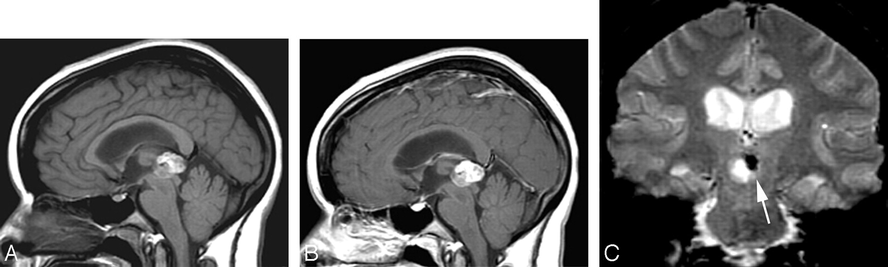
A 27-year-old woman presented with headache and hydrocephalus. MR imaging at 1.5T revealed a mass centered between the posterior commissure and pineal region, which compressed the tectum and aqueduct (Fig 1). The mass was hyperintense on noncontrast T1-weighted images and enhanced heterogeneously after administration of gadolinium.

Case 1. A 27-year-old woman with a headache and hydrocephalus. Sagittal noncontrast T1-weighted 1.5T image (A) demonstrates a heterogeneously hyperintense mass involving the posterior commissure and pineal region. Postcontrast sagittal T1-weighted image (B) demonstrates subtle enhancement of the mass. Gradient-echo series (C) highlights calcification within the leftward displaced pineal gland but not within the mass itself. An arrow depicts a normal displaced pineal gland. There are no additional susceptibility effects to indicate hemorrhage, melanin, or mineralization elsewhere within the brain.
The patient underwent ventriculostomy and surgical resection of the mass. Gross and histologic examinations of the mass did not display evidence of hemorrhage, calcification, melanin, keratin debris, or fat. Focal calcification was present in the adjacent pineal gland, which was displaced to the left. The pineal gland was normal on histologic examination.
Case 2
A 51-year-old woman presented with a headache and impairment of upward gaze. Imaging demonstrated a mass centered on the posterior commissure with mass effect on the pineal gland, aqueduct, and posterior third ventricle (Fig 2A). As in case 1, there was intrinsic hyperintensity on T1-weighted images throughout the mass on 1.5T unenhanced sequences. Small cystic areas were present in the mass, and the solid portions of the mass enhanced heterogeneously. Resection of the mass was accomplished by an infratentorial-supracerebellar approach, and immunohistochemical profiling confirmed the mass to be a PTPR.

Cases 2, 3, and 4. Three women (aged 51 years, 50 years, and 51 years, respectively) presented with headaches and/or Parinaud syndrome. All cases are depicted by noncontrast T1-weighted sequences from 1.5T units (case 2 [A], case 3 [B], case 4 [C]). All 3 cases demonstrated intrinsic T1 hyperintensity and were centered on the posterior commissure.
Case 3
A 50-year-old woman presented with a 3-year history of gait imbalance, fatigue, and short-term memory loss. She reported a recent episode of confusion associated with weakness of the right lower extremity and was taken to the emergency department, where a 1.5T MR imaging examination demonstrated a T1 hyperintense mass at the posterior commissure with associated hydrocephalus (Fig 2B). Physical examination revealed upward gaze paresis but was otherwise unremarkable. A right occipital ventriculostomy was placed, and gross total resection was performed via an infratentorial-supracerebellar approach.
Case 4
A 51-year-old woman presented after an episode of severe headache and memory loss. She denied other neurologic symptoms, and physical examination was unremarkable. MR imaging at 1.5T revealed a T1 hyperintense mass centered on the posterior commissure with associated hydrocephalus (Fig 2C). A right parietal ventriculostomy was placed, and a frontal craniotomy was performed for gross total resection through a transcallosal-transventricular approach.
Summary of Imaging Findings
One of the lesions was centered between the posterior commissure and pineal region (Fig 1), and 3 were centered on the posterior commissure (Fig 2). All 4 lesions were heterogeneously T1 hyperintense on precontrast images and demonstrated mild enhancement (Fig 1). All 4 lesions demonstrated cystic components, and 3 compressed the tectum and cerebral aqueduct causing hydrocephalus. There was no indication of chemical shift artifact or hemorrhagic products within the lesions or elsewhere in the brain. Gradient-echo sequences obtained in 2 patients did not demonstrate susceptibility effects to designate underlying blood products, melanin, or calcification.
Discussion
Neuropathologic descriptions of a PTPR have recently appeared, culminating in formal recognition as a distinct entity in the 2007 WHO classification.1 The name is a neuropathologic description of the tumor manifested by papillary features, rosettes, and pseudorosettes.7 Other tumors of the pineal region that may exhibit papillary features include choroid plexus papillomas and carcinomas, papillary ependymoma, and metastatic papillary carcinomas. Pineal parenchymal tumors, meningiomas, and germ cell tumors may rarely display papillary features.3,7 The histologic appearance of a PTPR is less papillary than choroid plexus papilloma and more epithelial than ependymoma, without the fibrillary background.7 The recent neuropathology literature clearly defines the morphologic features and immunophenotypic profiles (Fig 3) that distinguish PTPRs from the other papillary-type masses that occur in the pineal region.2,4–7

Neuropathologic examination. Case 3, a patient with PTPR. All cases showed similar histologic and immunophenotypic features, including mixed solid and papillary areas (A). Cells in the solid areas displayed clear, vacuolated cytoplasm and tended to rosette around blood vessels. In the papillary areas (B), the tumor cells exhibited clear-to-eosinophilic cytoplasm and assumed a pseudostratified columnar-to-cuboidal epithelial appearance. The interface with adjacent brain parenchyma was sharp and pushing. All tumors exhibited an immunophenotype characteristic of PTPR, with strong expression of cytokeratin (C) and S-100 protein (D) (A and B, hematoxylin and eosin; C and D, immunoperoxidase with hematoxylin counter stain; original magnifications: A, x40; B, x200; C, x400; D, x400).
One recent report described a series of papillary tumors that were initially diagnosed as choroid plexus papilloma, papillary ependymoma, or papillary pineal parenchymal tumor and were subsequently reclassified as a primary PTPR after re-examination and immunohistochemical staining.4 Therefore, it is likely that other previous reports of unusual posterior third ventricle choroid plexus papillomas, papillary pineal parenchymal tumors, or papillary ependymomas of the pineal region may actually represent early examples of a PTPR.
The proper differentiation of papillary tumors has management implications because treatment response of PTPRs is less well documented than other tumors in the pineal region. An understanding of the biologic behavior of a PTPR is evolving as more cases are documented, and local recurrence of a PTPR has been described.4,7,10
With regard to a possible explanation of imaging characteristics of a PTPR, electron microscopic findings support a secretory function of the PTPR similar to choroid plexus-like tumors and some ependymal tumors. PTPRs appear to have well-differentiated secretory functions that may predispose to the secretion of proteins, glycoproteins, or other T1-shortening products. The secretory inclusions of PTPRs have been noted to contain proteins and glycoproteins.7,11 Concentration of proteins in the small cystic spaces seen in these masses may explain the intrinsic hyperintensity on T1-weighted sequences.
Before suggesting the diagnosis of a PTPR, other masses in the pineal region that may demonstrate intrinsic T1 hyperintensity should be excluded. This can be accomplished with routine MR imaging techniques. For example, a fat-saturated T1-weighted sequence may exclude a teratoma, dermoid, or lipoma. A partially thrombosed aneurysm or venous malformation with subacute blood products can be excluded with MR or CT angiography and absence of pulsation artifacts in the phase-encoding direction on routine sequences. A hemorrhagic metastasis from a renal, thyroid, or melanoma primary may be excluded by the lack of susceptibility effects on gradient-echo sequences. Melanotic melanomas will also demonstrate susceptibility effects.
The limited MR imaging reports of PTPRs in the literature have described a heterogeneous enhancing mass centered in the pineal region.4,7,8 The intrinsic hyperintensity on noncontrast T1-weighted sequences depicted in our series has not been reported previously and may not have been noticed as a trend because of the nature of isolated case reports. On the contrary, 1 published case reportedly did not show T1 hyperintensity.9 Therefore, PTPRs may not universally demonstrate this appearance or may do so with varying degrees of intensity. However, the fact that all 4 known cases at our 2 institutions demonstrated intrinsic T1 hyperintensity suggests that this may be a common imaging appearance for a PTPR. It will be interesting to see if this observation holds true as the imaging findings of known cases are reviewed and the proportion of cases with intrinsic T1 hyperintensity is better defined.
Conclusion
Our findings suggest that intrinsic T1 hyperintensity may be a characteristic imaging appearance for a PTPR. In the imaging absence of fat, hemorrhage, melanin, or calcification in a mass of the posterior commissure or pineal region, the diagnosis of a PTPR may be suggested so that specific immunohistochemical studies can be performed for a definitive diagnosis.
References
- Received May 15, 2007.
- Accepted after revision July 2, 2007.
- Copyright © American Society of Neuroradiology


