Advertisement
  • Main menu
  • User menu
  • Search
  • English ▼
    • English
    • Afrikaans
    • Albanian
    • Amharic
    • Arabic
    • Armenian
    • Azerbaijani
    • Basque
    • Belarusian
    • Bengali
    • Bosnian
    • Bulgarian
    • Catalan
    • Cebuano
    • Chichewa
    • Chinese (Simplified)
    • Chinese (Traditional)
    • Corsican
    • Croatian
    • Czech
    • Danish
    • Dutch
    • Esperanto
    • Estonian
    • Filipino
    • Finnish
    • French
    • Frisian
    • Galician
    • Georgian
    • German
    • Greek
    • Gujarati
    • Haitian Creole
    • Hausa
    • Hawaiian
    • Hebrew
    • Hindi
    • Hmong
    • Hungarian
    • Icelandic
    • Igbo
    • Indonesian
    • Irish
    • Italian
    • Japanese
    • Javanese
    • Kannada
    • Kazakh
    • Khmer
    • Korean
    • Kurdish (Kurmanji)
    • Kyrgyz
    • Lao
    • Latin
    • Latvian
    • Lithuanian
    • Luxembourgish
    • Macedonian
    • Malagasy
    • Malay
    • Malayalam
    • Maltese
    • Maori
    • Marathi
    • Mongolian
    • Myanmar (Burmese)
    • Nepali
    • Norwegian
    • Pashto
    • Persian
    • Polish
    • Portuguese
    • Punjabi
    • Romanian
    • Russian
    • Samoan
    • Scottish Gaelic
    • Serbian
    • Sesotho
    • Shona
    • Sindhi
    • Sinhala
    • Slovak
    • Slovenian
    • Somali
    • Spanish
    • Sudanese
    • Swahili
    • Swedish
    • Tajik
    • Tamil
    • Telugu
    • Thai
    • Turkish
    • Ukrainian
    • Urdu
    • Uzbek
    • Vietnamese
    • Welsh
    • Xhosa
    • Yiddish
    • Yoruba
    • Zulu

Main menu

  • Home
  • Content
    • Current Issue
    • Accepted Manuscripts
    • Article Preview
    • Past Issue Archive
    • Video Articles
    • AJNR Case Collection
    • Case of the Week Archive
    • Case of the Month Archive
    • Classic Case Archive
  • Special Collections
    • AJNR Awards
    • Low-Field MRI
    • Alzheimer Disease
    • ASNR Foundation Special Collection
    • Photon-Counting CT
    • View All
  • Multimedia
    • AJNR Podcasts
    • AJNR SCANtastic
    • Trainee Corner
    • MRI Safety Corner
    • Imaging Protocols
  • For Authors
    • Submit a Manuscript
    • Submit a Video Article
    • Submit an eLetter to the Editor/Response
    • Manuscript Submission Guidelines
    • Statistical Tips
    • Fast Publishing of Accepted Manuscripts
    • Graphical Abstract Preparation
    • Imaging Protocol Submission
    • Author Policies
  • About Us
    • About AJNR
    • Editorial Board
    • Editorial Board Alumni
  • More
    • Become a Reviewer/Academy of Reviewers
    • Subscribers
    • Permissions
    • Alerts
    • Feedback
    • Advertisers
    • ASNR Home

User menu

  • Alerts
  • Log in

Search

  • Advanced search
American Journal of Neuroradiology

American Journal of Neuroradiology

ASHNR American Society of Functional Neuroradiology ASHNR American Society of Pediatric Neuroradiology ASSR
  • Alerts
  • Log in

Advanced Search

English ▼
  • English
  • Afrikaans
  • Albanian
  • Amharic
  • Arabic
  • Armenian
  • Azerbaijani
  • Basque
  • Belarusian
  • Bengali
  • Bosnian
  • Bulgarian
  • Catalan
  • Cebuano
  • Chichewa
  • Chinese (Simplified)
  • Chinese (Traditional)
  • Corsican
  • Croatian
  • Czech
  • Danish
  • Dutch
  • Esperanto
  • Estonian
  • Filipino
  • Finnish
  • French
  • Frisian
  • Galician
  • Georgian
  • German
  • Greek
  • Gujarati
  • Haitian Creole
  • Hausa
  • Hawaiian
  • Hebrew
  • Hindi
  • Hmong
  • Hungarian
  • Icelandic
  • Igbo
  • Indonesian
  • Irish
  • Italian
  • Japanese
  • Javanese
  • Kannada
  • Kazakh
  • Khmer
  • Korean
  • Kurdish (Kurmanji)
  • Kyrgyz
  • Lao
  • Latin
  • Latvian
  • Lithuanian
  • Luxembourgish
  • Macedonian
  • Malagasy
  • Malay
  • Malayalam
  • Maltese
  • Maori
  • Marathi
  • Mongolian
  • Myanmar (Burmese)
  • Nepali
  • Norwegian
  • Pashto
  • Persian
  • Polish
  • Portuguese
  • Punjabi
  • Romanian
  • Russian
  • Samoan
  • Scottish Gaelic
  • Serbian
  • Sesotho
  • Shona
  • Sindhi
  • Sinhala
  • Slovak
  • Slovenian
  • Somali
  • Spanish
  • Sudanese
  • Swahili
  • Swedish
  • Tajik
  • Tamil
  • Telugu
  • Thai
  • Turkish
  • Ukrainian
  • Urdu
  • Uzbek
  • Vietnamese
  • Welsh
  • Xhosa
  • Yiddish
  • Yoruba
  • Zulu
  • Home
  • Content
    • Current Issue
    • Accepted Manuscripts
    • Article Preview
    • Past Issue Archive
    • Video Articles
    • AJNR Case Collection
    • Case of the Week Archive
    • Case of the Month Archive
    • Classic Case Archive
  • Special Collections
    • AJNR Awards
    • Low-Field MRI
    • Alzheimer Disease
    • ASNR Foundation Special Collection
    • Photon-Counting CT
    • View All
  • Multimedia
    • AJNR Podcasts
    • AJNR SCANtastic
    • Trainee Corner
    • MRI Safety Corner
    • Imaging Protocols
  • For Authors
    • Submit a Manuscript
    • Submit a Video Article
    • Submit an eLetter to the Editor/Response
    • Manuscript Submission Guidelines
    • Statistical Tips
    • Fast Publishing of Accepted Manuscripts
    • Graphical Abstract Preparation
    • Imaging Protocol Submission
    • Author Policies
  • About Us
    • About AJNR
    • Editorial Board
    • Editorial Board Alumni
  • More
    • Become a Reviewer/Academy of Reviewers
    • Subscribers
    • Permissions
    • Alerts
    • Feedback
    • Advertisers
    • ASNR Home
  • Follow AJNR on Twitter
  • Visit AJNR on Facebook
  • Follow AJNR on Instagram
  • Join AJNR on LinkedIn
  • RSS Feeds

AJNR Awards, New Junior Editors, and more. Read the latest AJNR updates

OtherBrain

Subarachnoid Hemosiderosis and Superficial Cortical Hemosiderosis in Cerebral Amyloid Angiopathy

J. Linn, J. Herms, M. Dichgans, H. Brückmann, G. Fesl, T. Freilinger and M. Wiesmann
American Journal of Neuroradiology January 2008, 29 (1) 184-186; DOI: https://doi.org/10.3174/ajnr.A0783
J. Linn
  • Find this author on Google Scholar
  • Find this author on PubMed
  • Search for this author on this site
J. Herms
  • Find this author on Google Scholar
  • Find this author on PubMed
  • Search for this author on this site
M. Dichgans
  • Find this author on Google Scholar
  • Find this author on PubMed
  • Search for this author on this site
H. Brückmann
  • Find this author on Google Scholar
  • Find this author on PubMed
  • Search for this author on this site
G. Fesl
  • Find this author on Google Scholar
  • Find this author on PubMed
  • Search for this author on this site
T. Freilinger
  • Find this author on Google Scholar
  • Find this author on PubMed
  • Search for this author on this site
M. Wiesmann
  • Find this author on Google Scholar
  • Find this author on PubMed
  • Search for this author on this site
  • Article
  • Figures & Data
  • Info & Metrics
  • Responses
  • References
  • PDF
Loading

Article Figures & Data

Figures

  • Fig 1.
    • Download figure
    • Open in new tab
    • Download powerpoint
    Fig 1.

    Patient 1, a 70-year-old woman with global aphasia and severe apraxia. T2*-weighted image demonstrates superficial cortical hemosiderosis in the left parietal cortex (arrows).

  • Fig 2.
    • Download figure
    • Open in new tab
    • Download powerpoint
    Fig 2.

    Patient 2, a 69-year-old man with severe headache, visual disturbances, and nausea. A,B, T2*-weighted images depict left frontal subarachnoid hemosiderosis (dotted arrows) and left parietal superficial cortical hemosiderosis (arrows).

  • Fig 3.
    • Download figure
    • Open in new tab
    • Download powerpoint
    Fig 3.

    Patient 3, a 72-year-old woman with histopathologically proved CAA and a large hyperacute right frontal intracerebral macrohemorrhage (thick arrows). A,B, T2*-weighted images demonstrate the hyperacute lobar hemorrhage (thick arrows), subarachnoid hemosiderosis (dotted lines), and superficial cortical hemosiderosis (thin arrows).
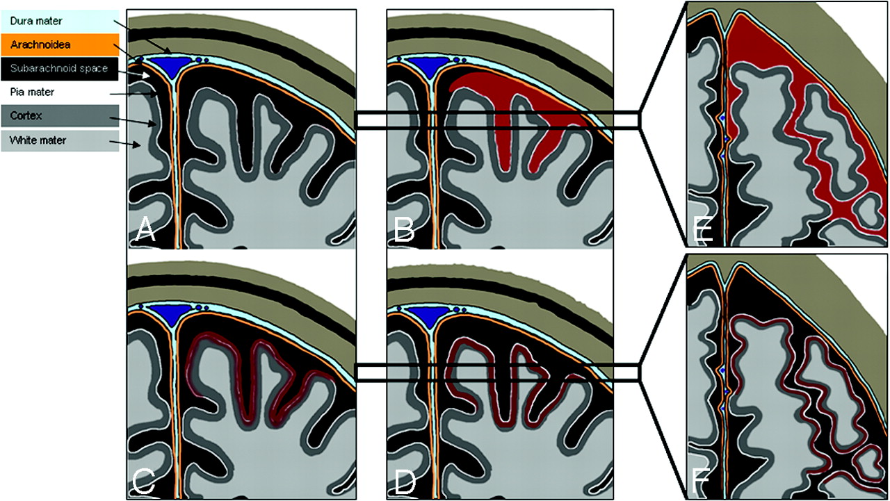
  • Fig 4.
    • Download figure
    • Open in new tab
    • Download powerpoint
    Fig 4.

    A–F, Schematic drawings illustrating subarachnoid hemosiderosis and superficial cortical hemosiderosis. A–D, Coronal schematic drawings, illustrating the time-dependent development of subarachnoid hemosiderosis and superficial cortical hemosiderosis (black, subarachnoid space; white line, pia mater; orange line, arachnoid layer; light blue, dura mater). E,F, Horizontal schematic drawings corresponding to the areas marked with black bars in B and C, demonstrating the visual appearance of subarachnoid hemosiderosis and superficial cortical hemosiderosis on axial sections. A, Normal appearance of the subarachnoid space. B,E, Subarachnoid hemorrhage (red) presenting as a linear signal intensity in the subarachnoid space (E). C, Residues of blood penetrating the pia mater and deposit in the superficial layers of the cerebral cortex. D,F, Superficial cortical hemosiderosis, defined as linear residues of blood in the superficial layers of the cerebral cortex (dark red). Superficial cortical hemosiderosis typically has a bilinear “tracklike” appearance on axial sections, caused by the signal intensity of the normal-appearing subarachnoid space in the middle, which is bordered bilaterally by linear deposits of hemosiderin in the superficial layers of the adjacent cortex (F).

PreviousNext
Back to top

In this issue

American Journal of Neuroradiology: 29 (1)
American Journal of Neuroradiology
Vol. 29, Issue 1
January 2008
  • Table of Contents
  • Index by author
Advertisement
Print
Download PDF
Email Article
Cite this article
0 Responses
Respond to this article
Share
Bookmark this article
Subarachnoid Hemosiderosis and Superficial Cortical Hemosiderosis in Cerebral Amyloid Angiopathy
J. Linn, J. Herms, M. Dichgans, H. Brückmann, G. Fesl, T. Freilinger, M. Wiesmann
American Journal of Neuroradiology Jan 2008, 29 (1) 184-186; DOI: 10.3174/ajnr.A0783
del.icio.us logo Twitter logo Facebook logo Mendeley logo
  • Tweet Widget
Purchase

Jump to section

  • Article
    • Abstract
    • Case Reports
    • Discussion
    • Acknowledgments
    • References
  • Figures & Data
  • Info & Metrics
  • Responses
  • References
  • PDF

Related Articles

  • No related articles found.
  • PubMed
  • Google Scholar

Cited By...

  • Cerebral amyloid angiopathy: an underdiagnosed cause of recurrent neurological symptoms
  • Spontaneous Intracranial Hypotension: Atypical Radiologic Appearances, Imaging Mimickers, and Clinical Look-Alikes
  • Cortical superficial siderosis and bleeding risk in cerebral amyloid angiopathy: A meta-analysis
  • Cortical superficial siderosis: A prospective observational cohort study
  • Innovative Magnetic Resonance Imaging Markers of Hereditary Cerebral Amyloid Angiopathy at 7 Tesla
  • Atrophy patterns in cerebral amyloid angiopathy with and without cortical superficial siderosis
  • Acute convexity subarachnoid haemorrhage and cortical superficial siderosis in probable cerebral amyloid angiopathy without lobar haemorrhage
  • Prevalence and Natural History of Superficial Siderosis: A Population-Based Study
  • Florbetapir imaging in cerebral amyloid angiopathy-related hemorrhages
  • Republished: Aneurysmal wall imaging in a case of cortical superficial siderosis and multiple unruptured aneurysms
  • Cortical superficial siderosis and first-ever cerebral hemorrhage in cerebral amyloid angiopathy
  • Early Magnetic Resonance Imaging and Cognitive Markers of Hereditary Cerebral Amyloid Angiopathy
  • Aneurysmal wall imaging in a case of cortical superficial siderosis and multiple unruptured aneurysms
  • Cortical superficial siderosis predicts early recurrent lobar hemorrhage
  • Cerebral Amyloid Angiopathy as an Etiology for Cortical Superficial Siderosis: An Unproven Hypothesis
  • Diagnostic Significance of Cortical Superficial Siderosis for Alzheimer Disease in Patients with Cognitive Impairment
  • Cerebral amyloid angiopathy with and without hemorrhage: Evidence for different disease phenotypes
  • Cortical superficial siderosis: A marker of vascular amyloid in patients with cognitive impairment
  • White Matter Perivascular Spaces Are Related to Cortical Superficial Siderosis in Cerebral Amyloid Angiopathy
  • Prevalence of cortical superficial siderosis in a memory clinic population
  • Cortical superficial siderosis and intracerebral hemorrhage risk in cerebral amyloid angiopathy
  • Prevalence and mechanisms of cortical superficial siderosis in cerebral amyloid angiopathy
  • Sporadic cerebral amyloid angiopathy revisited: recent insights into pathophysiology and clinical spectrum
  • In vivo amyloid imaging in cortical superficial siderosis
  • Prevalence of Superficial Siderosis in Patients with Cerebral Amyloid Angiopathy
  • Isolated Acute Nontraumatic Cortical Subarachnoid Hemorrhage
  • Prevalence of superficial siderosis in patients with cerebral amyloid angiopathy
  • Superficial Siderosis in Cerebral Amyloid Angiopathy
  • Neuroimaging in Superficial Siderosis: An In-Depth Look
  • Descriptive Analysis of the Boston Criteria Applied to a Dutch-Type Cerebral Amyloid Angiopathy Population
  • Superficial siderosis in the general population
  • Crossref
  • Google Scholar

This article has not yet been cited by articles in journals that are participating in Crossref Cited-by Linking.

More in this TOC Section

  • Evaluating the Effects of White Matter Multiple Sclerosis Lesions on the Volume Estimation of 6 Brain Tissue Segmentation Methods
  • Quiet PROPELLER MRI Techniques Match the Quality of Conventional PROPELLER Brain Imaging Techniques
  • Predictors of Reperfusion in Patients with Acute Ischemic Stroke
Show more Brain

Similar Articles

Advertisement

Indexed Content

  • Current Issue
  • Accepted Manuscripts
  • Article Preview
  • Past Issues
  • Editorials
  • Editor's Choice
  • Fellows' Journal Club
  • Letters to the Editor
  • Video Articles

Cases

  • Case Collection
  • Archive - Case of the Week
  • Archive - Case of the Month
  • Archive - Classic Case

More from AJNR

  • Trainee Corner
  • Imaging Protocols
  • MRI Safety Corner
  • Book Reviews

Multimedia

  • AJNR Podcasts
  • AJNR Scantastics

Resources

  • Turnaround Time
  • Submit a Manuscript
  • Submit a Video Article
  • Submit an eLetter to the Editor/Response
  • Manuscript Submission Guidelines
  • Statistical Tips
  • Fast Publishing of Accepted Manuscripts
  • Graphical Abstract Preparation
  • Imaging Protocol Submission
  • Evidence-Based Medicine Level Guide
  • Publishing Checklists
  • Author Policies
  • Become a Reviewer/Academy of Reviewers
  • News and Updates

About Us

  • About AJNR
  • Editorial Board
  • Editorial Board Alumni
  • Alerts
  • Permissions
  • Not an AJNR Subscriber? Join Now
  • Advertise with Us
  • Librarian Resources
  • Feedback
  • Terms and Conditions
  • AJNR Editorial Board Alumni

American Society of Neuroradiology

  • Not an ASNR Member? Join Now

© 2025 by the American Society of Neuroradiology All rights, including for text and data mining, AI training, and similar technologies, are reserved.
Print ISSN: 0195-6108 Online ISSN: 1936-959X

Powered by HighWire
Email this Article

Thank you for your interest in spreading the word on American Journal of Neuroradiology.

NOTE: We only request your email address so that the person you are recommending the page to knows that you wanted them to see it, and that it is not junk mail. We do not capture any email address.

Enter multiple addresses on separate lines or separate them with commas.
Subarachnoid Hemosiderosis and Superficial Cortical Hemosiderosis in Cerebral Amyloid Angiopathy
(Your Name) has sent you a message from American Journal of Neuroradiology
(Your Name) thought you would like to see the American Journal of Neuroradiology web site.
CAPTCHA
This question is for testing whether or not you are a human visitor and to prevent automated spam submissions.
Cite this article
J. Linn, J. Herms, M. Dichgans, H. Brückmann, G. Fesl, T. Freilinger, M. Wiesmann
Subarachnoid Hemosiderosis and Superficial Cortical Hemosiderosis in Cerebral Amyloid Angiopathy
American Journal of Neuroradiology Jan 2008, 29 (1) 184-186; DOI: 10.3174/ajnr.A0783

Citation Manager Formats

  • BibTeX
  • Bookends
  • EasyBib
  • EndNote (tagged)
  • EndNote 8 (xml)
  • Medlars
  • Mendeley
  • Papers
  • RefWorks Tagged
  • Ref Manager
  • RIS
  • Zotero

We use cookies on this site to enhance your user experience. By clicking any link on this page you are giving your consent for us to set cookies.