
Article Figures & Data
Figures
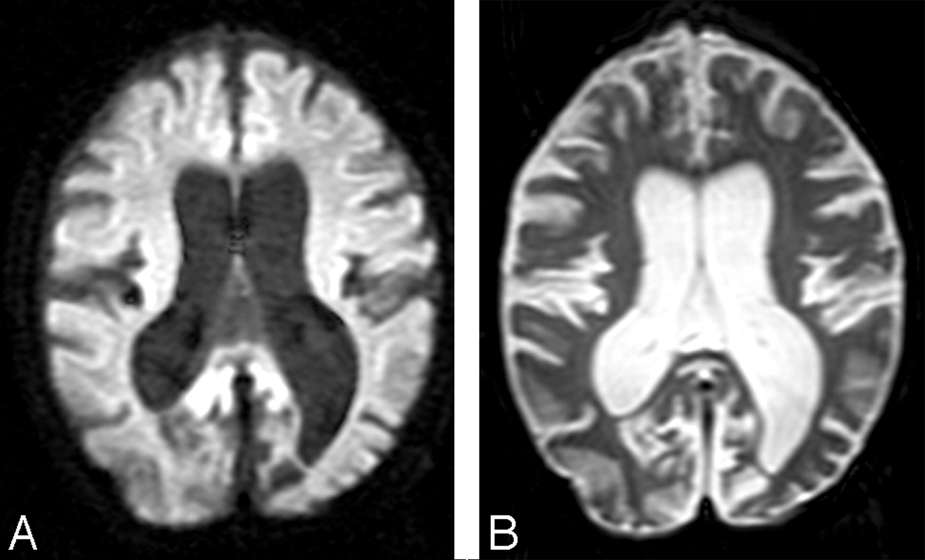
- Fig 1.
A, A diffusion-weighted image (b = 1000 s/mm2) and an image acquired without diffusion weighting (B) show enlarged sulci and dilated ventricles consistent with atrophic changes from a representative patient with LINCL.
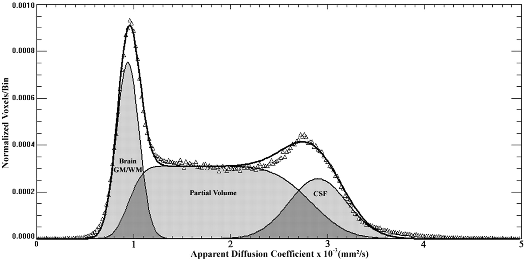
- Fig 2.
A whole-brain ADC histogram from a representative patient with LINCL shows the dual Gaussian and partial volume functions that were used to fit the whole brain, partial volume, and CSF compartments.
- Fig 3.
Voxels associated with specific ranges of ADC values provide visual confirmation that compartments identified as whole brain, partial volume, and CSF correspond with specific regions as shown from a representative section of a patient with LINCL. ADC ranges displayed are described below.
A, 1.0 ± 0.2 × 10⁻³ mm2/s.
B, 1.5 ± 0.2 × 10⁻³ mm2/s.
C, 2.0 ± 0.2 × 10⁻³ mm2/s.
D, 2.5 ± 0.2 × 10⁻³ mm2/s.
E, 3.0 ± 0.2 × 10⁻³ mm2/s.
F, 3.5 ± 0.2 × 10⁻³ mm2/s.
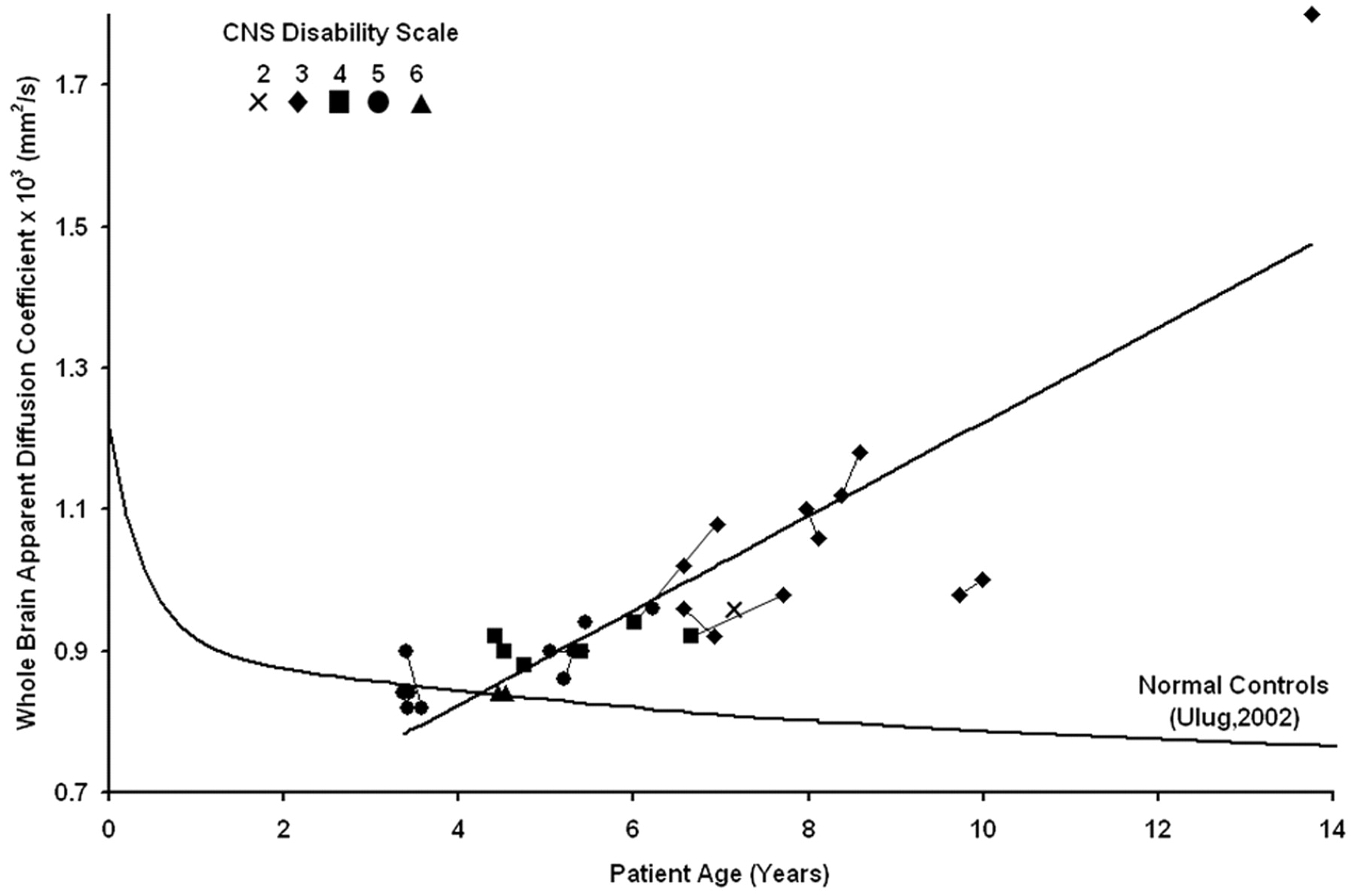
- Fig 4.
The whole-brain ADC value is plotted versus age for all of the patients showing an increasing trend over time. Serial studies are connected by lines, and the CNS disability scale is shown in the legend. Previously published whole-brain ADC values from age-matched control subjects are plotted for comparison.


Tables
Whole-brain ADC values for 18 patients with LINCL
Patient Age at Diagnosis, y Scan Age at Scan, y CNS Score ADC × 103, mm2/s BD-01 5.3 1 8.4 3 1.12 2 8.6 3 1.18 BD-02 4.8 3 9.7 3 0.98 4 10.0 3 1.00 BD-03 3.5 5 6.6 3 0.96 6 6.9 3 0.92 BD-04 4.5 7 13.8 3 1.80 BD-05 4.8 8 6.2 5 0.96 9 7.1 2 0.96 BD-06 4.4 10 8.0 3 1.10 11 8.1 3 1.06 BD-07 3.3 12 6.6 3 1.02 BD-08 4.7 13 6.0 4 0.94 14 7.0 3 1.08 BD-09 3.3 15 6.7 4 0.92 16 7.7 3 0.98 BD-10 3.8 17 5.0 5 0.90 18 5.4 5 0.90 BD-11 4.4 19 5.4 5 0.94 BD-12 2.8 20 4.4 4 0.92 21 4.5 4 0.90 BD-13 4.3 22 4.4 6 0.84 23 4.5 6 0.84 BD-14 1.9 24 3.4 5 0.90 25 3.6 5 0.82 BD-15 4.4 26 4.7 4 0.88 BD-16 4.8 27 5.2 5 0.86 28 5.3 5 0.90 BD-17 4.8 29 5.4 4 0.90 BD-18 2.8 30 3.4 5 0.84 31 3.4 5 0.82 32 3.4 5 0.84 Note:—ADC indicates apparent diffusion coefficient; LINCL, late infantile neuronal ceroid lipofuscinosis; CNS, central nervous system.
In this issue


Jump to section
Related Articles
Cited By...
- Global and Widespread Local White Matter Abnormalities in Juvenile Neuronal Ceroid Lipofuscinosis
- Volumetric Description of Brain Atrophy in Neuronal Ceroid Lipofuscinosis 2: Supratentorial Gray Matter Shows Uniform Disease Progression
- Gray Matter Growth Is Accompanied by Increasing Blood Flow and Decreasing Apparent Diffusion Coefficient during Childhood
- Differential Diagnosis of Normal Pressure Hydrocephalus by MRI Mean Diffusivity Histogram Analysis
- Assessment of Disease Severity in Late Infantile Neuronal Ceroid Lipofuscinosis Using Multiparametric MR Imaging
- Arterial spin labeling and altered cerebral blood flow patterns in the minimally conscious state