
Article Figures & Data
Figures
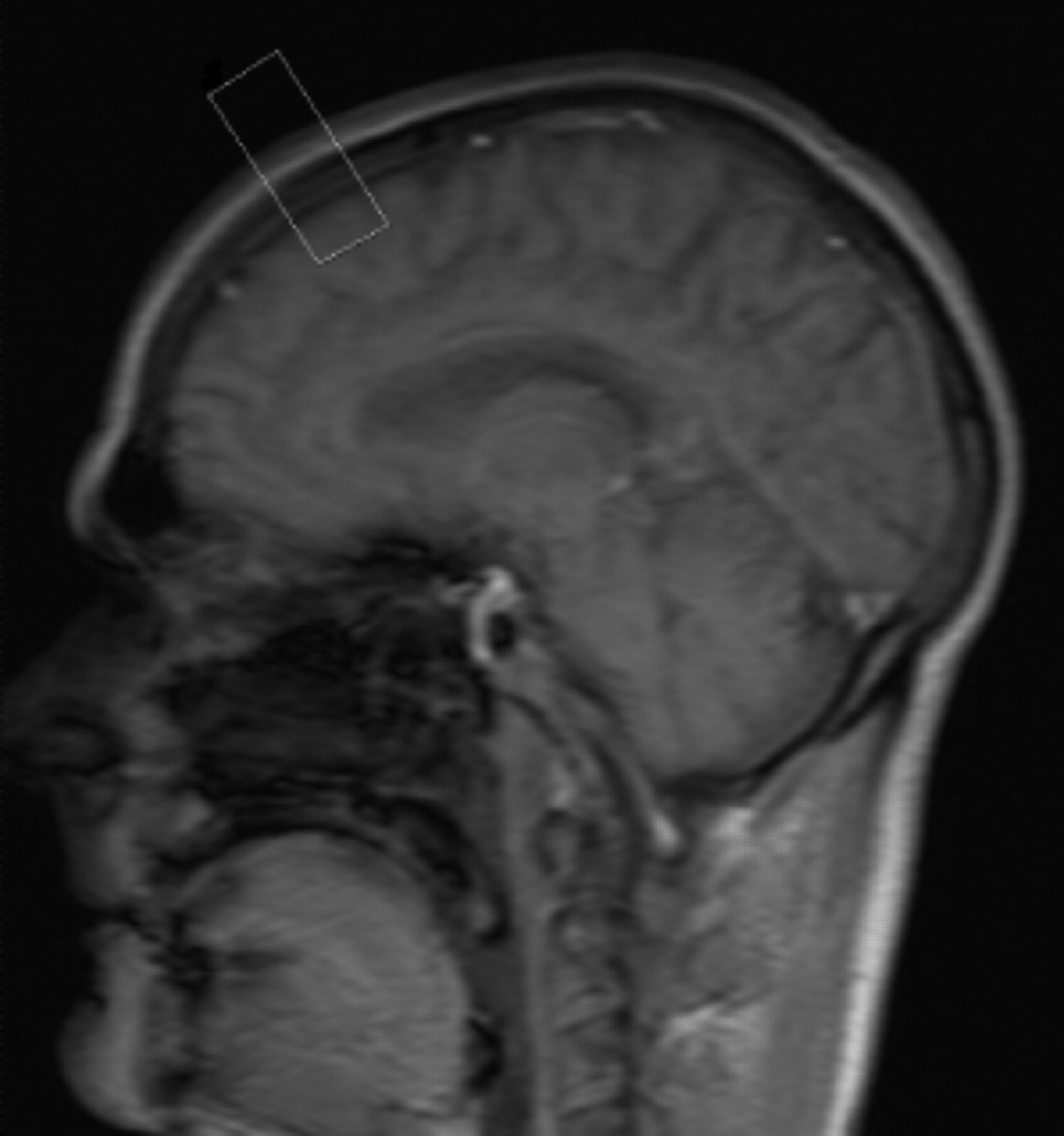
- Fig 1.
The navigator box is positioned midsagittal just posterior to the bregma.
- Fig 2.
A, Sagittal T1 FSE (TR, 429 ms; TE, 10 ms; acquisition time, 2 minutes) demonstrates considerable motion artifacts in our patients undergoing mechanical ventilation.
B, Sagittal T1 FSE (TR, 429 ms; TE, 10 ms) with the navigator pulse to gate the same as patient's head rocking motion. Although the acquisition time for this sequence was 3.5 minutes, it is still far superior in quality to the original FSE (A).
- Fig 3.
A, Axial T2 FSE (TR, 5890 ms; TE, 104 ms; echo train, 21; acquisition time, 2 minutes) performed without gating and resulting in a nondiagnostic image due to head rocking secondary to high-pressure ventilation.
B, Axial T2 FSE (TR, 6635 ms; TE, 89 ms; echo train, 21; acquisition time, 5 min). This is the same sequence and patient seen in A but with the addition of the navigator pulse.


Tables
Acquisition Time (min:sec) TR (ms) TE (ms) Echo Train Resolution IPAT Acceleration Factor Comments T1 SAG FSE 2:08 429 10 3 256 × 205 2 Nondiagnostic due to motion T2 Ax FSE 2:22 5890 104 21 256 × 205 2 Nondiagnostic due to motion T1 GRE, 3 planes 0:32 102 4.8 NA 256 × 156 2 Nondiagnostic due to motion T1 FSE with navigator, 3 planes 3:30–4:00 429 10 3 256 × 205 2 Diagnostic T2 Ax FSE 3:00 5890 104 21 256 × 205 2 Nondiagnostic T2 Ax FSE navigator 5:00 6635 89 21 256 × 205 2 Diagnostic Note:—IPAT indicates integrated parallel acquisition technique; SAG, sagittal; FSE, fast spin-echo; Ax, axial; GRE, gradient-echo; NA, not applicable. Both the TR and the acquisition time will be a variable of the frequency of patient motion. They should be adjusted inversely with concatenations to keep the effective TR short for T1 sequences.
Acquisition Time (min:sec) TR (ms) TE (ms) Echo Train Resolution IPAT AccelerationFactor Comments T1 SAG and Cor FSE 2:18 475 16 3 256 × 256 2 Nondiagnostic due to motion T2 Ax FSE 2:22 5890 104 21 256 × 205 2 Nondiagnostic due to motion T1 GRE, 3 planes 0:32 102 4.8 NA 256 × 156 2 Nondiagnostic due to motion T1 FSE with navigator, 3 planes 4:00–6:00 429 10 3 256 × 205 2 Diagnostic T2 Ax FSE 4:30 5890 104 21 256 × 205 2 Diagnostic Note:—IPAT indicates integrated parallel acquisition technique; SAG, sagittal; Cor, coronal; FSE, fast spin-echo; Ax, axial; GRE, gradient-echo; NA, not applicable. Both the TR and the acquisition time will be a variable of the frequency of patient motion. They should be adjusted inversely with concatenations to keep the effective TR short for T1 sequences.


