Advertisement

Main menu

  • Home
  • Content
    • Current Issue
    • Accepted Manuscripts
    • Article Preview
    • Past Issue Archive
    • Video Articles
    • AJNR Case Collection
    • Case of the Week Archive
    • Case of the Month Archive
    • Classic Case Archive
  • Special Collections
    • AJNR Awards
    • Low-Field MRI
    • Alzheimer Disease
    • ASNR Foundation Special Collection
    • Photon-Counting CT
    • View All
  • Multimedia
    • AJNR Podcasts
    • AJNR SCANtastic
    • Trainee Corner
    • MRI Safety Corner
    • Imaging Protocols
  • For Authors
    • Submit a Manuscript
    • Submit a Video Article
    • Submit an eLetter to the Editor/Response
    • Manuscript Submission Guidelines
    • Statistical Tips
    • Fast Publishing of Accepted Manuscripts
    • Graphical Abstract Preparation
    • Imaging Protocol Submission
    • Author Policies
  • About Us
    • About AJNR
    • Editorial Board
    • Editorial Board Alumni
  • More
    • Become a Reviewer/Academy of Reviewers
    • Subscribers
    • Permissions
    • Alerts
    • Feedback
    • Advertisers
    • ASNR Home

User menu

  • Alerts
  • Log in

Search

  • Advanced search
American Journal of Neuroradiology

American Journal of Neuroradiology

ASHNR American Society of Functional Neuroradiology ASHNR American Society of Pediatric Neuroradiology ASSR
  • Alerts
  • Log in

Advanced Search

  • Home
  • Content
    • Current Issue
    • Accepted Manuscripts
    • Article Preview
    • Past Issue Archive
    • Video Articles
    • AJNR Case Collection
    • Case of the Week Archive
    • Case of the Month Archive
    • Classic Case Archive
  • Special Collections
    • AJNR Awards
    • Low-Field MRI
    • Alzheimer Disease
    • ASNR Foundation Special Collection
    • Photon-Counting CT
    • View All
  • Multimedia
    • AJNR Podcasts
    • AJNR SCANtastic
    • Trainee Corner
    • MRI Safety Corner
    • Imaging Protocols
  • For Authors
    • Submit a Manuscript
    • Submit a Video Article
    • Submit an eLetter to the Editor/Response
    • Manuscript Submission Guidelines
    • Statistical Tips
    • Fast Publishing of Accepted Manuscripts
    • Graphical Abstract Preparation
    • Imaging Protocol Submission
    • Author Policies
  • About Us
    • About AJNR
    • Editorial Board
    • Editorial Board Alumni
  • More
    • Become a Reviewer/Academy of Reviewers
    • Subscribers
    • Permissions
    • Alerts
    • Feedback
    • Advertisers
    • ASNR Home
  • Follow AJNR on Twitter
  • Visit AJNR on Facebook
  • Follow AJNR on Instagram
  • Join AJNR on LinkedIn
  • RSS Feeds

AJNR Awards, New Junior Editors, and more. Read the latest AJNR updates

OtherBRAIN

Utility of Navigator-Prospective Acquisition Correction Technique (PACE) for Reducing Motion in Brain MR Imaging Studies

J.D. Barnwell, J.K. Smith and M. Castillo
American Journal of Neuroradiology April 2007, 28 (4) 790-791;
J.D. Barnwell
  • Find this author on Google Scholar
  • Find this author on PubMed
  • Search for this author on this site
J.K. Smith
  • Find this author on Google Scholar
  • Find this author on PubMed
  • Search for this author on this site
M. Castillo
  • Find this author on Google Scholar
  • Find this author on PubMed
  • Search for this author on this site
  • Article
  • Figures & Data
  • Info & Metrics
  • Responses
  • References
  • PDF
Loading

Abstract

SUMMARY: In this retrospective review, we demonstrate the utility of using a commercially available gated navigator sequence (prospective acquisition correction technique [PACE]) to reduce rhythmic breathing motion on brain MR images. For purposes of this report, we studied 2 sedated patients who had marked head rocking, one due to deep breathing and snoring and the other due to ventilator support. Motion degraded the routine images, which, despite a short acquisition time, were nondiagnostic. After application of PACE, a technique commonly used in abdominal studies, the brain images in both patients were judged to be of acceptable diagnostic quality. The use of PACE to diminish head motion is now routine at our institution when sedated or ventilated patients degrade images with involuntary head rocking.

Prospective acquisition correction technique (PACE) effectively removes breathing motion during abdominal MR imaging studies (including cholangiopancreatography) using a real-time gradient navigator pulse to trigger acquisition at a specified portion of the respiratory cycle.1,2 The real-time pulse is a 2-dimensional section that can produce a continuous representation of tissue location as it moves. Therefore, as the tissue fills more and less of the slab volume, the navigator image produces a continuous wave-like image representing the tissue location over time. It is then possible to identify a spot on the wave (the accept position) that represents a specific location of the tissue at one point during its movement and selectively acquire phase encoding steps only at that location. Thus, PACE may be used to remove extreme patient motion from MR imaging studies when the motion is 1-dimensional and rhythmic. At our institution, we have extended the routine use of PACE to diminish motion-related artifacts in brain MR imaging. To demonstrate the utility of this technique in brain MR imaging, we retrospectively reviewed 2 sedated patients, one snoring and the other on a ventilator, in whom conventional sequences were degraded. The application of PACE, however, yielded images of diagnostic quality.

Technique and Representative Cases

The study was performed under an Institutional Review Board exemption letter that allows for retrospective review of medical records, as long as no patient identifying information is retained or reported. Two patients, one under conscious sedation and the other being mechanically ventilated, underwent brain MR imaging to rule out pathologic processes. Studies were performed on a 1.5T system with a 12-channel head coil. In the first patient, motion was due to high-pressure ventilator support; in the second, head rocking was secondary to deep snoring. Both patients underwent basic T1 and T2 fast spin-echo (FSE), T1 gradient-echo, and navigator PACE T1 and T2 FSE sequences with integrated parallel acquisition technique (IPAT) on an Avanto device (Siemens, Erlangen, Germany).

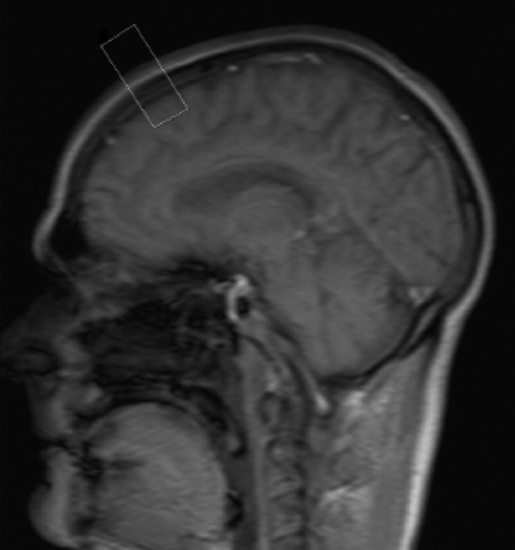
The first patient's sequences are reflected in Table 1. The patient presented with exaggerated breathing motion in response to high-pressure ventilator support. Initial FSE sequences were run with immobilization devices but were deemed nondiagnostic. We then switched to our standard noncooperative protocol that includes gradient-echo sequences, but fell short of producing a diagnostic result. At this point, we repeated the initial T1 and T2 FSE sequences with the addition of the navigator PACE correction pulse. The 2-dimensional PACE navigator pulse was positioned at a 90° angle to the sagittal suture just posterior to the bregma as demonstrated in Fig 1. The navigator's acceptance window was set to 32 ± 2 mm, TR to 150 ms, and the accept position was determined. Scans were run to completion by increasing scan time by 50%–150% depending on patient movement. Acquisition times for T1 and T2 sequences were 3.5 and 5 minutes, respectively, and resulted in images that were independently deemed by 2 neuroradiologists to be of diagnostic quality.

Fig 1.
  • Download figure
  • Open in new tab
  • Download powerpoint
Fig 1.

The navigator box is positioned midsagittal just posterior to the bregma.

View this table:
  • View inline
  • View popup
Table 1:

Sequences run on patient 1

The second patient (Table 2) was sedated for claustrophobia and discomfort before undergoing brain MR imaging to rule out cancerous processes. The initial scans demonstrated exaggerated head rocking secondary to snoring and deep breathing. Immobilization devices and noncooperative images failed to produce a diagnostic result, and the navigator sequence was once again used. By gating the patient's head movements, the scan time was increased by a factor of 3, but the resulting images were deemed diagnostic by 2 independent neuroradiologists.

View this table:
  • View inline
  • View popup
Table 2:

Sequences run on patient 2

Discussion

Image degradation is occasionally seen secondary to exaggerated respiratory patterns in ventilated and heavily sedated patients. In both of our patients, 1-dimensional, involuntary head movement occurred in the anterior-to-posterior dimension. Standard FSE sequences failed to produce diagnostic images despite external immobilization, as seen in Figs 2 and 3.

Fig 2.
  • Download figure
  • Open in new tab
  • Download powerpoint
Fig 2.

A, Sagittal T1 FSE (TR, 429 ms; TE, 10 ms; acquisition time, 2 minutes) demonstrates considerable motion artifacts in our patients undergoing mechanical ventilation.

B, Sagittal T1 FSE (TR, 429 ms; TE, 10 ms) with the navigator pulse to gate the same as patient's head rocking motion. Although the acquisition time for this sequence was 3.5 minutes, it is still far superior in quality to the original FSE (A).

Fig 3.
  • Download figure
  • Open in new tab
  • Download powerpoint
Fig 3.

A, Axial T2 FSE (TR, 5890 ms; TE, 104 ms; echo train, 21; acquisition time, 2 minutes) performed without gating and resulting in a nondiagnostic image due to head rocking secondary to high-pressure ventilation.

B, Axial T2 FSE (TR, 6635 ms; TE, 89 ms; echo train, 21; acquisition time, 5 min). This is the same sequence and patient seen in A but with the addition of the navigator pulse.

The free breather PACE navigator pulse is currently used in abdominal MR imaging and MR cholangiopancreatography to remove breathing motion, producing diagnostic images of higher resolution than those achievable with a standard breath-holding sequence.1,2 The navigator's low TR and flip angle produce real-time gating that is superior to other gating methods.1,2 Because of the image clarity associated with the navigator, fine structures, such as infant bile ducts, can be accurately evaluated.3 When the navigator is used in conjunction with IPAT, the acquisition times remain in a reasonable range.4,5 In addition, this technique can be adapted to brain MR imaging when 1-dimensional, rhythmic motion significantly degrades images.

We have previously studied this type of patient using gradient-echo and echo-planar imaging with short acquisition times in an attempt to reduce the motion artifacts. These sequences produce images of lower resolution and lower contrast and are less sensitive to smaller or low contrast abnormalities. The navigated pulse sequence, on the other hand, allows one to obtain standard resolution FSE images by acquiring phase encoding steps at the same location throughout the patient's motion. The result is better resolution and demonstration of anatomy as seen in Figs 2 and 3. The navigator's selectivity results in a significant increase in scan times, as seen in Tables 1 and 2. It should also be noted that this navigator sequence only gates 1-dimensional motion and is not useful for complex motion. It has been our experience that patients benefiting from this approach are those on high-pressure ventilators and obese patients who require sedation for imaging but experience deep breathing and snoring that rocks the head with inspiration and expiration. We now routinely use this technique for brain imaging in patients with simple 1-dimensional motion when immobilization fails to eliminate head motion. This sequence is not a substitute for those used in the imaging of nonsedated claustrophobic patients, because these take longer to acquire. They are, however, a simple way to considerably improve motion-degraded images, as we demonstrated in the patients described herein. In addition, the PACE navigator is easy to use and is commercially available. This technique should be particularly helpful to centers imaging patients undergoing mechanical ventilation and conscious sedation.

References

  1. ↵
    Klessen C, Asbach P, Kroencke TJ, et al. Magnetic resonance imaging of the upper abdomen using a free-breathing T2-weighted turbo spin-echo sequence with navigator-triggered prospective acquisition correction. Magn Reson Imaging 2005;21:576–82
  2. ↵
    Asbach P, Klessen C, Kroencke TJ, et al. Magnetic resonance cholangiopancreatography using a free-breathing T2-weighted turbo spin-echo sequence with navigator-triggered prospective acquisition correction. Magn Reson Imaging 2005;23:939–45
    CrossRefPubMed
  3. ↵
    Schaefer JF, Kirshner HJ, Lichy M, et al. Highly resolved free-breathing magnetic resonance cholangiopancreatography in the diagnostic workup of pancreaticobiliary diseases in infants and young children–initial experiences. J Pediatr Surg 2006;41:1645–51
    CrossRefPubMed
  4. ↵
    Asbach P, Klessen C, Stemmer A, et al. Respiratory-triggered MRCP applying parallel acquisition techniques. J Magn Reson Imaging 2006;24:1095–100
    CrossRefPubMed
  5. ↵
    Zech CJ, Herrmann KA, Huber A, et al. High-resolution MR-imaging of the liver with T2-weighted sequences using integrated parallel imaging: comparison of prospective motion correction and respiratory triggering. J Magn Reson Imaging 2004;20:443–50
    CrossRefPubMed
  • Received August 2, 2006.
  • Accepted after revision October 21, 2006.
  • Copyright © American Society of Neuroradiology
View Abstract
PreviousNext
Back to top

In this issue

American Journal of Neuroradiology: 28 (4)
American Journal of Neuroradiology
Vol. 28, Issue 4
April 2007
  • Table of Contents
  • Index by author
Advertisement
Print
Download PDF
Email Article

Thank you for your interest in spreading the word on American Journal of Neuroradiology.

NOTE: We only request your email address so that the person you are recommending the page to knows that you wanted them to see it, and that it is not junk mail. We do not capture any email address.

Enter multiple addresses on separate lines or separate them with commas.
Utility of Navigator-Prospective Acquisition Correction Technique (PACE) for Reducing Motion in Brain MR Imaging Studies
(Your Name) has sent you a message from American Journal of Neuroradiology
(Your Name) thought you would like to see the American Journal of Neuroradiology web site.
CAPTCHA
This question is for testing whether or not you are a human visitor and to prevent automated spam submissions.
Cite this article
J.D. Barnwell, J.K. Smith, M. Castillo
Utility of Navigator-Prospective Acquisition Correction Technique (PACE) for Reducing Motion in Brain MR Imaging Studies
American Journal of Neuroradiology Apr 2007, 28 (4) 790-791;

Citation Manager Formats

  • BibTeX
  • Bookends
  • EasyBib
  • EndNote (tagged)
  • EndNote 8 (xml)
  • Medlars
  • Mendeley
  • Papers
  • RefWorks Tagged
  • Ref Manager
  • RIS
  • Zotero
0 Responses
Respond to this article
Share
Bookmark this article
Utility of Navigator-Prospective Acquisition Correction Technique (PACE) for Reducing Motion in Brain MR Imaging Studies
J.D. Barnwell, J.K. Smith, M. Castillo
American Journal of Neuroradiology Apr 2007, 28 (4) 790-791;
del.icio.us logo Twitter logo Facebook logo Mendeley logo
  • Tweet Widget
  • Facebook Like
  • Google Plus One

Jump to section

  • Article
    • Abstract
    • Technique and Representative Cases
    • Discussion
    • References
  • Figures & Data
  • Info & Metrics
  • Responses
  • References
  • PDF

Related Articles

  • No related articles found.
  • PubMed
  • Google Scholar

Cited By...

  • Motion-Compensation Techniques in Neonatal and Fetal MR Imaging
  • Crossref
  • Google Scholar

This article has not yet been cited by articles in journals that are participating in Crossref Cited-by Linking.

More in this TOC Section

  • Multimodal CT Provides Improved Performance for Lacunar Infarct Detection
  • Optimal MRI Sequence for Identifying Occlusion Location in Acute Stroke: Which Value of Time-Resolved Contrast-Enhanced MRA?
  • Evaluating the Effects of White Matter Multiple Sclerosis Lesions on the Volume Estimation of 6 Brain Tissue Segmentation Methods
Show more BRAIN

Similar Articles

Advertisement

Indexed Content

  • Current Issue
  • Accepted Manuscripts
  • Article Preview
  • Past Issues
  • Editorials
  • Editor's Choice
  • Fellows' Journal Club
  • Letters to the Editor
  • Video Articles

Cases

  • Case Collection
  • Archive - Case of the Week
  • Archive - Case of the Month
  • Archive - Classic Case

More from AJNR

  • Trainee Corner
  • Imaging Protocols
  • MRI Safety Corner
  • Book Reviews

Multimedia

  • AJNR Podcasts
  • AJNR Scantastics

Resources

  • Turnaround Time
  • Submit a Manuscript
  • Submit a Video Article
  • Submit an eLetter to the Editor/Response
  • Manuscript Submission Guidelines
  • Statistical Tips
  • Fast Publishing of Accepted Manuscripts
  • Graphical Abstract Preparation
  • Imaging Protocol Submission
  • Evidence-Based Medicine Level Guide
  • Publishing Checklists
  • Author Policies
  • Become a Reviewer/Academy of Reviewers
  • News and Updates

About Us

  • About AJNR
  • Editorial Board
  • Editorial Board Alumni
  • Alerts
  • Permissions
  • Not an AJNR Subscriber? Join Now
  • Advertise with Us
  • Librarian Resources
  • Feedback
  • Terms and Conditions
  • AJNR Editorial Board Alumni

American Society of Neuroradiology

  • Not an ASNR Member? Join Now

© 2025 by the American Society of Neuroradiology All rights, including for text and data mining, AI training, and similar technologies, are reserved.
Print ISSN: 0195-6108 Online ISSN: 1936-959X

Powered by HighWire