Advertisement

Main menu

  • Home
  • Content
    • Current Issue
    • Accepted Manuscripts
    • Article Preview
    • Past Issue Archive
    • Video Articles
    • AJNR Case Collection
    • Case of the Week Archive
    • Case of the Month Archive
    • Classic Case Archive
  • Special Collections
    • Advancing NeuroMRI with High-Relaxivity Contrast Agents
    • AJNR Awards
    • Low-Field MRI
    • Alzheimer Disease
    • ASNR Foundation Special Collection
    • Photon-Counting CT
    • View All
  • Multimedia
    • AJNR Podcasts
    • AJNR SCANtastic
    • Trainee Corner
    • MRI Safety Corner
    • Imaging Protocols
  • For Authors
    • Submit a Manuscript
    • Submit a Video Article
    • Submit an eLetter to the Editor/Response
    • Manuscript Submission Guidelines
    • Statistical Tips
    • Fast Publishing of Accepted Manuscripts
    • Graphical Abstract Preparation
    • Imaging Protocol Submission
    • Author Policies
  • About Us
    • About AJNR
    • Editorial Board
    • Editorial Board Alumni
  • More
    • Become a Reviewer/Academy of Reviewers
    • Subscribers
    • Permissions
    • Alerts
    • Feedback
    • Advertisers
    • ASNR Home

User menu

  • Alerts
  • Log in

Search

  • Advanced search
American Journal of Neuroradiology

American Journal of Neuroradiology

ASHNR American Society of Functional Neuroradiology ASHNR American Society of Pediatric Neuroradiology ASSR
  • Alerts
  • Log in

Advanced Search

  • Home
  • Content
    • Current Issue
    • Accepted Manuscripts
    • Article Preview
    • Past Issue Archive
    • Video Articles
    • AJNR Case Collection
    • Case of the Week Archive
    • Case of the Month Archive
    • Classic Case Archive
  • Special Collections
    • Advancing NeuroMRI with High-Relaxivity Contrast Agents
    • AJNR Awards
    • Low-Field MRI
    • Alzheimer Disease
    • ASNR Foundation Special Collection
    • Photon-Counting CT
    • View All
  • Multimedia
    • AJNR Podcasts
    • AJNR SCANtastic
    • Trainee Corner
    • MRI Safety Corner
    • Imaging Protocols
  • For Authors
    • Submit a Manuscript
    • Submit a Video Article
    • Submit an eLetter to the Editor/Response
    • Manuscript Submission Guidelines
    • Statistical Tips
    • Fast Publishing of Accepted Manuscripts
    • Graphical Abstract Preparation
    • Imaging Protocol Submission
    • Author Policies
  • About Us
    • About AJNR
    • Editorial Board
    • Editorial Board Alumni
  • More
    • Become a Reviewer/Academy of Reviewers
    • Subscribers
    • Permissions
    • Alerts
    • Feedback
    • Advertisers
    • ASNR Home
  • Follow AJNR on Twitter
  • Visit AJNR on Facebook
  • Follow AJNR on Instagram
  • Join AJNR on LinkedIn
  • RSS Feeds

AJNR Awards, New Junior Editors, and more. Read the latest AJNR updates


Improved Turnaround Times | Median time to first decision: 12 days

Research ArticleSpine Imaging and Spine Image-Guided Interventions

Plasma-Mediated Radiofrequency Ablation Assisted Percutaneous Cement Injection for Treating Advanced Malignant Vertebral Compression Fractures

B.A. Georgy and W. Wong
American Journal of Neuroradiology April 2007, 28 (4) 700-705;
B.A. Georgy
  • Find this author on Google Scholar
  • Find this author on PubMed
  • Search for this author on this site
W. Wong
  • Find this author on Google Scholar
  • Find this author on PubMed
  • Search for this author on this site
  • Article
  • Figures & Data
  • Info & Metrics
  • Responses
  • References
  • PDF
Loading

Article Figures & Data

Figures

  • Tables
  • Fig 1.
    • Download figure
    • Open in new tab
    • Download powerpoint
    Fig 1.

    A, Axial CT scans show a large degree of cortical disruption in a 70-year-old man with metastatic colon cancer.

    B and C, The SpineWand device was inserted into the cannula to perform the tissue ablation; the clinician is capable of ablating tissue in a superior or inferior lateral direction.

    D, In this patient, 2 10-mm Kyphon balloons were inserted to obtain hemostasis while preparing the bone cement. A myelogram was performed to clearly delineate the posterior cortical margin.

    E, The axial CT scans collected immediately after the procedure showed that cement was cleanly deposited in the ablated tumor void with no posterior extraosseous extension of cement.
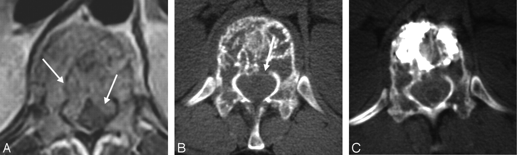
  • Fig 2.
    • Download figure
    • Open in new tab
    • Download powerpoint
    Fig 2.

    A, An axial T1-weighted image through the T11 level showing prominent epidural involvement in a 34-year-old woman with multiple myeloma.

    B, The axial CT image showed associated cortical disruption.

    C, The axial CT images obtained after the procedure, showing adequate cement filling with no epidural extension. Note the tight thecal sac as evident by using myelographic contrast agent.

  • Fig 3.
    • Download figure
    • Open in new tab
    • Download powerpoint
    Fig 3.

    A, The preprocedure axial CT examination showing almost complete absence of the posterior cortex at the L1 level in a 76-year-old man with metastatic hepatoma to the spine.

    B, The postprocedural CT axial images showed well-bounded deposition of cement in the anterior part of the vertebral body with no extension into the compromised posterior aspect.

Tables

  • Figures
  • Patient demographics

    PatientAgeSexTreated Level(s)Pathologic ConditionsAnatomy of Metastatic LesionOther ProceduresPain Score (Before–After)Postprocedure CT
    Group 1
        1*75FL2 (Vp)Plasmacytoma, history of breast cancer, lupus erythematosus, chronic steroid usePosterior cortical defectVp, T6, T8, T9, T10 (benign compression fractures)9/10–2/10Cement confined to the lytic lesions
        2*71ML1 (Vp)Lung cancerEpidural extension and posterior cortical defect9/10–3/10Cement confined to the lytic lesions
        327MT11 (Vp)Undifferentiated testicular malignancyEpidural extensionVp, T12, L18/10–5/10, deceasedNo postprocedure CT; no EE on plain film
        4*82ML5 (Vp)Lung squamous cell carcinomaPosterior cortical defect and epidural extension; tumor extension into right neural foramen9/10–5/10Cement on periphery of the metastatic lesion
        552FT9 (Vp)Breast cancerAnterior cortical defectVp, T5, T7, T108/10–5/10Cement around the lytic lesions
        6*70ML2 (Kp)Colon cancerPosterior cortical defect9/10–5/10, deceasedCement confined to the lytic lesions
        7*34FT12 (Kp)Multiple myelomaPosterior cortical defect and epidural extensionKp, T7,T8, T11Cannot tell differenceCement confined to the lytic lesions
        858FT12 (Kp)Breast cancerPosterior cortical disruption and epidural extensionCervical, sacral, and bilateral hip metastases9/10–5/10, deceasedCement confined to the lytic lesions
        980FL4 (Kp)Stomach & breast cancerEpidural extension and complete absence of posterior cortex on CTKp, L2; Vp, L3; followed by Kp, T7, T8, T117/10–3/10Cement confined to the lytic lesions
        1076ML1 (Kp)HepatomaPosterior cortical disruptionKp, L310/10–5/10, deceasedCement confirmed in normal bone anterior to the lytic lesion
        11*72ML1 (Kp)Multiple myelomaEpidural extension; cortical defectVp, L26/10–2/10Cement confined to the lytic lesions
    Group 2
        1256ML1 (Vp)Renal cell metastasisParavertebral extension and lateral cortical disruption2 stage coblation for vertebral & paravertebral components; arterial embolization8/10–2/10Cement confined to the lytic lesions; EE through anterolateral cortex
        1336FL2 (Kp)Cervical cancerAnterior and posterior cortical defect; paraspinal mass extended into left L2–L3 NRBRecurrent lesion after radiation; left L2–L3 NRB9/10–9/10Cement confined to the lytic lesions; some anterior EE
        1466FL3 (Kp right; Vp left)Lung cancerPosterior cortical defect; epidural extension; extension into right neural foramenRight NRB10/10–0/10Cement confined to the lytic lesions; EE anteriorly and into right neural foramen
        1568ML5 (Kp)Urethral cancerEpidural extension and posterior cortical defect; extension into the right pedicle and neural foramenS1 sacroplasty, NRB9/10–7/10Cement on periphery of lesion, extending into the right pedicle
    • Note:—EE indicates extraosseous extension of cement; Group 1, bone cement confined to lytic lesions and confined to vertebral body; Group 2, bone cement with extraosseous extension outside vertebral body; Vp, vertebroplasty; Kp, kyphoplasty; NRB, nerve root block.

    • * Myelogram performed.

PreviousNext
Back to top

In this issue

American Journal of Neuroradiology: 28 (4)
American Journal of Neuroradiology
Vol. 28, Issue 4
April 2007
  • Table of Contents
  • Index by author
Advertisement
Print
Download PDF
Email Article

Thank you for your interest in spreading the word on American Journal of Neuroradiology.

NOTE: We only request your email address so that the person you are recommending the page to knows that you wanted them to see it, and that it is not junk mail. We do not capture any email address.

Enter multiple addresses on separate lines or separate them with commas.
Plasma-Mediated Radiofrequency Ablation Assisted Percutaneous Cement Injection for Treating Advanced Malignant Vertebral Compression Fractures
(Your Name) has sent you a message from American Journal of Neuroradiology
(Your Name) thought you would like to see the American Journal of Neuroradiology web site.
CAPTCHA
This question is for testing whether or not you are a human visitor and to prevent automated spam submissions.
Cite this article
B.A. Georgy, W. Wong
Plasma-Mediated Radiofrequency Ablation Assisted Percutaneous Cement Injection for Treating Advanced Malignant Vertebral Compression Fractures
American Journal of Neuroradiology Apr 2007, 28 (4) 700-705;

Citation Manager Formats

  • BibTeX
  • Bookends
  • EasyBib
  • EndNote (tagged)
  • EndNote 8 (xml)
  • Medlars
  • Mendeley
  • Papers
  • RefWorks Tagged
  • Ref Manager
  • RIS
  • Zotero
0 Responses
Respond to this article
Share
Bookmark this article
Plasma-Mediated Radiofrequency Ablation Assisted Percutaneous Cement Injection for Treating Advanced Malignant Vertebral Compression Fractures
B.A. Georgy, W. Wong
American Journal of Neuroradiology Apr 2007, 28 (4) 700-705;
del.icio.us logo Twitter logo Facebook logo Mendeley logo
  • Tweet Widget
  • Facebook Like
  • Google Plus One

Jump to section

  • Article
    • Abstract
    • Materials and Methods
    • Results
    • Discussion
    • Conclusion
    • Acknowledgments
    • Footnotes
    • References
  • Figures & Data
  • Info & Metrics
  • Responses
  • References
  • PDF

Related Articles

  • No related articles found.
  • PubMed
  • Google Scholar

Cited By...

  • Vertebral Augmentation for Neoplastic Lesions with Posterior Wall Erosion and Epidural Mass
  • Percutaneous plasma mediated radiofrequency ablation of spinal osteoid osteomas
  • The surgical management of metastatic epidural compression of the spinal cord
  • Bone Cement Deposition Patterns with Plasma-Mediated Radio-Frequency Ablation and Cement Augmentation for Advanced Metastatic Spine Lesions
  • Crossref
  • Google Scholar

This article has not yet been cited by articles in journals that are participating in Crossref Cited-by Linking.

More in this TOC Section

  • Dynamic CT Myelography: Patient Positioning
  • Evaluation of SIH MR Scoring Systems in Normals
  • Management Outcomes For VO Spine Biopsy
Show more SPINE IMAGING AND SPINE IMAGE-GUIDED INTERVENTIONS

Similar Articles

Advertisement

Indexed Content

  • Current Issue
  • Accepted Manuscripts
  • Article Preview
  • Past Issues
  • Editorials
  • Editor's Choice
  • Fellows' Journal Club
  • Letters to the Editor
  • Video Articles

Cases

  • Case Collection
  • Archive - Case of the Week
  • Archive - Case of the Month
  • Archive - Classic Case

More from AJNR

  • Trainee Corner
  • Imaging Protocols
  • MRI Safety Corner
  • Book Reviews

Multimedia

  • AJNR Podcasts
  • AJNR Scantastics

Resources

  • Turnaround Time
  • Submit a Manuscript
  • Submit a Video Article
  • Submit an eLetter to the Editor/Response
  • Manuscript Submission Guidelines
  • Statistical Tips
  • Fast Publishing of Accepted Manuscripts
  • Graphical Abstract Preparation
  • Imaging Protocol Submission
  • Evidence-Based Medicine Level Guide
  • Publishing Checklists
  • Author Policies
  • Become a Reviewer/Academy of Reviewers
  • News and Updates

About Us

  • About AJNR
  • Editorial Board
  • Editorial Board Alumni
  • Alerts
  • Permissions
  • Not an AJNR Subscriber? Join Now
  • Advertise with Us
  • Librarian Resources
  • Feedback
  • Terms and Conditions
  • AJNR Editorial Board Alumni

American Society of Neuroradiology

  • Not an ASNR Member? Join Now

© 2025 by the American Society of Neuroradiology All rights, including for text and data mining, AI training, and similar technologies, are reserved.
Print ISSN: 0195-6108 Online ISSN: 1936-959X

Powered by HighWire