
Article Figures & Data
Figures
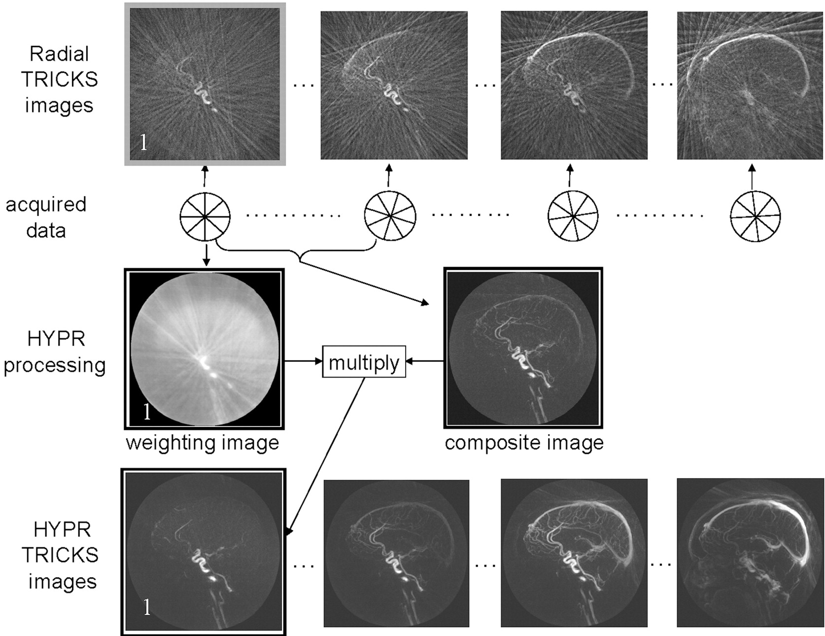
- Fig 1.
Schematic diagram of the HYPR reconstruction algorithm. Data were acquired by using undersampled radial trajectories. Images in the top row were reconstructed by using filtered backprojection. Images in the bottom row were reconstructed by using the HYPR method. All processing was performed on the source images, and the resulting maximum-intensity-projection images are shown. The low attenuation of vessels (high sparsity) in the source images makes them amenable to HYPR processing. For the HYPR processing, note that the sagittal sinus that appears in the composite image is suppressed following multiplication by the weighting image for frame 1.
- Fig 2.
Comparison of in-plane resolution and artifacts for Cartesian TRICKS (0.94 × 1.5 mm) (A, B) and HYPR TRICKS (0.47 × 0.47 mm) (C, D). Arrows in the Cartesian TRICKS images show the ghosting and black band artifacts resulting from fluctuations in signal intensity occurring during acquisition of data for a single frame, caused by changes in the concentration of contrast material. More rapid and larger fluctuations lead to more severe artifacts.
- Fig 3.
Comparison of Cartesian TRICKS timeframes (the 3 images with the black frames) and HYPR TRICKS timeframes from a patient with AVM. The frame update times are 0.4 seconds for HYPR TRICKS and 2.4 seconds for Cartesian TRICKS.


Tables
Acceleration comparison of HYPR TRICKS with different 3D imaging techniques
3D Hybrid 3D CART 512-TRICKS CLTRICKS HYPRTRICKS Npa 512 × π/2 512 512 120b 10 Nz Nz Nz Nz/3 2 × Nz / 4 Nz/3 fa 80 × 3 = 240 51 × 3 = 153 51 29c 1 Note:—Nz indicates the number of sections; fa, the acceleration factor of HYPR TRICKS versus the corresponding technique, which is ratio of Np × Nz from the 2 techniques; 3D Hybrid, 3D radial in-plane and Cartesian through-plane technique; 3D CART, 3D Cartesian technique full Nyquist sampling; 512-TRICKS, 3D Cartesian TRICKS with the same spatial resolution as HYPR TRICKS without partial Fourier, rectangular FOV, etc; CLTRICKS, clinically used 3D Cartesian TRICKS that is described in the article. Parameters chosen in the article were based on the clinical protocol.
a Where Np is the number of encodings per timeframe in the kx-ky plane. For radial imaging, it is the number of projections per timeframe, and for the Cartesian imaging, it is the number of phased-encoding lines per timeframe.
b Partial Fourier and rectangular FOV techniques were applied, given the in-plane pixel size 6 times larger than that achieved by the HYPR TRICKS.
c This factor was calculated on the basis of the ratio of the actual frame update time (2.4 seconds versus 0.26 seconds) together with the ratio of the voxel size (2.8 mm3 versus 0.88 mm3).


