
Article Figures & Data
Figures
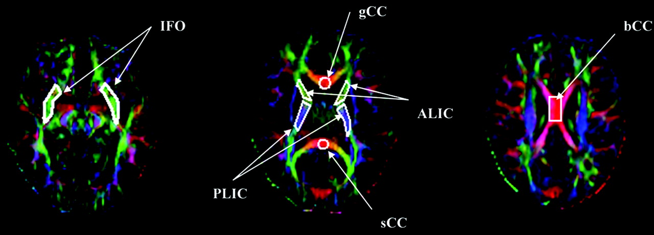
- Fig 1.
Color-coded FA map from a control participant. ROI include gCC, sCC, bCC, ALIC, PLIC, and IFO.
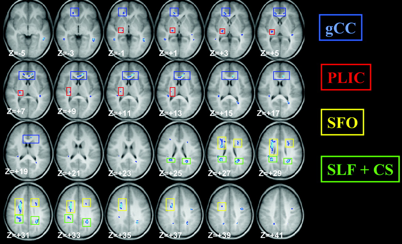
- Fig 2.
Z-score map for voxel-based FA group comparison. TBI versus control, nominal z = 6, cluster size = 20, corrected P < .001. Voxels in light blue color represent higher statistical significance that those in dark blue color. The position in z direction is also provided at the lower left corner in each section (in MNI space).
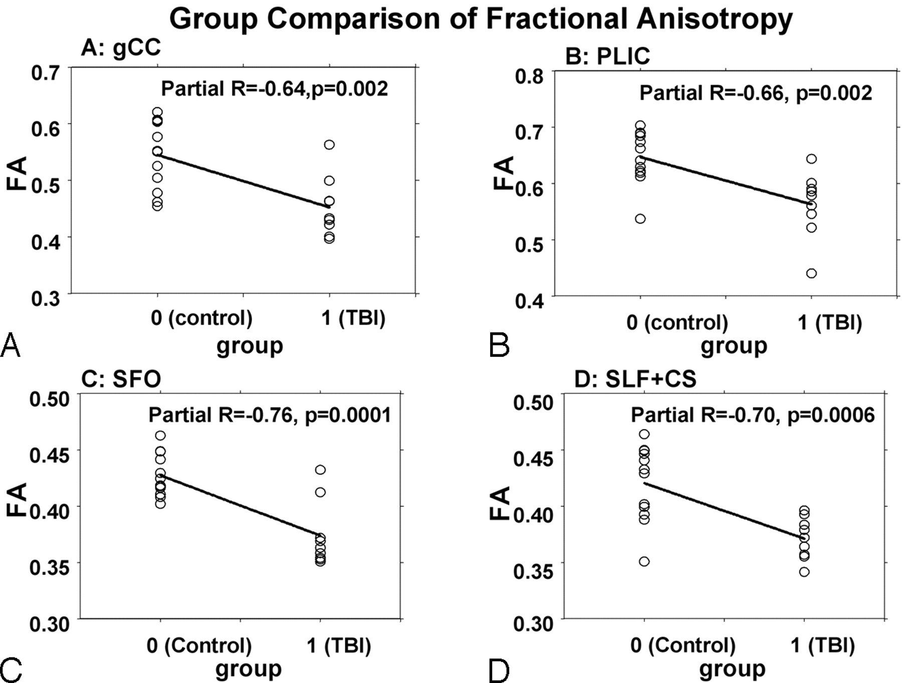
- Fig 3.
Group comparison of FA values between TBI and control participant, with sex difference controlled for, in (A) gCC; (B) right posterior limb of IC; (C) Fronto-occipital fasciculus; and (D) superior longitudinal fasciculus and other CS regions.
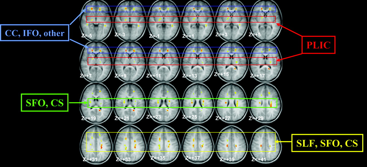
- Fig 4.
Composite z-score map demonstrating correlation between FA values and GCS scores in TBI group. The z value is calculated based on the R value from the corresponding pixel. Norminal z = 6, cluster size = 20, corrected P < .01. The voxels with significant correlation are coded with color and overlaid on an anatomic image. Some of these voxels are contiguous in space and represent specific WM structures. Four such groups of voxels are marked with rectangles in different colors. These 4 groups (clusters) are (blue) CC and IFO; (red) PLIC; (green) SFO and CS; and (yellow) SLF, SFO, and CS.
- Fig 5.
Linear regression of mean FA values adjusted for sex in various focal WM areas vs GCS score. Sex factor is used as covariate. Four subplots A through D correspond with 4 contiguous brain regions as marked with different colored rectangles in Fig 4.


Tables
Mann-Whitney U test results of ROI-based group comparison
ROIs FA in TBI (n = 9) FA in Control (n = 12) P sCC 0.69 ± 0.11 0.77 ± 0.06 .06 gCC 0.73 ± 0.04 0.77 ± 0.04 <.05* bCC 0.69 ± 0.12 0.75 ± 0.05 .22 ALIC 0.53 ± 0.03 0.58 ± 0.04 <.05* PLIC 0.58 ± 0.02 0.63 ± 0.04 <.01* SLF 0.49 ± 0.05 0.50 ± 0.05 .42 IFO 0.45 ± 0.06 0.45 ± 0.05 .75 Note:—ROI indicates regions of interest; FA, fractional anisotropy; TBI, traumatic brain injury; sCC, splenium of corpus callosum; gCC, genu of corpus callosum; bCC, body of corpus callosum; ALIC, anterior limb of interior capsule; PLIC, posterior limb of interior capsule; SLF, superior longitudinal fasciculus; IFO, inferior fronto-occipital fasciculus.
* Statistically significant at P < 0.05.
In this issue


Jump to section
Related Articles
Cited By...
- White Matter Disruption in Pediatric Traumatic Brain Injury: Results from ENIGMA Pediatric msTBI
- Diffusion Measures Indicate Fight Exposure-Related Damage to Cerebral White Matter in Boxers and Mixed Martial Arts Fighters
- Long-Term White Matter Changes after Severe Traumatic Brain Injury: A 5-Year Prospective Cohort
- A Decade of DTI in Traumatic Brain Injury: 10 Years and 100 Articles Later
- Aberrant Diffusion and Geometric Properties in the Left Arcuate Fasciculus of Developmentally Delayed Children: A Diffusion Tensor Imaging Study
- Voxel-Based Analysis of Diffusion Tensor Imaging in Mild Traumatic Brain Injury in Adolescents
- Anisotropic Diffusion Properties in Infants with Hydrocephalus: A Diffusion Tensor Imaging Study
- Evaluation of Delayed Neuronal and Axonal Damage Secondary to Moderate and Severe Traumatic Brain Injury Using Quantitative MR Imaging Techniques