Advertisement
  • Main menu
  • User menu
  • Search
  • English ▼
    • English
    • Afrikaans
    • Albanian
    • Amharic
    • Arabic
    • Armenian
    • Azerbaijani
    • Basque
    • Belarusian
    • Bengali
    • Bosnian
    • Bulgarian
    • Catalan
    • Cebuano
    • Chichewa
    • Chinese (Simplified)
    • Chinese (Traditional)
    • Corsican
    • Croatian
    • Czech
    • Danish
    • Dutch
    • Esperanto
    • Estonian
    • Filipino
    • Finnish
    • French
    • Frisian
    • Galician
    • Georgian
    • German
    • Greek
    • Gujarati
    • Haitian Creole
    • Hausa
    • Hawaiian
    • Hebrew
    • Hindi
    • Hmong
    • Hungarian
    • Icelandic
    • Igbo
    • Indonesian
    • Irish
    • Italian
    • Japanese
    • Javanese
    • Kannada
    • Kazakh
    • Khmer
    • Korean
    • Kurdish (Kurmanji)
    • Kyrgyz
    • Lao
    • Latin
    • Latvian
    • Lithuanian
    • Luxembourgish
    • Macedonian
    • Malagasy
    • Malay
    • Malayalam
    • Maltese
    • Maori
    • Marathi
    • Mongolian
    • Myanmar (Burmese)
    • Nepali
    • Norwegian
    • Pashto
    • Persian
    • Polish
    • Portuguese
    • Punjabi
    • Romanian
    • Russian
    • Samoan
    • Scottish Gaelic
    • Serbian
    • Sesotho
    • Shona
    • Sindhi
    • Sinhala
    • Slovak
    • Slovenian
    • Somali
    • Spanish
    • Sudanese
    • Swahili
    • Swedish
    • Tajik
    • Tamil
    • Telugu
    • Thai
    • Turkish
    • Ukrainian
    • Urdu
    • Uzbek
    • Vietnamese
    • Welsh
    • Xhosa
    • Yiddish
    • Yoruba
    • Zulu

Main menu

  • Home
  • Content
    • Current Issue
    • Accepted Manuscripts
    • Article Preview
    • Past Issue Archive
    • Video Articles
    • AJNR Case Collection
    • Case of the Week Archive
    • Case of the Month Archive
    • Classic Case Archive
  • Special Collections
    • Advancing NeuroMRI with High-Relaxivity Contrast Agents
    • AJNR Awards
    • Low-Field MRI
    • Alzheimer Disease
    • ASNR Foundation Special Collection
    • Photon-Counting CT
    • View All
  • Multimedia
    • AJNR Podcasts
    • AJNR SCANtastic
    • Trainee Corner
    • MRI Safety Corner
    • Imaging Protocols
  • For Authors
    • Submit a Manuscript
    • Submit a Video Article
    • Submit an eLetter to the Editor/Response
    • Manuscript Submission Guidelines
    • Statistical Tips
    • Fast Publishing of Accepted Manuscripts
    • Graphical Abstract Preparation
    • Imaging Protocol Submission
    • Author Policies
  • About Us
    • About AJNR
    • Editorial Board
    • Editorial Board Alumni
  • More
    • Become a Reviewer/Academy of Reviewers
    • Subscribers
    • Permissions
    • Alerts
    • Feedback
    • Advertisers
    • ASNR Home

User menu

  • Alerts
  • Log in

Search

  • Advanced search
American Journal of Neuroradiology

American Journal of Neuroradiology

ASHNR American Society of Functional Neuroradiology ASHNR American Society of Pediatric Neuroradiology ASSR
  • Alerts
  • Log in

Advanced Search

English ▼
  • English
  • Afrikaans
  • Albanian
  • Amharic
  • Arabic
  • Armenian
  • Azerbaijani
  • Basque
  • Belarusian
  • Bengali
  • Bosnian
  • Bulgarian
  • Catalan
  • Cebuano
  • Chichewa
  • Chinese (Simplified)
  • Chinese (Traditional)
  • Corsican
  • Croatian
  • Czech
  • Danish
  • Dutch
  • Esperanto
  • Estonian
  • Filipino
  • Finnish
  • French
  • Frisian
  • Galician
  • Georgian
  • German
  • Greek
  • Gujarati
  • Haitian Creole
  • Hausa
  • Hawaiian
  • Hebrew
  • Hindi
  • Hmong
  • Hungarian
  • Icelandic
  • Igbo
  • Indonesian
  • Irish
  • Italian
  • Japanese
  • Javanese
  • Kannada
  • Kazakh
  • Khmer
  • Korean
  • Kurdish (Kurmanji)
  • Kyrgyz
  • Lao
  • Latin
  • Latvian
  • Lithuanian
  • Luxembourgish
  • Macedonian
  • Malagasy
  • Malay
  • Malayalam
  • Maltese
  • Maori
  • Marathi
  • Mongolian
  • Myanmar (Burmese)
  • Nepali
  • Norwegian
  • Pashto
  • Persian
  • Polish
  • Portuguese
  • Punjabi
  • Romanian
  • Russian
  • Samoan
  • Scottish Gaelic
  • Serbian
  • Sesotho
  • Shona
  • Sindhi
  • Sinhala
  • Slovak
  • Slovenian
  • Somali
  • Spanish
  • Sudanese
  • Swahili
  • Swedish
  • Tajik
  • Tamil
  • Telugu
  • Thai
  • Turkish
  • Ukrainian
  • Urdu
  • Uzbek
  • Vietnamese
  • Welsh
  • Xhosa
  • Yiddish
  • Yoruba
  • Zulu
  • Home
  • Content
    • Current Issue
    • Accepted Manuscripts
    • Article Preview
    • Past Issue Archive
    • Video Articles
    • AJNR Case Collection
    • Case of the Week Archive
    • Case of the Month Archive
    • Classic Case Archive
  • Special Collections
    • Advancing NeuroMRI with High-Relaxivity Contrast Agents
    • AJNR Awards
    • Low-Field MRI
    • Alzheimer Disease
    • ASNR Foundation Special Collection
    • Photon-Counting CT
    • View All
  • Multimedia
    • AJNR Podcasts
    • AJNR SCANtastic
    • Trainee Corner
    • MRI Safety Corner
    • Imaging Protocols
  • For Authors
    • Submit a Manuscript
    • Submit a Video Article
    • Submit an eLetter to the Editor/Response
    • Manuscript Submission Guidelines
    • Statistical Tips
    • Fast Publishing of Accepted Manuscripts
    • Graphical Abstract Preparation
    • Imaging Protocol Submission
    • Author Policies
  • About Us
    • About AJNR
    • Editorial Board
    • Editorial Board Alumni
  • More
    • Become a Reviewer/Academy of Reviewers
    • Subscribers
    • Permissions
    • Alerts
    • Feedback
    • Advertisers
    • ASNR Home
  • Follow AJNR on Twitter
  • Visit AJNR on Facebook
  • Follow AJNR on Instagram
  • Join AJNR on LinkedIn
  • RSS Feeds

AJNR Awards, New Junior Editors, and more. Read the latest AJNR updates


Improved Turnaround Times | Median time to first decision: 12 days

Research ArticleBrain

Reversed Discrepancy between CT and Diffusion-Weighted MR Imaging in Acute Ischemic Stroke

E.Y. Kim, J.W. Ryoo, H.G. Roh, K.H. Lee, S.S. Kim, I.C. Song, K.-H. Chang and D.G. Na
American Journal of Neuroradiology October 2006, 27 (9) 1990-1995;
E.Y. Kim
  • Find this author on Google Scholar
  • Find this author on PubMed
  • Search for this author on this site
J.W. Ryoo
  • Find this author on Google Scholar
  • Find this author on PubMed
  • Search for this author on this site
H.G. Roh
  • Find this author on Google Scholar
  • Find this author on PubMed
  • Search for this author on this site
K.H. Lee
  • Find this author on Google Scholar
  • Find this author on PubMed
  • Search for this author on this site
S.S. Kim
  • Find this author on Google Scholar
  • Find this author on PubMed
  • Search for this author on this site
I.C. Song
  • Find this author on Google Scholar
  • Find this author on PubMed
  • Search for this author on this site
K.-H. Chang
  • Find this author on Google Scholar
  • Find this author on PubMed
  • Search for this author on this site
D.G. Na
  • Find this author on Google Scholar
  • Find this author on PubMed
  • Search for this author on this site
  • Article
  • Figures & Data
  • Info & Metrics
  • Responses
  • References
  • PDF
Loading

Article Figures & Data

Figures

  • Tables
  • Fig 1.
    • Download figure
    • Open in new tab
    • Download powerpoint
    Fig 1.

    Patient 7. A, ROIs for parenchymal hypoattenuation and DWI hyperintensity were placed manually on unenhanced CT and DWI (TR, 10,000 ms; TE, 71.7 ms). The 2 ROIs for CT lesion and DWI hyperintensity provide an ROI for the lesion with reversed discrepancy showing CT parenchymal hypoattenuation and no obvious DWI hyperintensity.

    B, Initial unenhanced CT shows subtle hypoattenuation of lentiform nucleus, caudate, and insula in the left MCA territory (white arrows). A hyperattenuated artery sign of the left inferior M2 branch (black arrow) is also seen. Maximum intensity projection CT angiography (CTA) shows occlusion of the left inferior M2 (arrow). DWI shows hyperintense lesions in the left temporal lobe and posterior insula, but DWI hyperintensity seems very subtle and not obvious in basal ganglia and is not seen in most areas of insula. ADC map shows cytotoxic edema in the left temporal lobe. Perfusion maps show an increased TTP delay and decreased rCBF only in the left temporal lobe, and no obvious perfusion abnormality is seen in the left basal ganglia. Initial MRA shows occlusion of the left inferior M2 (arrow).

    C, Follow-up MR images at day 1 show delayed DWI hyperintensity and decreased ADC in the left basal ganglia (arrow). Perfusion maps and MRA demonstrate reperfusion in the left MCA territory and recanalization of the left M2 occlusion.

    D, Follow-up CT and fluid-attenuated inversion recovery (FLAIR) images (TR, 10,000 ms; TE 133 ms; TI, 2200 ms) 7 days later show infarction in the RD lesion of basal ganglia and insula, and DWI shows increased extent of DWI hyperintense lesion in the left basal ganglia. FLAIR and T2-weighted MR images (TR, 3683 ms; TE, 104 ms) obtained 9 months after the initial CT show atrophy of the left basal ganglia and insula. T1 hyperintensity is seen in the left striatum at long-term follow-up. In this patient, there was a spontaneous rapid improvement of aphasia and motor weakness within 1 hour after symptom onset.
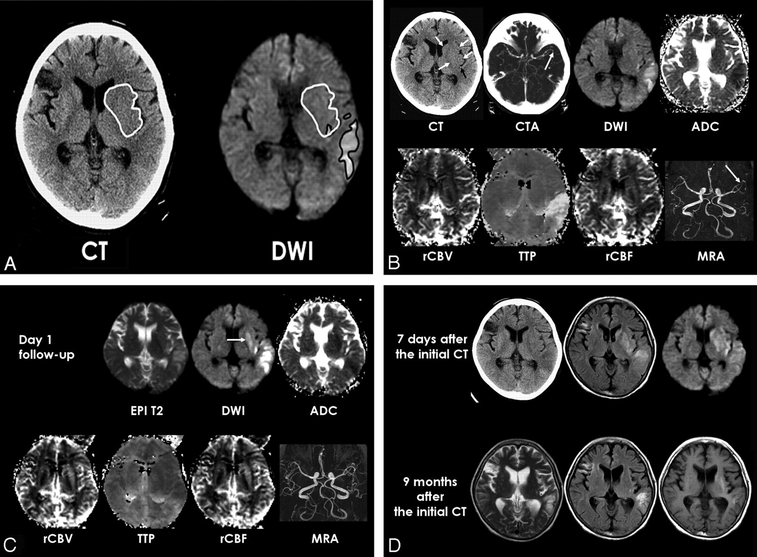
  • Fig 2.
    • Download figure
    • Open in new tab
    • Download powerpoint
    Fig 2.

    Patient 8. A, Initial unenhanced CT shows parenchymal hypoattenuation and loss of gray-white matter distinction in the left frontal lobe of ACA and MCA territory (arrows). Echo-planar spin-echo T2-weighted image (EPI T2, DWI of b value of 0, TR, 6500 ms; TE, 96.8 ms) shows no hyperintensity in the left frontal lobe. DWI (6500/96.8) and ADC map show hyperintensity and reduced ADC only in the medial frontal cortex and precentral gyrus that are only a part of hypoattenuated ischemic lesion depicted on CT. Perfusion maps show a TTP delay in the DWI lesion but no significant perfusion abnormality in the left frontal lobe lesion with reversed discrepancy on CT and DWI (arrowheads). MRA shows decreased signal intensity of the left distal MCA branches (arrow) and there was occlusion of the left A2 on MRA (not shown).

    B, Follow-up MR images at day 1 show delayed DWI hyperintensity and ADC decrease in the left frontal lobe lesion with reversed discrepancy (arrows) and more severe cytotoxic edema in the medial frontal cortex and precentral gyrus of the left frontal lobe. Follow-up MRA shows recanalization of distal MCA branches but persistent perfusion abnormality in the ischemic lesion found on the initial DWI.

    C, Follow-up CT image obtained 7 days after the initial CT demonstrates ischemic edema with heterogeneous hypoattenuation in the left frontal lobe. CT image obtained 11 months after the initial CT shows overt infarction with necrosis in the lesion with reversed discrepancy.

Tables

  • Figures
    • View popup
    Table 1:

    Clinical data of 9 patients with reversed discrepancy (RD)

    Patient No./Age (y)/SexTime to CT (h)Time to MRA (h)Arterior Occlusion on Initial MRAASPECTSLocation of RDMR Findings at Day 1 Follow-upNIHSSmRS at Day 90
    CTDWIRecanalization on MRADelayed DWI Hyperintensity of RD lesionInitialDay 1
    1/F/752.14.3ICA35Left BGAbsentPresent21224
    2/M/864.05.8ICA35Right BGAbsentPresent14154
    3/M/645.35.8ICA88Left BGAbsentPresent661
    4/M/761.74.3M143Right BGPresentPresent15176
    5/F/840.92.3M115Right BGAbsentAbsent12114
    6/F/801.82.5M279Right BGPresentPresent8135
    7/F/602.33.3M277Left BGPresentPresent992
    8/M/742.55.3MCA distal branch, A268Left insula, frontal lobePresentPresent18163
    9/M/502.24.0*24Right BGNo occlusionPresent1382
    • Note:—CT indicates computed tomography; MRA, magnetic resonance angiography; DWI, diffusion-weighted MR imaging; ASPECTS, Alberta Stroke Program early CT score; mRS, modified Rankin Scale; ICA, internal carotid artery; MCA, middle cerebral artery; BG, basal ganglia.

    • * Site of arterial occlusion was not accurately determined due to poor image quality of MRA.

    • View popup
    Table 2:

    Mean values of computed tomographic (CT) attenuation, apparent diffusion coefficient (ADC), diffusion-weighted MR imaging (DWI)*

    Initial CT and MRIFollow-up MRI at day 1
    RD Lesion (ROIRD)DWI Lesion 1 (ROIDWII)DWI Lesion 2 (ROIDW12)RD Lesion (ROIRD)Whole DWI Lesion (ROIDW)
    ΔHU2.33 ± 0.742.90 ± 1.340.18 ± 0.28——
    ADC (10−6 mm2/s)830 ± 91658 ± 108665 ± 112677 ± 163614 ± 184
    ADC ratio0.97 ± 0.070.74 ± 0.090.76 ± 0.100.76 ± 0.140.68 ± 0.15
    DWSI ratio1.08 ± 0.111.41 ± 0.171.40 ± 0.171.39 ± 0.321.97 ± 0.41
    T2SI ratio1.10 ± 0.081.11 ± 0.111.10 ± 0.111.19 ± 0.181.45 ± 0.20
    TTP delay (second)2.56 ± 1.669.09 ± 5.379.01 ± 5.390.06 ± 1.815.63 ± 5.02
    Relative CBV1.05 ± 0.240.98 ± 0.350.09 ± 0.351.16 ± 0.370.79 ± 0.31
    Relative CBF0.87 ± 0.200.59 ± 0.310.59 ± 0.311.31 ± 0.410.75 ± 0.37
    • Note:—Values are expressed as mean ± SD. ROI indicates region of interest; ΔHU indicates the difference between the Hounsfield unit of the lesion and the control; relative cerebral blood volume (CBV) or cerebral blood flow (CBF) indicates the ratio of lesion value to that of contralateral hemisphere; DWSI, signal intensity on diffusion-weighted MR image; T2SI, signal intensity on T2-weighted image.

    • * Perfusion weighted MR imaging (PWI) data of 1 patient was not included for the quantitative analysis of PWI values due to poor enhancement of the initial PWI.

PreviousNext
Back to top

In this issue

American Journal of Neuroradiology: 27 (9)
American Journal of Neuroradiology
Vol. 27, Issue 9
October 2006
  • Table of Contents
  • Index by author
Advertisement
Print
Download PDF
Email Article
Cite this article
0 Responses
Respond to this article
Share
Bookmark this article
Reversed Discrepancy between CT and Diffusion-Weighted MR Imaging in Acute Ischemic Stroke
E.Y. Kim, J.W. Ryoo, H.G. Roh, K.H. Lee, S.S. Kim, I.C. Song, K.-H. Chang, D.G. Na
American Journal of Neuroradiology Oct 2006, 27 (9) 1990-1995;
del.icio.us logo Twitter logo Facebook logo Mendeley logo
  • Tweet Widget

Jump to section

  • Article
    • Abstract
    • Materials and Methods
    • Results
    • Discussion
    • References
  • Figures & Data
  • Info & Metrics
  • Responses
  • References
  • PDF

Related Articles

  • No related articles found.
  • PubMed
  • Google Scholar

Cited By...

  • Recent Administration of Iodinated Contrast Renders Core Infarct Estimation Inaccurate Using RAPID Software
  • Diagnosis of DWI-negative acute ischemic stroke: A meta-analysis
  • Diffusion-Weighted Magnetic Resonance Imaging May Underestimate Acute Ischemic Lesions: Cautions on Neglecting a Computed Tomography-Diffusion-Weighted Imaging Discrepancy
  • Early Ischemic Change on CT Versus Diffusion-Weighted Imaging for Patients With Stroke Receiving Intravenous Recombinant Tissue-Type Plasminogen Activator Therapy: Stroke Acute Management With Urgent Risk-factor Assessment and Improvement (SAMURAI) rt-PA Registry
  • Correlation between CT and Diffusion-Weighted Imaging of Acute Cerebral Ischemia in a Rat Model
  • Crossref
  • Google Scholar

This article has not yet been cited by articles in journals that are participating in Crossref Cited-by Linking.

More in this TOC Section

  • Evaluating the Effects of White Matter Multiple Sclerosis Lesions on the Volume Estimation of 6 Brain Tissue Segmentation Methods
  • Quiet PROPELLER MRI Techniques Match the Quality of Conventional PROPELLER Brain Imaging Techniques
  • Predictors of Reperfusion in Patients with Acute Ischemic Stroke
Show more Brain

Similar Articles

Advertisement

Indexed Content

  • Current Issue
  • Accepted Manuscripts
  • Article Preview
  • Past Issues
  • Editorials
  • Editor's Choice
  • Fellows' Journal Club
  • Letters to the Editor
  • Video Articles

Cases

  • Case Collection
  • Archive - Case of the Week
  • Archive - Case of the Month
  • Archive - Classic Case

More from AJNR

  • Trainee Corner
  • Imaging Protocols
  • MRI Safety Corner
  • Book Reviews

Multimedia

  • AJNR Podcasts
  • AJNR Scantastics

Resources

  • Turnaround Time
  • Submit a Manuscript
  • Submit a Video Article
  • Submit an eLetter to the Editor/Response
  • Manuscript Submission Guidelines
  • Statistical Tips
  • Fast Publishing of Accepted Manuscripts
  • Graphical Abstract Preparation
  • Imaging Protocol Submission
  • Evidence-Based Medicine Level Guide
  • Publishing Checklists
  • Author Policies
  • Become a Reviewer/Academy of Reviewers
  • News and Updates

About Us

  • About AJNR
  • Editorial Board
  • Editorial Board Alumni
  • Alerts
  • Permissions
  • Not an AJNR Subscriber? Join Now
  • Advertise with Us
  • Librarian Resources
  • Feedback
  • Terms and Conditions
  • AJNR Editorial Board Alumni

American Society of Neuroradiology

  • Not an ASNR Member? Join Now

© 2025 by the American Society of Neuroradiology All rights, including for text and data mining, AI training, and similar technologies, are reserved.
Print ISSN: 0195-6108 Online ISSN: 1936-959X

Powered by HighWire
Email this Article

Thank you for your interest in spreading the word on American Journal of Neuroradiology.

NOTE: We only request your email address so that the person you are recommending the page to knows that you wanted them to see it, and that it is not junk mail. We do not capture any email address.

Enter multiple addresses on separate lines or separate them with commas.
Reversed Discrepancy between CT and Diffusion-Weighted MR Imaging in Acute Ischemic Stroke
(Your Name) has sent you a message from American Journal of Neuroradiology
(Your Name) thought you would like to see the American Journal of Neuroradiology web site.
CAPTCHA
This question is for testing whether or not you are a human visitor and to prevent automated spam submissions.
Cite this article
E.Y. Kim, J.W. Ryoo, H.G. Roh, K.H. Lee, S.S. Kim, I.C. Song, K.-H. Chang, D.G. Na
Reversed Discrepancy between CT and Diffusion-Weighted MR Imaging in Acute Ischemic Stroke
American Journal of Neuroradiology Oct 2006, 27 (9) 1990-1995;

Citation Manager Formats

  • BibTeX
  • Bookends
  • EasyBib
  • EndNote (tagged)
  • EndNote 8 (xml)
  • Medlars
  • Mendeley
  • Papers
  • RefWorks Tagged
  • Ref Manager
  • RIS
  • Zotero

We use cookies on this site to enhance your user experience. By clicking any link on this page you are giving your consent for us to set cookies.