
Article Figures & Data
Figures
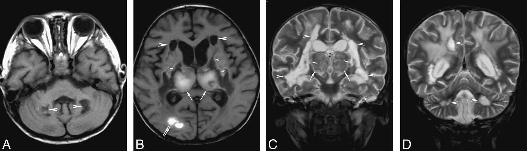
- Fig 1.
A 1.3-year-old boy (patient 1) left with spasticity and decerebrate posture. A, coronal T2-weighted imaging (4000 ms/90 ms, repetition time [TR]/echo time [TE]) shows symmetrical hyperintensity in the thalami (arrows), the centrum semiovale (arrowheads), and the brain stem, including the midbrain (double arrowheads) and the pons (double arrows). Note swelling of the thalami.
B, Postcontrast coronal T1-weighted imaging (630 ms/20 ms, TR/TE) shows irregular ringlike enhancement in the thalami (arrows).
- Fig 2.
A 2.3-year-old boy (patient 12) with severe sequelae.
A, Axial T1-weighted imaging (449 ms/12 ms, TR/TE) shows sharply marginated hypointensity in the cerebellum (arrowheads).
B, Axial T1-weighted imaging shows hyperintensity in the thalami (arrows) and the right occipital lobe (double arrows). Sharply marginated hypointense areas are found in the cerebral white matter (arrowheads) and the internal capsules (small arrowheads). The lentiform nuclei also show mixed intensity (double arrowheads).
C and D, Coronal T2-weighted imaging show mixed intensity in the thalami (arrows), and hyperintensity in the cerebral white matter, internal capsules (small arrowheads), and the cerebellum (double arrowheads). There are 2 types of white matter lesions: those in the central white matter and internal capsules showing hyperintensity comparable with the CSF (arrowheads) and those in the subcortical regions being less hyperintense (double arrows).
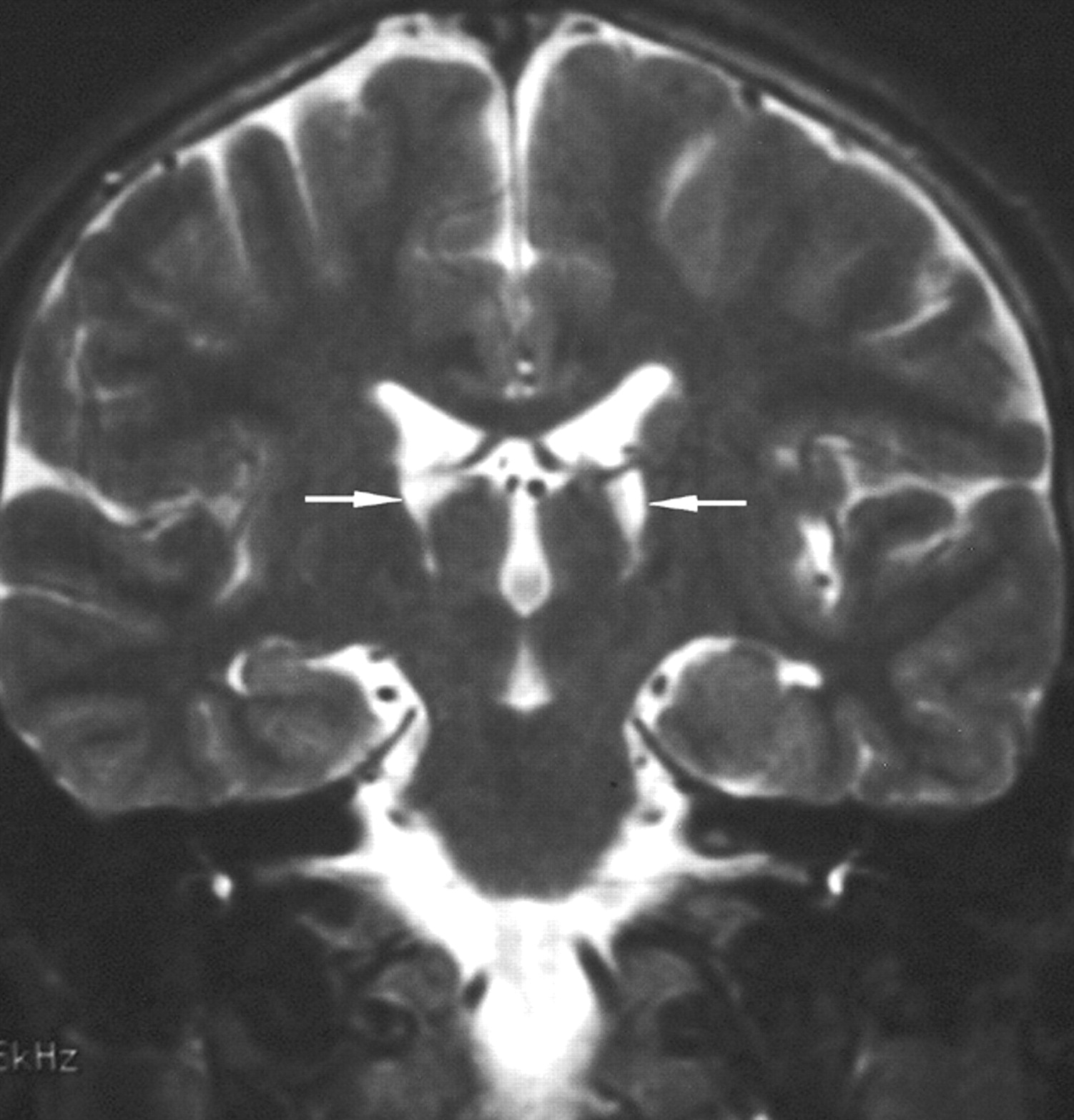
- Fig 3.
A 10-year-old boy (patient 5) recovered with hand tremor. Follow-up coronal T2-weighted imaging shows shrunken bilateral thalami with cavitation (arrows).


Tables
- Table 1:
Clinical features of 12 patients with acute necrotizing encephalopathy of childhood
Patient No./Age/Sex Clinical Features Positive Viral Study Outcome HUI:1 Outcome Category 1/1 y 4 mo/M Fever, conscious disturbance, seizure Influenza B Spasticity with decerebrate posture 3 2/3 y 3 mo/M Fever, conscious disturbance, seizure Mycoplasma Spasticity 2 3/12 y/F Fever, conscious disturbance, decerebrate posture, spasticity, urine and stool incontinence Influenza B, mycoplasma (possible) Spasticity, urine and stool incontinence 2 4/7 mo/M Conscious disturbance N/A Fully recovered 1 5/10 y/M Fever, conscious disturbance, seizure Influenza A Bilateral hand tremor; left-sided dystonia 2 6/1 y/M Fever, lower extremity rigidity N/A Fully recovered 1 7/3 y 5 mo/M Fever, conscious disturbance, decerebrate posture Influenza B Spasticity, speech disorder 2 8/2 mo/M Fever, conscious disturbance, seizure, motor weakness with cogwheel rigidity HHV-6 Speech disorder; full motor recovery 2 9/6 y/F Fever, skin rash, conscious disturbance, seizure, lower extremity paralysis, speech disability Adenovirus Neurogenic bladder, lower extremity weakness 2 10/2 y/M Fever, coma, seizure N/A Lower extremity weakness with rigidity 2 11/2 y 6 mo/F Fever, conscious disturbance, seizure, lower extremity paralysis Negative Lower extremity weakness 2 12/2 y 4 mo/F Fever, conscious disturbance 1 mo, vomiting, seizure Enterovirus Conscious disturbance, diplegia, spasticity 3 Note:—NUI:1 indicates Health Utilities Index Mark 1; HHV-6, human herpes virus 6; N/A, not available.
- Table 2:
Neuroimaging findings in the initial MR and the follow-up MR study in 12 patients with acute necrotizing encepalopathy of childhood
Patient No. Duration from Onset to Initial and to Follow-up MR Studies (d) Location of Involvement Hemorrhage (1) Cavitation (1) MR Score Brain Stem (1) White Matter (1) (Cerebral, cerebellar) A B 1 0; 43 + +, + + + 4 3 2 0; 103 − +, + − − 1 0 3 10; 47 + −, + + − 3 2 4 3; 83 + +, − − − 2 1 5 2; 182 + −, − − + 2 2 6 9; 76 − +, − − − 1 0 7 0; 19 + +, − + − 3 2 8 7; 15 + +, − − + 3 2 9 18; 134 + −, − − − 1 1 10 0; 16 + +, − − − 2 1 11 3; 16 + −, − + − 2 2 12 10; 29 + +, + + + 4 3
In this issue


Jump to section
Related Articles
Cited By...
- A Closer Investigation of the Synchronous Bilateral Pattern of MRI Lesions in Acute Necrotizing Encephalopathy Type 1
- Serial Imaging of Virus-Associated Necrotizing Disseminated Acute Leukoencephalopathy (VANDAL) in COVID-19
- Clinical and Radiologic Findings of Acute Necrotizing Encephalopathy in Young Adults
- Neurologic complications of COVID-19
- Pearls & Oy-sters: Leukoencephalopathy in critically ill patients with COVID-19
- Pearls & Oy-sters: Bilateral globus pallidus lesions in a patient with COVID-19
- COVID-19-associated acute necrotising encephalopathy successfully treated with steroids and polyvalent immunoglobulin with unusual IgG targeting the cerebral fibre network
- Influenza A-associated acute necrotising encephalopathy in a 10-year-old child
- Hemorrhagic Posterior Reversible Encephalopathy Syndrome as a Manifestation of COVID-19 Infection
- COVID-19-related acute necrotizing encephalopathy with brain stem involvement in a patient with aplastic anemia
- Clinical Reasoning: Acute onset facial droop in a 36-year-old pregnant woman
- Acute necrotising encephalopathy in a child with H1N1 influenza infection: a clinicoradiological diagnosis and follow-up
- Acute encephalopathy with bilateral thalamotegmental involvement and a benign course: a case report from Brazil
- Acute Necrotizing Encephalopathy in a Child during the 2009 Influenza A(H1N1) Pandemia: MR Imaging in Diagnosis and Follow-Up