
Article Figures & Data
Figures
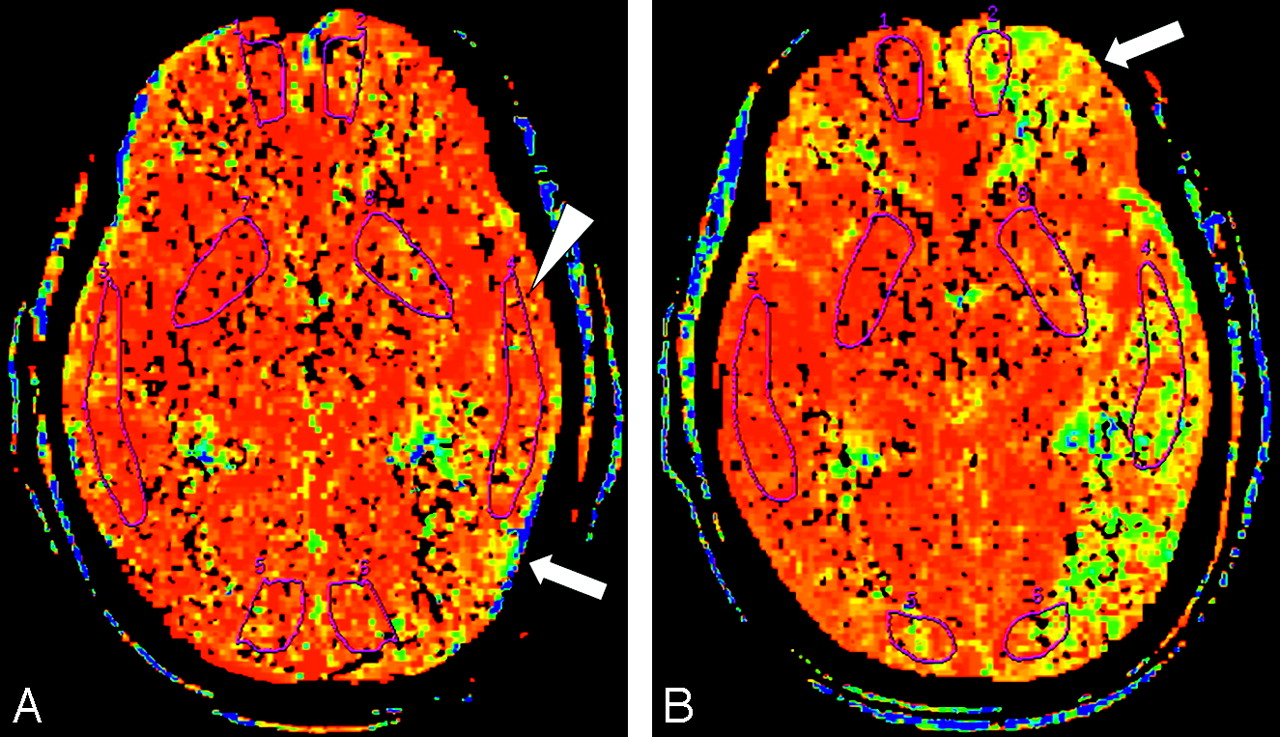
- Fig 1.
MTT maps in a 66-year-old woman.
A, MTT map before acetazolamide infusion shows prolonged MTT in the left hemisphere represented as areas displayed (yellow and green arrow). The ROI placed in the left hemisphere (arrowhead) does not include the areas of maximal MTT elevation.
B, MTT map obtained 15 minutes after infusion of acetazolamide, with use of the same section location and display parameters as those in A. Despite appropriate response on the right, no shortening in MTT is apparent in the left hemisphere. Instead, additional prolongation in MTT can be seen in areas in the left middle cerebral artery territory and the frontal lobe (arrow). By using ROI analysis, we measured a paradoxical prolongation in mean cortical MTT value of 93.7% on the left and interpreted it as evidence of hemodynamic impairment due to reduced perfusion pressure.

Tables
- Table 1:
Patient characteristics and hemodynamic impairment in hemispheres distal to stenoses
Patient No./Age (y)/Sex Cerebrovascular History Vascular Pathology Hemodynamic Impairment in Hemispheres Distal to Stenoses Defined by Normal Ranges Defined by % Change CBV (mL/100 g) MTT (s) CBF MTT 1/81/M Asymptomatic Stenotic right ICA, 95% 2/61/F Asymptomatic Stenotic left MCA, 70% 5.5, prolonged 3/83/M Asymptomatic Stenotic left ICA, 95% 6.1, increased 6.3, prolonged 4/54/M Asymptomatic Stenotic left ICA, 75% 5.1, prolonged 3.2 5/64/F Asymptomatic Stenotic left MCA, 90% 6/63/F TIA Occluded left MCA 7/49/M TIA Occluded left MCA 6.0 8/73/M TIA Occluded left ICA 5.5, prolonged 9/73/M TIA Occluded left MCA 6.8, prolonged 10/58/M TIA Occluded right MCA 11/75/M TIA Stenotic right ICA, 75% 12/68/M TIA Occluded right ICA 8.9, prolonged −20.8 14.3 13/82/F RIND Stenotic right ICA, 95% 19.8 14/74/F Minor stroke Occluded right MCA 6.2, prolonged −25.7 58.4 15/66/F Minor stroke Stenotic left MCA, 70% −31.6 93.7 Note:—CBV indicates cerebral blood volume; CBF, cerebral blood flow; MTT, mean transit time. TIA, transient ischemic attack; RIND, reversible ischemic neurologic deficits; ICA, internal carotid artery; MCA, middle cerebral artery.
- Table 2:
Values of perfusion CT parameters and percentage change before and after acetazolamide in the cerebral hemispheres ipsilateral and contralateral to the stenoses
Mean CBF (mL/100 g/min) Mean CBV (mL/100 g) Mean MTT (s) Hemispheres Ipsilateral to Stenotic Side Hemispheres Contralateral to Stenotic Side P Value Hemispheres Ipsilateral to Stenotic Side Hemispheres Contralateral to Stenotic Side P Value Hemispheres Ipsilateral to Stenotic Side Hemispheres Contralateral to Stenotic Side P Value Baseline 52.3 ± 18.8 69.8 ± 32.2 .003 3.9 ± 1.2 3.0 ± 1.3 .933 5.0 ± 1.7 3.4 ± 0.8 <.001 Acetazolamide 58.9 ± 26.3 97.4 ± 47.1 <.001 3.4 ± 1.7 3.5 ± 1.5 .643 5.4 ± 2.2 2.8 ± 0.7 <.001 P Value .137 <.001 .126 .003 .296 <.001 Mean CBF Mean CBV Mean MTT % change 12.53 ± 32.20 40.68 ± 26.98 <.001 12.69 ± 23.78 21.14 ± 34.45 .244 8.81 ± 30.06 −17.72 ± 10.38 .005 Note:—Values are mean ± SD. CBF indicates cerebral blood flow; CBV, cerebral blood volume; MTT, mean transit time.
Mean CBF Ratio Mean CBV Ratio Mean MTT Ratio Baseline 0.79 ± 0.16 1.06 ± 0.31 1.46 ± 0.31 Acetazolamide 0.63 ± 0.17 0.98 ± 0.15 1.91 ± 0.56 P value <.001 .200 .002 Note:—Values are mean ± SD. CBF indicates cerebral blood flow; CBV, cerebral blood volume; MTT, mean transit time.
In this issue


Jump to section
Related Articles
Cited By...
- Association Between Changes in Lipid Profiles and Progression of Symptomatic Intracranial Atherosclerotic Stenosis: A Prospective Multicenter Study
- Vasodilatory Capacity of the Cerebral Vasculature in Patients with Carotid Artery Stenosis
- Reply:
- The Acetazolamide Challenge: Techniques and Applications in the Evaluation of Chronic Cerebral Ischemia
- Tracer Delay-Insensitive Algorithm Can Improve Reliability of CT Perfusion Imaging for Cerebrovascular Steno-Occlusive Disease: Comparison with Quantitative Single-Photon Emission CT