
Article Figures & Data
Figures
- Fig 1.
Small right MCA aneurysm treated with balloon assistance (patient 51).
A, CT obtained 80 minutes after the treatment reveals increased cortical attenuation (arrows).
B–C, Follow-up CT examinations with a 4-hour interval; each shows partial (B) and total (C) resolution of the cortical increased attenuation.
D, DWI image of the MR image obtained immediately after the first CT (A) shows no corresponding abnormality.
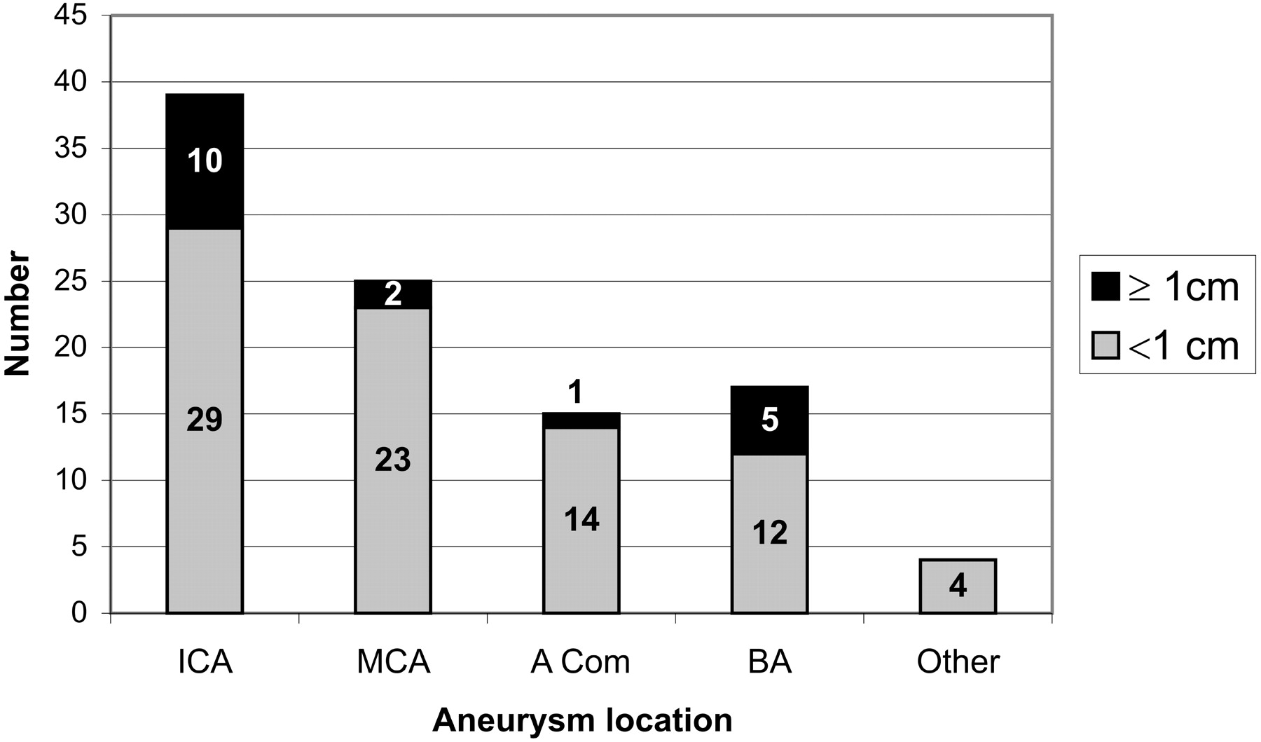
- Fig 2.
Graph shows the distribution of aneurysms according to their location and size. ICA indicates internal carotid artery; MCA, middle cerebral artery; Acom, anterior communicating artery; BA, basilar artery.
- Fig 3.
Posttreatment angiogram (A) shows the completely occluded small aneurysm (arrows) at the vertebrobasilar junction of the fenestrated basilar artery (patient 20). Posttreatment CT image (B) reveals increased attenuation in the posterior cerebral artery territory bilaterally (arrow). Control CT scan (C) obtained 5 hours after the first one shows complete resolution of the finding.
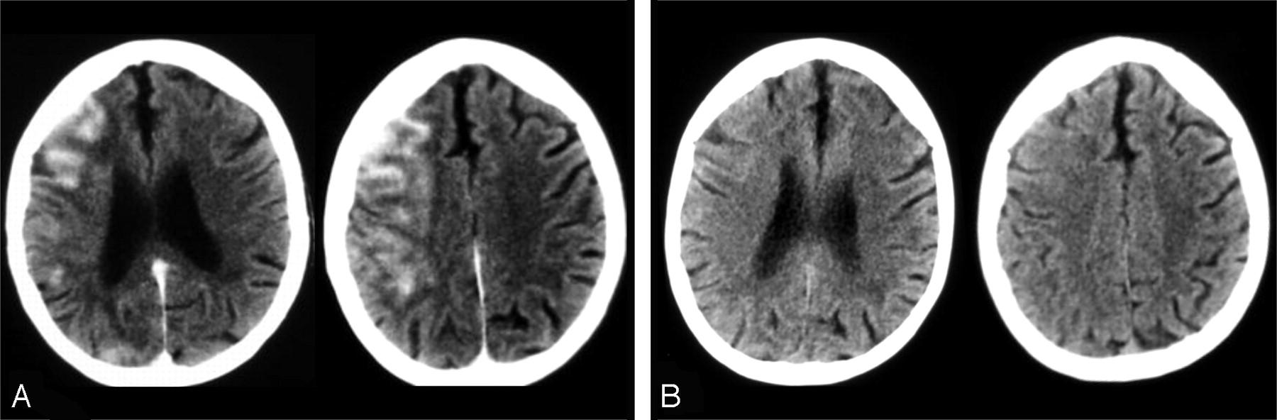
- Fig 4.
Small MCA aneurysm treated with no balloon assistance (patient 21). A, CT images obtained 38 minutes after the treatment reveal increased cortical attenuation in the left frontal region (arrows). Control CT examination (B) shows resolution of the finding.
- Fig 5.
A–B, CT examinations performed 53 minutes after the endovascular treatment of the right MCA aneurysm by using a remodeling technique. Patient 60 shows blood within the suprasellar cistern (A) in addition to increased cortical attenuation (arrows) in the right frontal region (B). C–D, Follow-up CT scans obtained after 6 hours demonstrated the persistence of blood appearance within the suprasellar cistern (C) and complete resolution of the cortical hyperattenuation (D). E–G, FLAIR (E and F) and DWI (G) of MR images obtained after the first CT examination confirm the presence of blood (arrows) within the cisterns (E) with no MR imaging abnormality in the region of cortical hyperattenuation (F and G).
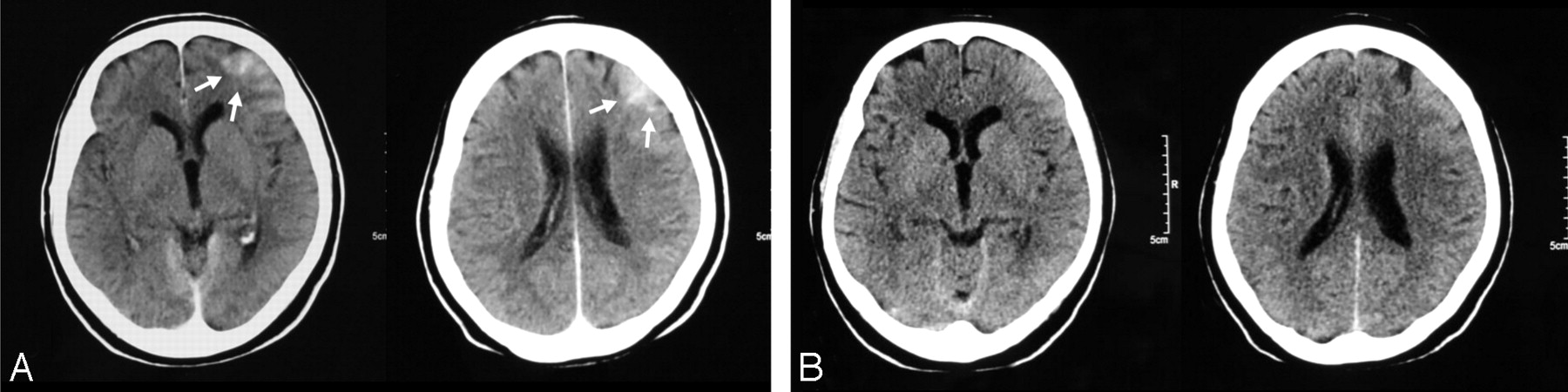
- Fig 6.
Early (A) and late (B) postembolization CT scans of the patient who had a large ICA aneurysm (patient 8). CT scan obtained 45 minutes after the procedure (A) shows very prominent hyperattenuation in the ipsilateral cortex, resolving totally in the control CT scan obtained after 6 hours (B).


Tables
Patient data including patient demographics, presence of subarachnoid hemorrhage (SAH) with or without vasospasm, collateral circulation, and procedural details in regard to presence of the cortical hyperdensity finding
Patient No./Sex/Age (y) Aneurysm Location Aneurysm Size SAH Vasospasm Balloon Remodeling Contrast Material (cc/kg) Heparin (u/kg) Total Microballoon Inflation Time (s) No. of Balloon Inflations Maximum Microballoon Inflation Time (s) Size of Micro-catheter (F) Micro-catheter Time (min) Elapsed Time Until CT Performed (min) Collateral AcomA PcomA 1/M/51 RICA Small + − + 2.6 158 440 3 158 2.5 45 80 + + RICA Small + − + 2.6 158 360 4 135 2.5 50 85 + + 2/F/50 RMCA Small + − + 4.4 143 900 8 87 1.7 53 97 + + 3/F/83 LICA Small + + + 6.7 183 300 5 90 2.5 60 60 + + 4/F/44 RICA Small − − + 3.9 104 588 8 187 1.9 90 30 + + 5/F/40 RMCA Small − − + 3.9 93,7 521 9 89 2.5 62 59 + + 6/F/30 RMCA Small + + + 4.6 170 292 5 90 1.7 105 40 + + 7/F/59 RMCA Small + + − 5.2 250 1.7 55 80 + + 8/F/50 RICA Large − − + 4.4 160 606 14 95 2.5 120 45 + − 9/F/74 RMCA Small + + − 2.0 80,6 2.5 30 68 − + 10/F/38 Basilar tip Large + + − 4.6 140 2.5 75 70 − − 11/M/46 LICA Small − − + 3.5 156 476 6 110 2.5 55 66 + + 12/M/48 LICA Small − − + 2.9 125 820 8 150 2.5 70 72 + + 13/F/34 RMCA Small + − + 4.2 154 260 3 115 2.5 40 46 + + 14/M/42 RMCA Small − − + 3.6 118 597 7 120 2.5 48 81 + + 15/F/63 AcomA Small − − − 3.3 139 2.5 50 80 + − 16/F/60 AcomA Small + + − 2.9 167 2.5 60 60 + + 17/M/62 RMCA Small + − − 3.3 139 1.7 65 80 + + 18/F/65 RMCA Small + + − 3.1 125 1.7 20 50 − + 19/M/27 RICA Small − − + 1.4 139 825 5 295 1.9 50 45 + + 20/F/52 Basilar fenest Small − − + 2.8 139 187 3 95 1.7 45 70 + − 21/F/60 LMCA Small + − − 4 200 2.5 28 38 − − 22/M/54 Basilar tip Small − − + 1.7 107 150 2 90 1.7 15 70 − − 23/F/48 Basilar tip Small + + − 1.5 149 2.5 40 75 − + 24/F/67 LICA Giant − − + 4.5 224 1552 17 141 2.5 120 64 + − 25/F/63 RPcomA Small − − − 2.4 105 1.7 45 40 − + 26/M/52 Basilar tip Small − − + 3.3 208 802 10 120 1.7 60 90 − − 27/M/52 Basilar tip Small + + + 1.2 149 440 10 50 1.7 70 45 + + 28/F/50 LICA Giant − − + 3.8 125 1488 7 323 1.9 60 40 + + 29/M/60 AcomA Small + + − 5.3 200 1.9 180 90 + + 30/F/53 LICA Small − − + 4.6 192 1287 15 210 1.7 115 45 + + 31/F/46 RMCA Small − − − 2.4 165 1.7 64 50 − + 32/F/65 RICA Large + + + 4.6 115 999 13 174 1.7 150 56 + + 33/F/67 RICA Small − − + 2.8 117 795 7 245 1.7 90 89 − − 34/M/70 RPcomA Small + − + 4.1 123 1322 15 139 1.7 120 70 − + 35/M/48 RMCA Small − − + 4.7 88,2 318 3 169 3 70 50 − + 36/F/70 AcomA Small + + − 2.3 100 1.7 35 120 + + 37/M/52 LICA Large − − + 3.3 175 2282 6 779 1.9 90 67 + + 38/M/32 RICA Small − − + 4.3 143 1235 5 417 1.9 56 135 + + 39/F/49 AcomA Small − − + 5 167 592 9 95 1.7 89 90 − + 40/F/59 AcomA Small − − + 2 125 300 4 106 1.7 60 58 + + 41/F/57 RICA Large − − + 3.8 93.7 711 6 172 1.7 43 90 + − 42/F/65 LICA Small − − + 4 152 1350 7 635 1.9 150 40 + + 43/F/52 LICA Small − − + 2 100 1705 10 301 1.9 80 70 + + 44/F/60 LICA Small − − + 3.4 269 612 7 160 1.9 68 75 + + 45/F/50 LMCA Large − − + 4 183 301 8 69 3 40 45 − − 46/F/41 RICA Small − − + 5 258 641 3 323 1.9 85 110 + + 47/F/68 RICA Small − − + 3 156 529 2 470 3 20 110 + − 48/F/70 AcomA Small − − + 4 24 504 6 131 1.7 90 70 + + 49/F/46 LPICA Small + − − 2.5 154 1.9 40 47 − − 50/M/57 LMCA Small − − + 2 159 680 7 170 1.7 45 85 − + 51/M/34 RMCA Small − − + 5.4 239 881 10 125 1.9 60 80 − + 52/M/68 Basilar tip Small − − + 3.5 165 3 15 180 + + AcomA Small − − + 3.5 165 323 6 77 1.7 60 125 + + 53/M/49 RACA Small + − − 3.8 93.8 3 65 200 + + 54/M/44 LPcomA Small − − + 6.1 296 1643 19 199 1.7 225 45 + + 55/M/18 AcomA Small + − + 3.6 242 498 4 121 1.7 45 68 + + 56/F/40 LPICA Small + − − 3.8 125 1.7 45 138 + + 57/F/41 RMCA Small + + − 3.3 250 1.7 105 70 + + 58/F/52 L Sup Cer Small + + − 3.8 119 1.7 70 90 − − RMCA Small + − + 3.8 119 355 6 117 3 40 90 − − 59/F/52 Basilar tip Small − − + 3.6 250 566 12 69 1.7 45 60 + + 60/F/45 RMCA Small − − + 5.9 192 302 5 85 1.7 30 53 + + 61/F/55 RPCA Large + − − 6.4 179 1.7 30 70 − + 62/M/23 Midbasilar Large − − + 3.9 212 820 8 176 1.7 45 180 − + 63/M/33 RMCA Small + − + 4.2 183 115 2 69 1.7 45 90 + + 64/F/54 LICA Small − − + 4.8 218 1652 7 300 1.9 70 148 + + 65/M/51 RPCA Small − − − 1.4 159 1.7 35 65 − + 66/F/43 LMCA Small + + + 6.9 254 276 3 150 1.7 40 120 + + RMCA Small + + + 6.9 254 229 3 96 1.7 15 120 + + 67/F/61 LICA Small − − − 6 500 1.7 100 160 − + 68/M/59 RICA Small + − + 7.9 283 471 6 169 3 195 49 − + 69/M/25 RPCA Large − − − 5 228 3 150 63 − + 70/F/43 RICA Small − − + 5 179 769 8 164 1.7 60 110 + − 71/M/52 Basilar tip Large − − + 5.1 179 723 8 150 3 130 90 − − 72/F/39 RICA Small − − + 6.7 117 547 7 114 3 45 37 + + 73/M/32 AcomA Small − − + 3.8 153 870 11 130 1.7 90 63 + + 74/F/51 LICA Small − − + 3.8 244 1912 6 530 1.9 60 90 + + 75/F/65 R Sup Cer Small + + − 6.2 149 − − − 1.7 45 110 − + 76/F/52 RICA Small + − + 5 188 587 6 110 1.7 47 63 + + 77/F/42 RMCA Small + − + 4.1 164 366 8 105 1.7 60 100 + − 78/F/49 RICA Large − − + 4 327 303 8 49 3 50 180 + − 79/F/32 LICA Small − − + 5.4 278 1189 5 434 1.9 76 50 + + 80/F/59 RICA Giant + + + 6.3 188 1298 10 181 3 195 84 + + 82/F/63 Basilar tip Small - - + 4.7 133 557 8 183 1.7 35 140 - + RMCA Small − − + 4.7 133 259 6 55 1.7 35 70 − − 83/F/65 RVA Small + + − 2.8 139 1.7 40 130 − − 84/F/50 LSupCer Small − − + 3.5 104 214 3 95 1.7 20 225 + + LICA Small − − + 3.5 104 673 4 490 1.9 40 164 + + 85/M/57 LMCA Small − − + 4 91,5 472 7 135 1.7 35 49 + + 86/M/50 ACom Small − − + 3.6 137 691 10 95 1.7 60 81 + − 87/M/60 AcomA Small + − + 7.4 219 975 16 133 1.7 145 130 + + 88/F/81 AcomA Large + + − 3 185 1.7 30 82 − + 89/M/63 AcomA Small − − + 3 122 686 8 148 1.7 60 60 + + 90/M/66 LICA Large − − + 4.3 185 486 10 80 1.7 40 78 + + RICA Small − − + 4.3 185 516 5 165 1.7 15 60 + − 91/F/70 LICA Small + − + 6 182 2047 10 420 1.9 50 42 + + 92/M/32 AcomA Small + − + 2.6 165 346 7 103 1.7 50 85 + − 93/F/55 LMCA Large − − + 3.1 147 801 11 105 3 65 75 − + Note:—L indicates left; R, right; ICA, internal carotid artery; MCA, middle cerebral artery; AcomA, anterior communicating artery; PcomA, posterior communicating artery; PICA, posterior inferior cerebellar artery; ACA, anterior cerebral artery; PCA, posterior cerebral artery; VA, vertebral artery; Sup Cer, superior cerebellar artery; Basilar Fenest, basilary fenestration; SAH, subarachnoid hemorrhage. *All patients in whom Onyx was used in the treatment (Onyx, Onyx + coil, Onyx + stent).
In this issue


Jump to section
Related Articles
Cited By...
- Risk factor analyses of contrast leakage and contrast-induced encephalopathy following coil embolization for unruptured intracranial aneurysm
- HARMless: Transient Cortical and Sulcal Hyperintensity on Gadolinium-Enhanced FLAIR after Elective Endovascular Coiling of Intracranial Aneurysms
- Outcome Differences between Intra-Arterial Iso- and Low-Osmolality Iodinated Radiographic Contrast Media in the Interventional Management of Stroke III Trial
- Flat Detector Angio-CT following Intra-Arterial Therapy of Acute Ischemic Stroke: Identification of Hemorrhage and Distinction from Contrast Accumulation due to Blood-Brain Barrier Disruption
- Predictive value of flat-panel CT for haemorrhagic transformations in patients with acute stroke treated with thrombectomy
- Subarachnoid Hyperattenuation on Flat Panel Detector-Based Conebeam CT Immediately after Uneventful Coil Embolization of Unruptured Intracranial Aneurysms