Advertisement

Main menu

  • Home
  • Content
    • Current Issue
    • Accepted Manuscripts
    • Article Preview
    • Past Issue Archive
    • Video Articles
    • AJNR Case Collection
    • Case of the Week Archive
    • Case of the Month Archive
    • Classic Case Archive
  • Special Collections
    • AJNR Awards
    • Low-Field MRI
    • Alzheimer Disease
    • ASNR Foundation Special Collection
    • Photon-Counting CT
    • View All
  • Multimedia
    • AJNR Podcasts
    • AJNR SCANtastic
    • Trainee Corner
    • MRI Safety Corner
    • Imaging Protocols
  • For Authors
    • Submit a Manuscript
    • Submit a Video Article
    • Submit an eLetter to the Editor/Response
    • Manuscript Submission Guidelines
    • Statistical Tips
    • Fast Publishing of Accepted Manuscripts
    • Graphical Abstract Preparation
    • Imaging Protocol Submission
    • Author Policies
  • About Us
    • About AJNR
    • Editorial Board
    • Editorial Board Alumni
  • More
    • Become a Reviewer/Academy of Reviewers
    • Subscribers
    • Permissions
    • Alerts
    • Feedback
    • Advertisers
    • ASNR Home

User menu

  • Alerts
  • Log in

Search

  • Advanced search
American Journal of Neuroradiology

American Journal of Neuroradiology

ASHNR American Society of Functional Neuroradiology ASHNR American Society of Pediatric Neuroradiology ASSR
  • Alerts
  • Log in

Advanced Search

  • Home
  • Content
    • Current Issue
    • Accepted Manuscripts
    • Article Preview
    • Past Issue Archive
    • Video Articles
    • AJNR Case Collection
    • Case of the Week Archive
    • Case of the Month Archive
    • Classic Case Archive
  • Special Collections
    • AJNR Awards
    • Low-Field MRI
    • Alzheimer Disease
    • ASNR Foundation Special Collection
    • Photon-Counting CT
    • View All
  • Multimedia
    • AJNR Podcasts
    • AJNR SCANtastic
    • Trainee Corner
    • MRI Safety Corner
    • Imaging Protocols
  • For Authors
    • Submit a Manuscript
    • Submit a Video Article
    • Submit an eLetter to the Editor/Response
    • Manuscript Submission Guidelines
    • Statistical Tips
    • Fast Publishing of Accepted Manuscripts
    • Graphical Abstract Preparation
    • Imaging Protocol Submission
    • Author Policies
  • About Us
    • About AJNR
    • Editorial Board
    • Editorial Board Alumni
  • More
    • Become a Reviewer/Academy of Reviewers
    • Subscribers
    • Permissions
    • Alerts
    • Feedback
    • Advertisers
    • ASNR Home
  • Follow AJNR on Twitter
  • Visit AJNR on Facebook
  • Follow AJNR on Instagram
  • Join AJNR on LinkedIn
  • RSS Feeds

AJNR Awards, New Junior Editors, and more. Read the latest AJNR updates

Research ArticleBrain

Does High-field MR Imaging Have an Influence on the Classification of Patients with Clinically Isolated Syndromes According to Current Diagnostic MR Imaging Criteria for Multiple Sclerosis?

M.P. Wattjes, M. Harzheim, C.K. Kuhl, J. Gieseke, S. Schmidt, L. Klotz, T. Klockgether, H.H. Schild and G.G. Lutterbey
American Journal of Neuroradiology September 2006, 27 (8) 1794-1798;
M.P. Wattjes
  • Find this author on Google Scholar
  • Find this author on PubMed
  • Search for this author on this site
M. Harzheim
  • Find this author on Google Scholar
  • Find this author on PubMed
  • Search for this author on this site
C.K. Kuhl
  • Find this author on Google Scholar
  • Find this author on PubMed
  • Search for this author on this site
J. Gieseke
  • Find this author on Google Scholar
  • Find this author on PubMed
  • Search for this author on this site
S. Schmidt
  • Find this author on Google Scholar
  • Find this author on PubMed
  • Search for this author on this site
L. Klotz
  • Find this author on Google Scholar
  • Find this author on PubMed
  • Search for this author on this site
T. Klockgether
  • Find this author on Google Scholar
  • Find this author on PubMed
  • Search for this author on this site
H.H. Schild
  • Find this author on Google Scholar
  • Find this author on PubMed
  • Search for this author on this site
G.G. Lutterbey
  • Find this author on Google Scholar
  • Find this author on PubMed
  • Search for this author on this site
  • Article
  • Figures & Data
  • Info & Metrics
  • Responses
  • References
  • PDF
Loading

Article Figures & Data

Figures

  • Tables
  • Fig 1.
    • Download figure
    • Open in new tab
    • Download powerpoint
    Fig 1.

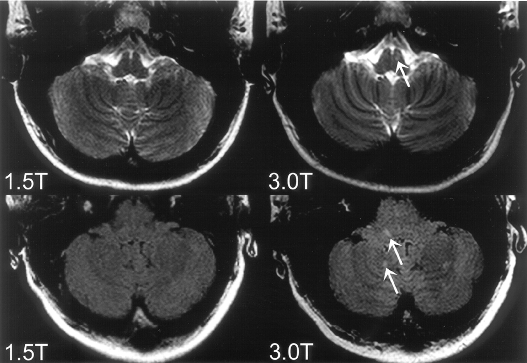
    Top row: T2-weighted images of a 19-year-old patient presenting with unilateral optic neuritis. One infratentorial lesion in the brain stem (arrow) could be identified on the 3T image but not on the corresponding 1.5T image. Bottom row: FLAIR images of a 30-year-old patient presenting with unilateral optic neuritis. The 3T image shows infratentorial lesions in the brain stem and the cerebellum (arrows) which were not prospectively identified on the corresponding 1.5T image.

  • Fig 2.
    • Download figure
    • Open in new tab
    • Download powerpoint
    Fig 2.

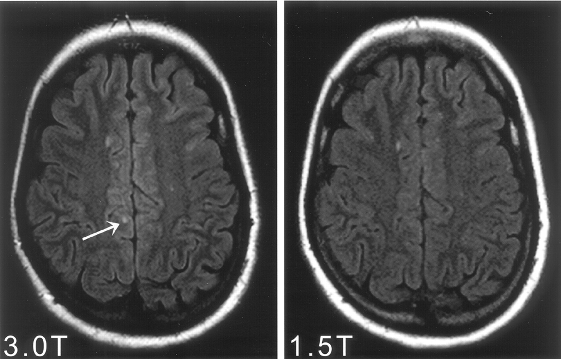
    Identical axial sections of FLAIR images within the supratentorial brain obtained at 3T (left) and 1.5T (right) MR imaging. One more juxtacortical lesion (arrow) could be identified on the 3T FLAIR images in comparison with the corresponding 1.5T examination.

Tables

  • Figures
    • View popup
    Table 1:

    MR imaging sequence parameters

    Pulse Sequence
    SystemFLAIRT2 TSET1 SE
    1.5T3T1.5T3T1.5T3T
    Field of view (mm)230230230230230230
    Matrix256256256256256256
    Section thickness (mm)555555
    Measured voxel size (mm)0.90/0.90/50.90/0.90/50.90/0.90/50.90/0.90/50.90/0.90/50.90/0.90/5
    Turbo factor29382316
    Number of signals averaged212121
    Repetition time (ms)600012 00035004100500500
    Echo time (ms)1101401001001212
    Inversion time (ms)20002850
    Acquisition time (min:s)3:004:002:272:193:343:27
    • Note:— FLAIR indicates fluid-attenuated inversion recovery; TSE, turbo spin-echo; SE, spin-echo.

    • View popup
    Table 2:

    Patients with discordant results between 1.5T and 3T according to Barkhof MR imaging criteria

    Patient No.Clinical PresentationNumber of Fulfilled MR Imaging CriteriaAdditional MR Imaging Criterion
    1.5T3T
    1Optic neuritis01≥1 juxtacortical lesion
    2Optic neuritis01≥1 infratentorial lesion
    3Brain stem syndrome12≥9 T2 hyperintense lesions
    4Brain stem syndrome12≥9 T2 hyperintense lesions
    5Brain stem syndrome12≥1 juxtacortical lesion
    6Optic neuritis12≥9 T2 hyperintense lesions
    7Optic neuritis12≥1 infratentorial lesion
    8*Optic neuritis23≥1 infratentorial lesion
    9*Optic neuritis23≥9 T2 hyperintense lesions
    10Optic neuritis34≥3 periventricular lesions
    11Spinal cord syndrome34≥1 infratentorial lesion
    • * Patient fulfilled 3 criteria instead of 2 at 3T, and they were therefore diagnosed of having lesion dissemination in space according to McDonald diagnostic criteria.

PreviousNext
Back to top

In this issue

American Journal of Neuroradiology: 27 (8)
American Journal of Neuroradiology
Vol. 27, Issue 8
September 2006
  • Table of Contents
  • Index by author
Advertisement
Print
Download PDF
Email Article

Thank you for your interest in spreading the word on American Journal of Neuroradiology.

NOTE: We only request your email address so that the person you are recommending the page to knows that you wanted them to see it, and that it is not junk mail. We do not capture any email address.

Enter multiple addresses on separate lines or separate them with commas.
Does High-field MR Imaging Have an Influence on the Classification of Patients with Clinically Isolated Syndromes According to Current Diagnostic MR Imaging Criteria for Multiple Sclerosis?
(Your Name) has sent you a message from American Journal of Neuroradiology
(Your Name) thought you would like to see the American Journal of Neuroradiology web site.
CAPTCHA
This question is for testing whether or not you are a human visitor and to prevent automated spam submissions.
Cite this article
M.P. Wattjes, M. Harzheim, C.K. Kuhl, J. Gieseke, S. Schmidt, L. Klotz, T. Klockgether, H.H. Schild, G.G. Lutterbey
Does High-field MR Imaging Have an Influence on the Classification of Patients with Clinically Isolated Syndromes According to Current Diagnostic MR Imaging Criteria for Multiple Sclerosis?
American Journal of Neuroradiology Sep 2006, 27 (8) 1794-1798;

Citation Manager Formats

  • BibTeX
  • Bookends
  • EasyBib
  • EndNote (tagged)
  • EndNote 8 (xml)
  • Medlars
  • Mendeley
  • Papers
  • RefWorks Tagged
  • Ref Manager
  • RIS
  • Zotero
0 Responses
Respond to this article
Share
Bookmark this article
Does High-field MR Imaging Have an Influence on the Classification of Patients with Clinically Isolated Syndromes According to Current Diagnostic MR Imaging Criteria for Multiple Sclerosis?
M.P. Wattjes, M. Harzheim, C.K. Kuhl, J. Gieseke, S. Schmidt, L. Klotz, T. Klockgether, H.H. Schild, G.G. Lutterbey
American Journal of Neuroradiology Sep 2006, 27 (8) 1794-1798;
del.icio.us logo Twitter logo Facebook logo Mendeley logo
  • Tweet Widget
  • Facebook Like
  • Google Plus One

Jump to section

  • Article
    • Abstract
    • Methods
    • Results
    • Discussion
    • Conclusion
    • Acknowledgments
    • Footnotes
    • References
  • Figures & Data
  • Info & Metrics
  • Responses
  • References
  • PDF

Related Articles

  • No related articles found.
  • PubMed
  • Google Scholar

Cited By...

  • Improving Detection of Multiple Sclerosis Lesions in the Posterior Fossa Using an Optimized 3D-FLAIR Sequence at 3T
  • Diffuse White Matter Damage Is Absent in Neuromyelitis Optica
  • Normal Findings on Brain Fluid-Attenuated Inversion Recovery MR Images at 3T
  • Clinical evaluation of a speed optimized T2 weighted fast spin echo sequence at 3.0 T using variable flip angle refocusing, half-Fourier acquisition and parallel imaging
  • Can imaging techniques measure neuroprotection and remyelination in multiple sclerosis?
  • Crossref
  • Google Scholar

This article has not yet been cited by articles in journals that are participating in Crossref Cited-by Linking.

More in this TOC Section

  • Optimal MRI Sequence for Identifying Occlusion Location in Acute Stroke: Which Value of Time-Resolved Contrast-Enhanced MRA?
  • Evaluating the Effects of White Matter Multiple Sclerosis Lesions on the Volume Estimation of 6 Brain Tissue Segmentation Methods
  • Quiet PROPELLER MRI Techniques Match the Quality of Conventional PROPELLER Brain Imaging Techniques
Show more Brain

Similar Articles

Advertisement

Indexed Content

  • Current Issue
  • Accepted Manuscripts
  • Article Preview
  • Past Issues
  • Editorials
  • Editor's Choice
  • Fellows' Journal Club
  • Letters to the Editor
  • Video Articles

Cases

  • Case Collection
  • Archive - Case of the Week
  • Archive - Case of the Month
  • Archive - Classic Case

More from AJNR

  • Trainee Corner
  • Imaging Protocols
  • MRI Safety Corner
  • Book Reviews

Multimedia

  • AJNR Podcasts
  • AJNR Scantastics

Resources

  • Turnaround Time
  • Submit a Manuscript
  • Submit a Video Article
  • Submit an eLetter to the Editor/Response
  • Manuscript Submission Guidelines
  • Statistical Tips
  • Fast Publishing of Accepted Manuscripts
  • Graphical Abstract Preparation
  • Imaging Protocol Submission
  • Evidence-Based Medicine Level Guide
  • Publishing Checklists
  • Author Policies
  • Become a Reviewer/Academy of Reviewers
  • News and Updates

About Us

  • About AJNR
  • Editorial Board
  • Editorial Board Alumni
  • Alerts
  • Permissions
  • Not an AJNR Subscriber? Join Now
  • Advertise with Us
  • Librarian Resources
  • Feedback
  • Terms and Conditions
  • AJNR Editorial Board Alumni

American Society of Neuroradiology

  • Not an ASNR Member? Join Now

© 2025 by the American Society of Neuroradiology All rights, including for text and data mining, AI training, and similar technologies, are reserved.
Print ISSN: 0195-6108 Online ISSN: 1936-959X

Powered by HighWire