Advertisement

Main menu

  • Home
  • Content
    • Current Issue
    • Accepted Manuscripts
    • Article Preview
    • Past Issue Archive
    • Video Articles
    • AJNR Case Collection
    • Case of the Week Archive
    • Case of the Month Archive
    • Classic Case Archive
  • Special Collections
    • AJNR Awards
    • Low-Field MRI
    • Alzheimer Disease
    • ASNR Foundation Special Collection
    • Photon-Counting CT
    • View All
  • Multimedia
    • AJNR Podcasts
    • AJNR SCANtastic
    • Trainee Corner
    • MRI Safety Corner
    • Imaging Protocols
  • For Authors
    • Submit a Manuscript
    • Submit a Video Article
    • Submit an eLetter to the Editor/Response
    • Manuscript Submission Guidelines
    • Statistical Tips
    • Fast Publishing of Accepted Manuscripts
    • Graphical Abstract Preparation
    • Imaging Protocol Submission
    • Author Policies
  • About Us
    • About AJNR
    • Editorial Board
    • Editorial Board Alumni
  • More
    • Become a Reviewer/Academy of Reviewers
    • Subscribers
    • Permissions
    • Alerts
    • Feedback
    • Advertisers
    • ASNR Home

User menu

  • Alerts
  • Log in

Search

  • Advanced search
American Journal of Neuroradiology

American Journal of Neuroradiology

ASHNR American Society of Functional Neuroradiology ASHNR American Society of Pediatric Neuroradiology ASSR
  • Alerts
  • Log in

Advanced Search

  • Home
  • Content
    • Current Issue
    • Accepted Manuscripts
    • Article Preview
    • Past Issue Archive
    • Video Articles
    • AJNR Case Collection
    • Case of the Week Archive
    • Case of the Month Archive
    • Classic Case Archive
  • Special Collections
    • AJNR Awards
    • Low-Field MRI
    • Alzheimer Disease
    • ASNR Foundation Special Collection
    • Photon-Counting CT
    • View All
  • Multimedia
    • AJNR Podcasts
    • AJNR SCANtastic
    • Trainee Corner
    • MRI Safety Corner
    • Imaging Protocols
  • For Authors
    • Submit a Manuscript
    • Submit a Video Article
    • Submit an eLetter to the Editor/Response
    • Manuscript Submission Guidelines
    • Statistical Tips
    • Fast Publishing of Accepted Manuscripts
    • Graphical Abstract Preparation
    • Imaging Protocol Submission
    • Author Policies
  • About Us
    • About AJNR
    • Editorial Board
    • Editorial Board Alumni
  • More
    • Become a Reviewer/Academy of Reviewers
    • Subscribers
    • Permissions
    • Alerts
    • Feedback
    • Advertisers
    • ASNR Home
  • Follow AJNR on Twitter
  • Visit AJNR on Facebook
  • Follow AJNR on Instagram
  • Join AJNR on LinkedIn
  • RSS Feeds

AJNR Awards, New Junior Editors, and more. Read the latest AJNR updates

OtherSpine Imaging and Spine Image-Guided Interventions

A Rare Cause of Flank Mass: Psoas Abscess due to Extensive Primary Thoracolumbar Tuberculous Spondylodiskitis

H.T. Sanal, M. Kocaoglu, A. Sehirlioglu and N. Bulakbasi
American Journal of Neuroradiology September 2006, 27 (8) 1735-1737;
H.T. Sanal
  • Find this author on Google Scholar
  • Find this author on PubMed
  • Search for this author on this site
M. Kocaoglu
  • Find this author on Google Scholar
  • Find this author on PubMed
  • Search for this author on this site
A. Sehirlioglu
  • Find this author on Google Scholar
  • Find this author on PubMed
  • Search for this author on this site
N. Bulakbasi
  • Find this author on Google Scholar
  • Find this author on PubMed
  • Search for this author on this site
  • Article
  • Figures & Data
  • Info & Metrics
  • Responses
  • References
  • PDF
Loading

Article Figures & Data

Figures

  • Fig 1.
    • Download figure
    • Open in new tab
    • Download powerpoint
    Fig 1.

    Transverse abdominal sonogram, obtained inferior to the right kidney, shows a cystic mass with fine internal echoes involving the right psoas muscle (arrows).

  • Fig 2.
    • Download figure
    • Open in new tab
    • Download powerpoint
    Fig 2.

    Abdominal MR imaging.

    A, Fat-saturated T2-weighted (TR/TE, 2100/90) axial MR image obtained through the iliac fossa shows a multilobulated cystic lesion (star) involving almost the whole of the right psoas muscle, extending to the right flank over the sacroiliac notch (arrowhead).

    B, Noncontrast T1-weighted (TR/TE, 614/16) and (C) contrast-enhanced T1-weighted axial images. The cystic lesion is low in signal intensity and shows rim enhancement after contrast media injection.
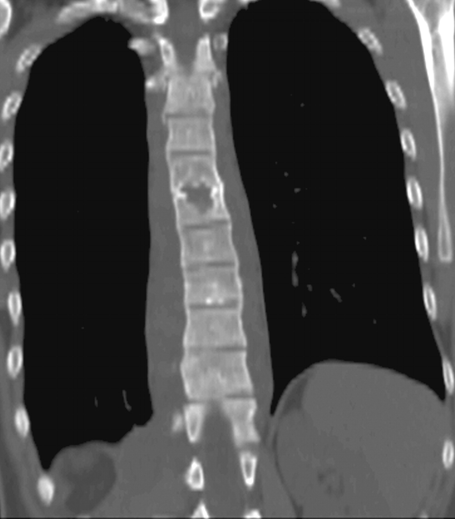
  • Fig 3.
    • Download figure
    • Open in new tab
    • Download powerpoint
    Fig 3.

    Coronal reconstructed noncontrast multidetector CT image of the chest demonstrates an extensive paravertebral soft-tissue mass and bone destruction at the adjacent endplates of the thoracic sixth and seventh vertebrae.

  • Fig 4.
    • Download figure
    • Open in new tab
    • Download powerpoint
    Fig 4.

    Spinal MR imaging.

    A, Coronal T2-weighted (TR/TE, 2500/120) thoracolumbar image shows widespread paravertebral soft tissue (arrowheads) and enlarged right psoas muscle (star) displacing the right kidney. The abscess has almost completely replaced the psoas muscle, though a small portion of it can still be noticed between the abscess and vertebral colon. High-signal-intensity patchy areas on vertebral bodies are consistent with osteomyelitis.

    B, T2-weighted and (C) contrast-enhanced T1-weighted (TR/TE, 580/14) sagittal scans. Enhancing prevertebral soft-tissue lesions (arrowheads) and osteomyelitis with high signal intensity on the T2-weighted scan, with patchy contrast enhancement on the T1-weighted scan, are seen. Subcutaneous flank mass reveals high T2 and low T1 signal intensity with rim enhancement (curved arrow). Midthoracic disk space narrowing suggesting diskitis and adjacent endplate bony destruction are also evident.

PreviousNext
Back to top

In this issue

American Journal of Neuroradiology: 27 (8)
American Journal of Neuroradiology
Vol. 27, Issue 8
September 2006
  • Table of Contents
  • Index by author
Advertisement
Print
Download PDF
Email Article

Thank you for your interest in spreading the word on American Journal of Neuroradiology.

NOTE: We only request your email address so that the person you are recommending the page to knows that you wanted them to see it, and that it is not junk mail. We do not capture any email address.

Enter multiple addresses on separate lines or separate them with commas.
A Rare Cause of Flank Mass: Psoas Abscess due to Extensive Primary Thoracolumbar Tuberculous Spondylodiskitis
(Your Name) has sent you a message from American Journal of Neuroradiology
(Your Name) thought you would like to see the American Journal of Neuroradiology web site.
CAPTCHA
This question is for testing whether or not you are a human visitor and to prevent automated spam submissions.
Cite this article
H.T. Sanal, M. Kocaoglu, A. Sehirlioglu, N. Bulakbasi
A Rare Cause of Flank Mass: Psoas Abscess due to Extensive Primary Thoracolumbar Tuberculous Spondylodiskitis
American Journal of Neuroradiology Sep 2006, 27 (8) 1735-1737;

Citation Manager Formats

  • BibTeX
  • Bookends
  • EasyBib
  • EndNote (tagged)
  • EndNote 8 (xml)
  • Medlars
  • Mendeley
  • Papers
  • RefWorks Tagged
  • Ref Manager
  • RIS
  • Zotero
0 Responses
Respond to this article
Share
Bookmark this article
A Rare Cause of Flank Mass: Psoas Abscess due to Extensive Primary Thoracolumbar Tuberculous Spondylodiskitis
H.T. Sanal, M. Kocaoglu, A. Sehirlioglu, N. Bulakbasi
American Journal of Neuroradiology Sep 2006, 27 (8) 1735-1737;
del.icio.us logo Twitter logo Facebook logo Mendeley logo
  • Tweet Widget
  • Facebook Like
  • Google Plus One

Jump to section

  • Article
    • Abstract
    • Case Report
    • Discussion
    • References
  • Figures & Data
  • Info & Metrics
  • Responses
  • References
  • PDF

Related Articles

  • No related articles found.
  • PubMed
  • Google Scholar

Cited By...

  • No citing articles found.
  • Crossref
  • Google Scholar

This article has not yet been cited by articles in journals that are participating in Crossref Cited-by Linking.

More in this TOC Section

  • Characteristics of SIH Type I Culprit Lesions
  • Management Outcomes For VO Spine Biopsy
  • Cone Beam CT Myelography
Show more SPINE IMAGING AND SPINE IMAGE-GUIDED INTERVENTIONS

Similar Articles

Advertisement

Indexed Content

  • Current Issue
  • Accepted Manuscripts
  • Article Preview
  • Past Issues
  • Editorials
  • Editor's Choice
  • Fellows' Journal Club
  • Letters to the Editor
  • Video Articles

Cases

  • Case Collection
  • Archive - Case of the Week
  • Archive - Case of the Month
  • Archive - Classic Case

More from AJNR

  • Trainee Corner
  • Imaging Protocols
  • MRI Safety Corner
  • Book Reviews

Multimedia

  • AJNR Podcasts
  • AJNR Scantastics

Resources

  • Turnaround Time
  • Submit a Manuscript
  • Submit a Video Article
  • Submit an eLetter to the Editor/Response
  • Manuscript Submission Guidelines
  • Statistical Tips
  • Fast Publishing of Accepted Manuscripts
  • Graphical Abstract Preparation
  • Imaging Protocol Submission
  • Evidence-Based Medicine Level Guide
  • Publishing Checklists
  • Author Policies
  • Become a Reviewer/Academy of Reviewers
  • News and Updates

About Us

  • About AJNR
  • Editorial Board
  • Editorial Board Alumni
  • Alerts
  • Permissions
  • Not an AJNR Subscriber? Join Now
  • Advertise with Us
  • Librarian Resources
  • Feedback
  • Terms and Conditions
  • AJNR Editorial Board Alumni

American Society of Neuroradiology

  • Not an ASNR Member? Join Now

© 2025 by the American Society of Neuroradiology All rights, including for text and data mining, AI training, and similar technologies, are reserved.
Print ISSN: 0195-6108 Online ISSN: 1936-959X

Powered by HighWire