Advertisement

Main menu

  • Home
  • Content
    • Current Issue
    • Accepted Manuscripts
    • Article Preview
    • Past Issue Archive
    • Video Articles
    • AJNR Case Collection
    • Case of the Week Archive
    • Case of the Month Archive
    • Classic Case Archive
  • Special Collections
    • AJNR Awards
    • Low-Field MRI
    • Alzheimer Disease
    • ASNR Foundation Special Collection
    • Photon-Counting CT
    • View All
  • Multimedia
    • AJNR Podcasts
    • AJNR SCANtastic
    • Trainee Corner
    • MRI Safety Corner
    • Imaging Protocols
  • For Authors
    • Submit a Manuscript
    • Submit a Video Article
    • Submit an eLetter to the Editor/Response
    • Manuscript Submission Guidelines
    • Statistical Tips
    • Fast Publishing of Accepted Manuscripts
    • Graphical Abstract Preparation
    • Imaging Protocol Submission
    • Author Policies
  • About Us
    • About AJNR
    • Editorial Board
    • Editorial Board Alumni
  • More
    • Become a Reviewer/Academy of Reviewers
    • Subscribers
    • Permissions
    • Alerts
    • Feedback
    • Advertisers
    • ASNR Home

User menu

  • Alerts
  • Log in

Search

  • Advanced search
American Journal of Neuroradiology

American Journal of Neuroradiology

ASHNR American Society of Functional Neuroradiology ASHNR American Society of Pediatric Neuroradiology ASSR
  • Alerts
  • Log in

Advanced Search

  • Home
  • Content
    • Current Issue
    • Accepted Manuscripts
    • Article Preview
    • Past Issue Archive
    • Video Articles
    • AJNR Case Collection
    • Case of the Week Archive
    • Case of the Month Archive
    • Classic Case Archive
  • Special Collections
    • AJNR Awards
    • Low-Field MRI
    • Alzheimer Disease
    • ASNR Foundation Special Collection
    • Photon-Counting CT
    • View All
  • Multimedia
    • AJNR Podcasts
    • AJNR SCANtastic
    • Trainee Corner
    • MRI Safety Corner
    • Imaging Protocols
  • For Authors
    • Submit a Manuscript
    • Submit a Video Article
    • Submit an eLetter to the Editor/Response
    • Manuscript Submission Guidelines
    • Statistical Tips
    • Fast Publishing of Accepted Manuscripts
    • Graphical Abstract Preparation
    • Imaging Protocol Submission
    • Author Policies
  • About Us
    • About AJNR
    • Editorial Board
    • Editorial Board Alumni
  • More
    • Become a Reviewer/Academy of Reviewers
    • Subscribers
    • Permissions
    • Alerts
    • Feedback
    • Advertisers
    • ASNR Home
  • Follow AJNR on Twitter
  • Visit AJNR on Facebook
  • Follow AJNR on Instagram
  • Join AJNR on LinkedIn
  • RSS Feeds

AJNR Awards, New Junior Editors, and more. Read the latest AJNR updates

OtherSpine Imaging and Spine Image-Guided Interventions

A Rare Cause of Flank Mass: Psoas Abscess due to Extensive Primary Thoracolumbar Tuberculous Spondylodiskitis

H.T. Sanal, M. Kocaoglu, A. Sehirlioglu and N. Bulakbasi
American Journal of Neuroradiology September 2006, 27 (8) 1735-1737;
H.T. Sanal
  • Find this author on Google Scholar
  • Find this author on PubMed
  • Search for this author on this site
M. Kocaoglu
  • Find this author on Google Scholar
  • Find this author on PubMed
  • Search for this author on this site
A. Sehirlioglu
  • Find this author on Google Scholar
  • Find this author on PubMed
  • Search for this author on this site
N. Bulakbasi
  • Find this author on Google Scholar
  • Find this author on PubMed
  • Search for this author on this site
  • Article
  • Figures & Data
  • Info & Metrics
  • Responses
  • References
  • PDF
Loading

Abstract

SUMMARY: Psoas abscess secondary to tuberculous spondylodiskitis is usually a complication of thoracolumbar vertebrae disease. The psoas abscess may be difficult clinically to diagnose because of its rarity, insidious onset of the disease, and nonspecific clinical presentation. We report multidetector CT and MR imaging findings of a psoas abscess secondary to primary tuberculous spondylodiskitis of the spine from the T3 to L2 vertebrae, which presented as a flank mass.

Spinal tuberculosis (Pott’s disease) is common in endemic regions.1-3 Vague symptoms and signs make the clinical diagnosis of psoas abscess with tuberculous spondylodiskitis a challenge.1,3 We report imaging findings of a case with extensive tuberculous spondylodiskitis accompanied by a psoas abscess, which presented as a soft-tissue flank mass beneath the skin.

Case Report

A 21-year-old man was admitted to the hospital with a 1-month history of a right-flank soft-tissue mass. His general condition was good, and he was not febrile. Initial investigations showed nothing remarkable with his blood count but an elevated erythrocyte sedimentation rate of 66. Sonography showed a huge lobulated retroperitoneal cystic mass with subcutaneous extension to the right flank (Fig. 1). To precisely define the borders of this lesion, we performed abdominal MR imaging, which revealed a cystic lesion, expanding and almost replacing the whole of the right psoas muscle, which was well-defined, lobulated, and hypointense on T1-weighted and hyperintense on T2-weighted images, with rim enhancement after paramagnetic contrast media administration (Fig 2A, -B). The lesion extended to the subcutaneous area in the right lumbar region over the iliac bone.

Fig 1.
  • Download figure
  • Open in new tab
  • Download powerpoint
Fig 1.

Transverse abdominal sonogram, obtained inferior to the right kidney, shows a cystic mass with fine internal echoes involving the right psoas muscle (arrows).

Fig 2.
  • Download figure
  • Open in new tab
  • Download powerpoint
Fig 2.

Abdominal MR imaging.

A, Fat-saturated T2-weighted (TR/TE, 2100/90) axial MR image obtained through the iliac fossa shows a multilobulated cystic lesion (star) involving almost the whole of the right psoas muscle, extending to the right flank over the sacroiliac notch (arrowhead).

B, Noncontrast T1-weighted (TR/TE, 614/16) and (C) contrast-enhanced T1-weighted axial images. The cystic lesion is low in signal intensity and shows rim enhancement after contrast media injection.

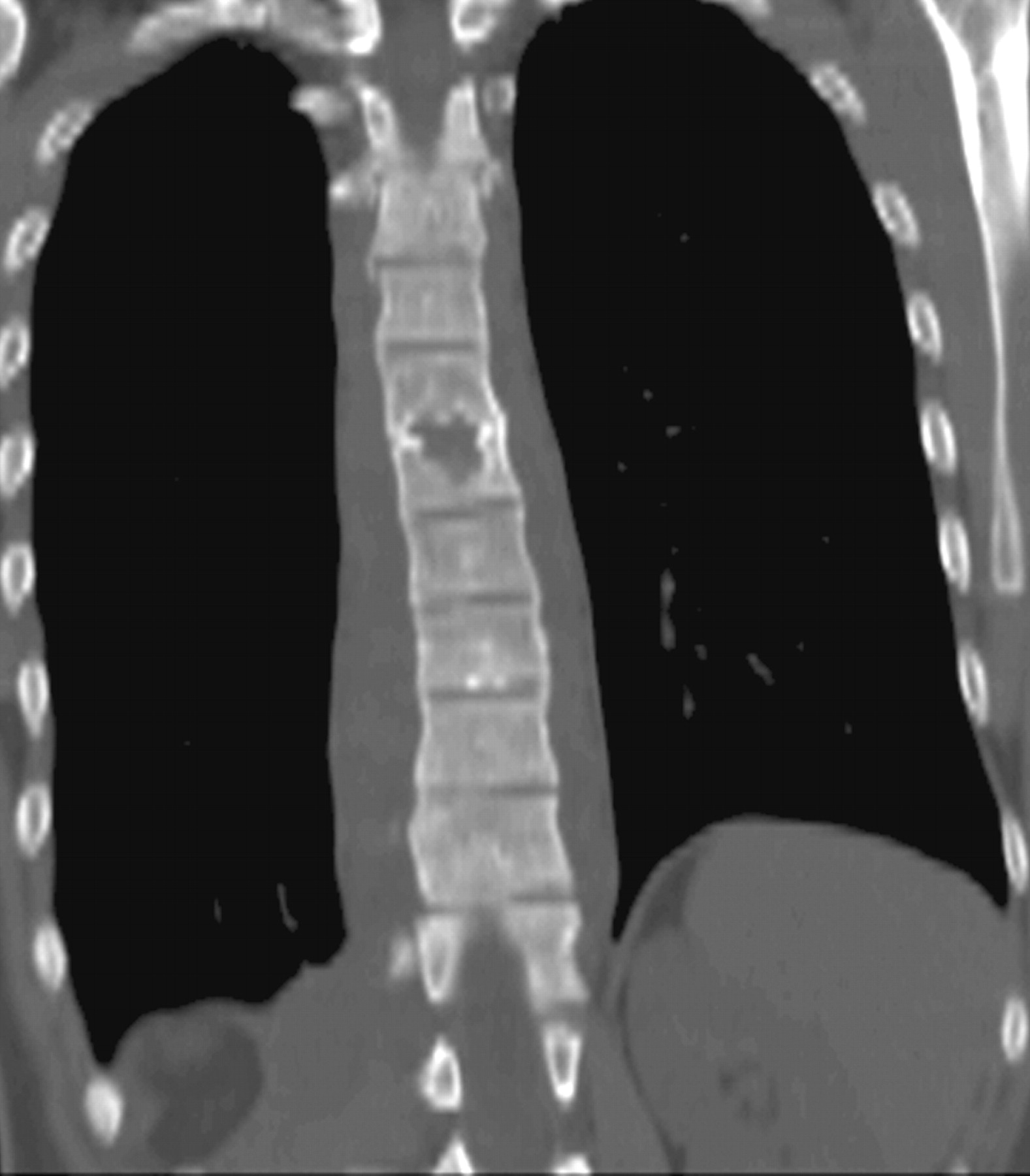
Because of a high index of suspected tuberculosis, radiologic examination was extended, including high-resolution chest CT and spinal MR imaging. On chest CT, there was no lung lesion suggesting tuberculous infection except a pre- and paravertebral soft-tissue attenuation (Fig 3). Between the levels of the third thoracic and second lumbar vertebrae, a paravertebral abscess formation, hypointense on T1-weighted and hyperintense on T2-weighted sequences with rim enhancement after contrast media injection, was seen. There was no MR imaging evidence of extension of the infection into the spinal canal. Signal intensity changes within the thoracic and upper lumbar vertebral body, such as low T1-weighted and high T2-weighted patchy areas with intravenous contrast enhancement, were compatible with osteomyelitis. The T6–7 disk space was narrowed, and bone destruction was also noted at adjacent endplates (Fig 4A-C). On the right side, the paravertebral abscess formation was in continuity with the right psoas muscle, causing psoas abscess, which was visualized in the first MR imaging. The abscess was drained at surgery; then, antituberculous therapy was started. The patient was discharged and asked to come for follow-up 1 month later.

Fig 3.
  • Download figure
  • Open in new tab
  • Download powerpoint
Fig 3.

Coronal reconstructed noncontrast multidetector CT image of the chest demonstrates an extensive paravertebral soft-tissue mass and bone destruction at the adjacent endplates of the thoracic sixth and seventh vertebrae.

Fig 4.
  • Download figure
  • Open in new tab
  • Download powerpoint
Fig 4.

Spinal MR imaging.

A, Coronal T2-weighted (TR/TE, 2500/120) thoracolumbar image shows widespread paravertebral soft tissue (arrowheads) and enlarged right psoas muscle (star) displacing the right kidney. The abscess has almost completely replaced the psoas muscle, though a small portion of it can still be noticed between the abscess and vertebral colon. High-signal-intensity patchy areas on vertebral bodies are consistent with osteomyelitis.

B, T2-weighted and (C) contrast-enhanced T1-weighted (TR/TE, 580/14) sagittal scans. Enhancing prevertebral soft-tissue lesions (arrowheads) and osteomyelitis with high signal intensity on the T2-weighted scan, with patchy contrast enhancement on the T1-weighted scan, are seen. Subcutaneous flank mass reveals high T2 and low T1 signal intensity with rim enhancement (curved arrow). Midthoracic disk space narrowing suggesting diskitis and adjacent endplate bony destruction are also evident.

Discussion

The skeletal system, which is involved in 1%–10% of patients with tuberculosis, is the most common extrapulmonary site for tuberculous infection. Approximately half of those cases manifest as spinal disease, and 75% are accompanied by paraspinal abscess.2,4 Psoas abscesses of tuberculous origin are infrequently caused by digestive, urologic, or genital tuberculosis.5 Generally, in tuberculous spondylodiskitis, a paraspinal abscess forms secondary to destruction of the cortical bone and elevation of the periosteum. In case of periosteum penetration with the inflammatory mass, a psoas abscess may form and may extend inferiorly as far as the groin and thigh under the psoas sheath on the muscle course.3 Subacute presentation along with nonspecific symptoms and signs makes the diagnosis of psoas abscess difficult.1 Patients may be referred to the department of rheumatology for back pain.4 Limping, a positive psoas sign, flexion deformity of the hip joint, fatigue, fever, night sweating, and weight loss also may be seen.6

Psoas abscess can be associated with disseminated tuberculous infection; however, in our patient, there was no accompanying lung lesion. Al-Shaikhi et al7 reported the CT findings of a 4-month-old girl who presented with a multiloculated cystic iliopsoas muscle mass extending through the flank muscle and diagnosed as primary psoas abscess; however, Staphylococcus aureus was confirmed as the causative microorganism.

Abscess formations occur more frequently in cases of tuberculosis infections than in cases of pyogenic infections. The chronic and insidious nature of tuberculous spondylitis causes late diagnosis of this disease; therefore, enough time passes for mass presentation of abscesses, which may be huge as in the present patient. On the other hand, early clinical presentation of pyogenic abscesses allows timely diagnosis and treatment, which decrease the incidence of mass formation secondary to pyogenic abscesses.1,3

In conclusion, in endemic regions, diagnosis of psoas abscess should not be simply put aside in patients with flank mass, but its possible associations with the thoracic cavity, as in our patient, must be investigated. Multiplanar imaging, especially MR imaging, is a useful diagnostic procedure in defining subtle diskovertebral lesions and in detecting unsuspected paravertebral soft-tissue extension.

References

  1. ↵
    Penado S, Espina B, Francisco Campo J. Abscess of the psoas muscle: description of a series of 23 cases. Enferm Infecc Microbiol Clin 2001;19:257–60
    PubMed
  2. ↵
    Levine MJ, Pedlow FX. Spinal infections. In: Garfin SR, Vaccaro AR, eds. Orthopaedic Knowledge Update: Spine. Rosemont, Ill: American Academy of Orthopaedic Surgeons;1997;257–271
  3. ↵
    Dinc H, Ahmetoglu A, Baykal S, et al. Image-guided percutaneous drainage of tuberculous iliopsoas and spondylodiskitic abscesses: midterm results. Radiology 2002;225:353–58
    PubMed
  4. ↵
    Lindahl S, Nyman RS, Brismar J, et al. Imaging of tuberculosis. IV. Spinal manifestations in 63 patients. Acta Radiol 1996;37:506–11
    CrossRefPubMed
  5. ↵
    Fitoz S, Atasoy C, Yagmurlu A, et al. Psoas abscess secondary to tuberculous lymphadenopathy: case report. Abdom Imaging 2001;26:323–24
    PubMed
  6. ↵
    Fam AG, Rubenstein J. Another look at spinal tuberculosis. J Rheumatol 1993;20:1731–40
    PubMed
  7. ↵
    Al-Shaikhi A, Shaw K, Laberge JM. A rapidly growing flank mass in an infant. J Pediatr Surg 2003;38:1415–17
    PubMed
  • Received September 20, 2005.
  • Accepted after revision September 28, 2005.
  • Copyright © American Society of Neuroradiology
View Abstract
PreviousNext
Back to top

In this issue

American Journal of Neuroradiology: 27 (8)
American Journal of Neuroradiology
Vol. 27, Issue 8
September 2006
  • Table of Contents
  • Index by author
Advertisement
Print
Download PDF
Email Article

Thank you for your interest in spreading the word on American Journal of Neuroradiology.

NOTE: We only request your email address so that the person you are recommending the page to knows that you wanted them to see it, and that it is not junk mail. We do not capture any email address.

Enter multiple addresses on separate lines or separate them with commas.
A Rare Cause of Flank Mass: Psoas Abscess due to Extensive Primary Thoracolumbar Tuberculous Spondylodiskitis
(Your Name) has sent you a message from American Journal of Neuroradiology
(Your Name) thought you would like to see the American Journal of Neuroradiology web site.
CAPTCHA
This question is for testing whether or not you are a human visitor and to prevent automated spam submissions.
Cite this article
H.T. Sanal, M. Kocaoglu, A. Sehirlioglu, N. Bulakbasi
A Rare Cause of Flank Mass: Psoas Abscess due to Extensive Primary Thoracolumbar Tuberculous Spondylodiskitis
American Journal of Neuroradiology Sep 2006, 27 (8) 1735-1737;

Citation Manager Formats

  • BibTeX
  • Bookends
  • EasyBib
  • EndNote (tagged)
  • EndNote 8 (xml)
  • Medlars
  • Mendeley
  • Papers
  • RefWorks Tagged
  • Ref Manager
  • RIS
  • Zotero
0 Responses
Respond to this article
Share
Bookmark this article
A Rare Cause of Flank Mass: Psoas Abscess due to Extensive Primary Thoracolumbar Tuberculous Spondylodiskitis
H.T. Sanal, M. Kocaoglu, A. Sehirlioglu, N. Bulakbasi
American Journal of Neuroradiology Sep 2006, 27 (8) 1735-1737;
del.icio.us logo Twitter logo Facebook logo Mendeley logo
  • Tweet Widget
  • Facebook Like
  • Google Plus One

Jump to section

  • Article
    • Abstract
    • Case Report
    • Discussion
    • References
  • Figures & Data
  • Info & Metrics
  • Responses
  • References
  • PDF

Related Articles

  • No related articles found.
  • PubMed
  • Google Scholar

Cited By...

  • No citing articles found.
  • Crossref
  • Google Scholar

This article has not yet been cited by articles in journals that are participating in Crossref Cited-by Linking.

More in this TOC Section

  • Cone Beam CT Myelography
  • Post-Procedural Brachial Neuritis Features
  • Diagnostic Value of Brain WMH in SIH
Show more Spine Imaging and Spine Image-Guided Interventions

Similar Articles

Advertisement

Indexed Content

  • Current Issue
  • Accepted Manuscripts
  • Article Preview
  • Past Issues
  • Editorials
  • Editor's Choice
  • Fellows' Journal Club
  • Letters to the Editor
  • Video Articles

Cases

  • Case Collection
  • Archive - Case of the Week
  • Archive - Case of the Month
  • Archive - Classic Case

More from AJNR

  • Trainee Corner
  • Imaging Protocols
  • MRI Safety Corner

Multimedia

  • AJNR Podcasts
  • AJNR Scantastics

Resources

  • Turnaround Time
  • Submit a Manuscript
  • Submit a Video Article
  • Submit an eLetter to the Editor/Response
  • Manuscript Submission Guidelines
  • Statistical Tips
  • Fast Publishing of Accepted Manuscripts
  • Graphical Abstract Preparation
  • Imaging Protocol Submission
  • Evidence-Based Medicine Level Guide
  • Publishing Checklists
  • Author Policies
  • Become a Reviewer/Academy of Reviewers
  • News and Updates

About Us

  • About AJNR
  • Editorial Board
  • Editorial Board Alumni
  • Alerts
  • Permissions
  • Not an AJNR Subscriber? Join Now
  • Advertise with Us
  • Librarian Resources
  • Feedback
  • Terms and Conditions
  • AJNR Editorial Board Alumni

American Society of Neuroradiology

  • Not an ASNR Member? Join Now

© 2025 by the American Society of Neuroradiology All rights, including for text and data mining, AI training, and similar technologies, are reserved.
Print ISSN: 0195-6108 Online ISSN: 1936-959X

Powered by HighWire