Advertisement

Main menu

  • Home
  • Content
    • Current Issue
    • Accepted Manuscripts
    • Article Preview
    • Past Issue Archive
    • Video Articles
    • AJNR Case Collection
    • Case of the Week Archive
    • Case of the Month Archive
    • Classic Case Archive
  • Special Collections
    • AJNR Awards
    • Low-Field MRI
    • Alzheimer Disease
    • ASNR Foundation Special Collection
    • Photon-Counting CT
    • View All
  • Multimedia
    • AJNR Podcasts
    • AJNR SCANtastic
    • Trainee Corner
    • MRI Safety Corner
    • Imaging Protocols
  • For Authors
    • Submit a Manuscript
    • Submit a Video Article
    • Submit an eLetter to the Editor/Response
    • Manuscript Submission Guidelines
    • Statistical Tips
    • Fast Publishing of Accepted Manuscripts
    • Graphical Abstract Preparation
    • Imaging Protocol Submission
    • Author Policies
  • About Us
    • About AJNR
    • Editorial Board
    • Editorial Board Alumni
  • More
    • Become a Reviewer/Academy of Reviewers
    • Subscribers
    • Permissions
    • Alerts
    • Feedback
    • Advertisers
    • ASNR Home

User menu

  • Alerts
  • Log in

Search

  • Advanced search
American Journal of Neuroradiology

American Journal of Neuroradiology

ASHNR American Society of Functional Neuroradiology ASHNR American Society of Pediatric Neuroradiology ASSR
  • Alerts
  • Log in

Advanced Search

  • Home
  • Content
    • Current Issue
    • Accepted Manuscripts
    • Article Preview
    • Past Issue Archive
    • Video Articles
    • AJNR Case Collection
    • Case of the Week Archive
    • Case of the Month Archive
    • Classic Case Archive
  • Special Collections
    • AJNR Awards
    • Low-Field MRI
    • Alzheimer Disease
    • ASNR Foundation Special Collection
    • Photon-Counting CT
    • View All
  • Multimedia
    • AJNR Podcasts
    • AJNR SCANtastic
    • Trainee Corner
    • MRI Safety Corner
    • Imaging Protocols
  • For Authors
    • Submit a Manuscript
    • Submit a Video Article
    • Submit an eLetter to the Editor/Response
    • Manuscript Submission Guidelines
    • Statistical Tips
    • Fast Publishing of Accepted Manuscripts
    • Graphical Abstract Preparation
    • Imaging Protocol Submission
    • Author Policies
  • About Us
    • About AJNR
    • Editorial Board
    • Editorial Board Alumni
  • More
    • Become a Reviewer/Academy of Reviewers
    • Subscribers
    • Permissions
    • Alerts
    • Feedback
    • Advertisers
    • ASNR Home
  • Follow AJNR on Twitter
  • Visit AJNR on Facebook
  • Follow AJNR on Instagram
  • Join AJNR on LinkedIn
  • RSS Feeds

AJNR Awards, New Junior Editors, and more. Read the latest AJNR updates

OtherHEAD & NECK

Biopsy of Deep-Seated Head and Neck Lesions under Intraoral Ultrasound Guidance

K.T. Wong, R.K.Y. Tsang, G.M.K. Tse, E.H.Y. Yuen and A.T. Ahuja
American Journal of Neuroradiology September 2006, 27 (8) 1654-1657;
K.T. Wong
  • Find this author on Google Scholar
  • Find this author on PubMed
  • Search for this author on this site
R.K.Y. Tsang
  • Find this author on Google Scholar
  • Find this author on PubMed
  • Search for this author on this site
G.M.K. Tse
  • Find this author on Google Scholar
  • Find this author on PubMed
  • Search for this author on this site
E.H.Y. Yuen
  • Find this author on Google Scholar
  • Find this author on PubMed
  • Search for this author on this site
A.T. Ahuja
  • Find this author on Google Scholar
  • Find this author on PubMed
  • Search for this author on this site
  • Article
  • Figures & Data
  • Info & Metrics
  • Responses
  • References
  • PDF
Loading

Article Figures & Data

Figures

  • Fig 1.
    • Download figure
    • Open in new tab
    • Download powerpoint
    Fig 1.

    A 63-year-old man with metastatic left retropharyngeal node from recurrent nasopharyngeal carcinoma.

    A, Postgadolinium enhanced, fat-suppressed T1-weighted (repetition time, 550 ms/echo time, 12 ms/number of signal averages, 2) axial MR image at the level of oropharynx shows an enlarged left retropharyngeal lymph node (arrow). Note the close relationship of the lymph node with the left internal carotid artery (arrowhead).

    B, Grayscale intraoral sonography in transverse plane reveals a well-defined, solid, round, hypoechoic left retropharyngeal lymph node (arrow) just medial to the left internal carotid artery (arrowhead). The sonographic appearances are suggestive of a malignant node.

  • Fig 2.
    • Download figure
    • Open in new tab
    • Download powerpoint
    Fig 2.

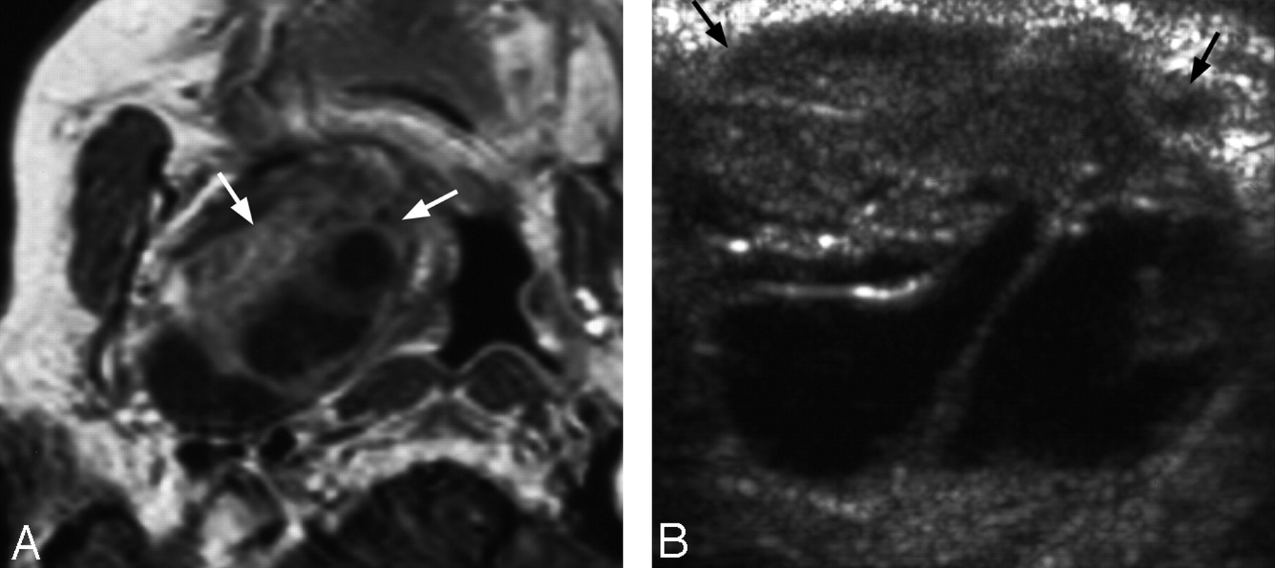
    An 82-year-old woman with peripheral nerve sheath tumor in the right parapharyngeal space.

    A, Postgadolinium T1-weighted (repetition time, 425 ms/echo time, 13 ms/number of signal averages, 2) axial MR image shows a well-circumscribed heterogeneous mass with both cystic and solid enhancing components in the right parapharyngeal space (arrows). The right lateral oropharyngeal wall is pushed toward the midline.

    B, Grayscale intraoral sonography in transverse plane confirms the presence of heterogeneous mixed cystic and solid mass (arrows) underneath the right oropharyngeal mucosa.

  • Fig 3.
    • Download figure
    • Open in new tab
    • Download powerpoint
    Fig 3.

    A 40-year-old woman with a pleomorphic adenoma in the deep left parotid space. T1-weighted (A) and postgadolinium enhanced T1-weighted (B) (repetition time, 450 ms/echo time, 13 ms/number of signal averages, 2) axial MR images show a well-circumscribed heterogeneously enhancing mass (arrows) arising from the left parotid space. Note that the left parapharyngeal fat is displaced anteromedially (arrowheads), which suggests parotid space origin of the mass.

    C, Grayscale intraoral sonography in the transverse plane shows a well-circumscribed, homogenous, hypoechoic left deep parotid space mass (arrows) underneath the left oropharyngeal mucosa. Note the deep portion of the lesion is not fully examined by this technique.
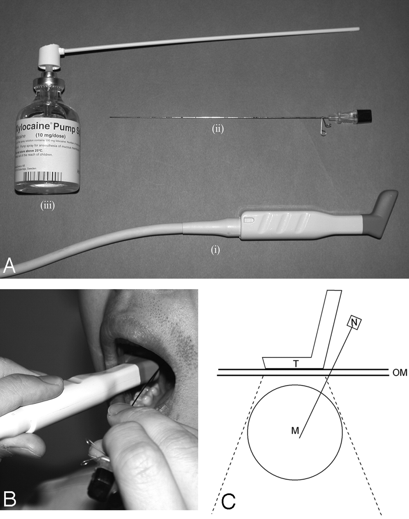
  • Fig 4.
    • Download figure
    • Open in new tab
    • Download powerpoint
    Fig 4.

    A, Basic equipment for intraoral sonography-guided biopsy: intraoral sonography transducer (i); 22-gauge Franseen needle (ii); 10% xylocaine spray (iii).

    B, Photograph shows the procedure of intraoral sonography-guided biopsy.

    C, Schematic diagram illustrates the biopsy needle pathway in relation to positions of the intraoral sonography transducer and the lesion. M = mass; N = needle; OM = oropharyngeal mucosa; T = transducer.

PreviousNext
Back to top

In this issue

American Journal of Neuroradiology: 27 (8)
American Journal of Neuroradiology
Vol. 27, Issue 8
September 2006
  • Table of Contents
  • Index by author
Advertisement
Print
Download PDF
Email Article

Thank you for your interest in spreading the word on American Journal of Neuroradiology.

NOTE: We only request your email address so that the person you are recommending the page to knows that you wanted them to see it, and that it is not junk mail. We do not capture any email address.

Enter multiple addresses on separate lines or separate them with commas.
Biopsy of Deep-Seated Head and Neck Lesions under Intraoral Ultrasound Guidance
(Your Name) has sent you a message from American Journal of Neuroradiology
(Your Name) thought you would like to see the American Journal of Neuroradiology web site.
CAPTCHA
This question is for testing whether or not you are a human visitor and to prevent automated spam submissions.
Cite this article
K.T. Wong, R.K.Y. Tsang, G.M.K. Tse, E.H.Y. Yuen, A.T. Ahuja
Biopsy of Deep-Seated Head and Neck Lesions under Intraoral Ultrasound Guidance
American Journal of Neuroradiology Sep 2006, 27 (8) 1654-1657;

Citation Manager Formats

  • BibTeX
  • Bookends
  • EasyBib
  • EndNote (tagged)
  • EndNote 8 (xml)
  • Medlars
  • Mendeley
  • Papers
  • RefWorks Tagged
  • Ref Manager
  • RIS
  • Zotero
0 Responses
Respond to this article
Share
Bookmark this article
Biopsy of Deep-Seated Head and Neck Lesions under Intraoral Ultrasound Guidance
K.T. Wong, R.K.Y. Tsang, G.M.K. Tse, E.H.Y. Yuen, A.T. Ahuja
American Journal of Neuroradiology Sep 2006, 27 (8) 1654-1657;
del.icio.us logo Twitter logo Facebook logo Mendeley logo
  • Tweet Widget
  • Facebook Like
  • Google Plus One

Jump to section

  • Article
    • Abstract
    • Case Reports
    • Discussion
    • References
  • Figures & Data
  • Info & Metrics
  • Responses
  • References
  • PDF

Related Articles

  • No related articles found.
  • PubMed
  • Google Scholar

Cited By...

  • No citing articles found.
  • Crossref
  • Google Scholar

This article has not yet been cited by articles in journals that are participating in Crossref Cited-by Linking.

More in this TOC Section

  • Chondrosarcoma vs Synovial Chondromatosis: Imaging
  • WHO Classification Update: Nasal&Skull Base Tumors
  • Peritumoral Signal in Vestibular Schwannomas
Show more Head & Neck

Similar Articles

Advertisement

Indexed Content

  • Current Issue
  • Accepted Manuscripts
  • Article Preview
  • Past Issues
  • Editorials
  • Editor's Choice
  • Fellows' Journal Club
  • Letters to the Editor
  • Video Articles

Cases

  • Case Collection
  • Archive - Case of the Week
  • Archive - Case of the Month
  • Archive - Classic Case

More from AJNR

  • Trainee Corner
  • Imaging Protocols
  • MRI Safety Corner
  • Book Reviews

Multimedia

  • AJNR Podcasts
  • AJNR Scantastics

Resources

  • Turnaround Time
  • Submit a Manuscript
  • Submit a Video Article
  • Submit an eLetter to the Editor/Response
  • Manuscript Submission Guidelines
  • Statistical Tips
  • Fast Publishing of Accepted Manuscripts
  • Graphical Abstract Preparation
  • Imaging Protocol Submission
  • Evidence-Based Medicine Level Guide
  • Publishing Checklists
  • Author Policies
  • Become a Reviewer/Academy of Reviewers
  • News and Updates

About Us

  • About AJNR
  • Editorial Board
  • Editorial Board Alumni
  • Alerts
  • Permissions
  • Not an AJNR Subscriber? Join Now
  • Advertise with Us
  • Librarian Resources
  • Feedback
  • Terms and Conditions
  • AJNR Editorial Board Alumni

American Society of Neuroradiology

  • Not an ASNR Member? Join Now

© 2025 by the American Society of Neuroradiology All rights, including for text and data mining, AI training, and similar technologies, are reserved.
Print ISSN: 0195-6108 Online ISSN: 1936-959X

Powered by HighWire