Advertisement

Main menu

  • Home
  • Content
    • Current Issue
    • Accepted Manuscripts
    • Article Preview
    • Past Issue Archive
    • Video Articles
    • AJNR Case Collection
    • Case of the Week Archive
    • Case of the Month Archive
    • Classic Case Archive
  • Special Collections
    • AJNR Awards
    • Low-Field MRI
    • Alzheimer Disease
    • ASNR Foundation Special Collection
    • Photon-Counting CT
    • View All
  • Multimedia
    • AJNR Podcasts
    • AJNR SCANtastic
    • Trainee Corner
    • MRI Safety Corner
    • Imaging Protocols
  • For Authors
    • Submit a Manuscript
    • Submit a Video Article
    • Submit an eLetter to the Editor/Response
    • Manuscript Submission Guidelines
    • Statistical Tips
    • Fast Publishing of Accepted Manuscripts
    • Graphical Abstract Preparation
    • Imaging Protocol Submission
    • Author Policies
  • About Us
    • About AJNR
    • Editorial Board
    • Editorial Board Alumni
  • More
    • Become a Reviewer/Academy of Reviewers
    • Subscribers
    • Permissions
    • Alerts
    • Feedback
    • Advertisers
    • ASNR Home

User menu

  • Alerts
  • Log in

Search

  • Advanced search
American Journal of Neuroradiology

American Journal of Neuroradiology

ASHNR American Society of Functional Neuroradiology ASHNR American Society of Pediatric Neuroradiology ASSR
  • Alerts
  • Log in

Advanced Search

  • Home
  • Content
    • Current Issue
    • Accepted Manuscripts
    • Article Preview
    • Past Issue Archive
    • Video Articles
    • AJNR Case Collection
    • Case of the Week Archive
    • Case of the Month Archive
    • Classic Case Archive
  • Special Collections
    • AJNR Awards
    • Low-Field MRI
    • Alzheimer Disease
    • ASNR Foundation Special Collection
    • Photon-Counting CT
    • View All
  • Multimedia
    • AJNR Podcasts
    • AJNR SCANtastic
    • Trainee Corner
    • MRI Safety Corner
    • Imaging Protocols
  • For Authors
    • Submit a Manuscript
    • Submit a Video Article
    • Submit an eLetter to the Editor/Response
    • Manuscript Submission Guidelines
    • Statistical Tips
    • Fast Publishing of Accepted Manuscripts
    • Graphical Abstract Preparation
    • Imaging Protocol Submission
    • Author Policies
  • About Us
    • About AJNR
    • Editorial Board
    • Editorial Board Alumni
  • More
    • Become a Reviewer/Academy of Reviewers
    • Subscribers
    • Permissions
    • Alerts
    • Feedback
    • Advertisers
    • ASNR Home
  • Follow AJNR on Twitter
  • Visit AJNR on Facebook
  • Follow AJNR on Instagram
  • Join AJNR on LinkedIn
  • RSS Feeds

AJNR Awards, New Junior Editors, and more. Read the latest AJNR updates

OtherHead and Neck Imaging

Palatal Enlargement in Chronic Lymphocytic Leukemia

P. Vibhute, E. Carneiro, E. Genden and P.M. Som
American Journal of Neuroradiology September 2006, 27 (8) 1649-1650;
P. Vibhute
  • Find this author on Google Scholar
  • Find this author on PubMed
  • Search for this author on this site
E. Carneiro
  • Find this author on Google Scholar
  • Find this author on PubMed
  • Search for this author on this site
E. Genden
  • Find this author on Google Scholar
  • Find this author on PubMed
  • Search for this author on this site
P.M. Som
  • Find this author on Google Scholar
  • Find this author on PubMed
  • Search for this author on this site
  • Article
  • Figures & Data
  • Info & Metrics
  • Responses
  • References
  • PDF
Loading

Abstract

SUMMARY: Palatal involvement in chronic lymphocytic leukemia (CLL) is rare and has only been reported 3 previous times in the non-radiology literature. To our knowledge this is the first imaging description of this entity. Based on our experience, when smoothly lobulated, homogeneous masses are identified on the oral surface of the hard palate, the diagnosis of CLL should be considered.

Chronic lymphocytic leukemia (CLL) is a B-lineage neoplasm of prefollicular center cells that is usually associated with circulating neoplastic small lymphocytes. From a morphologic and immunophenotypic perspective, the malignant cells of CLL are identical to those of nodal-based small lymphocytic lymphoma (SLL), and these 2 malignancies are thought to represent different manifestations of the same disease.1 CLL is the most common type of leukemia in the Western hemisphere; its prevalence in Europe and America ranges from 29% to 38% of all leukemias. Ninety percent of patients with CLL are older than 50 years of age.2 The most common oral manifestations of the leukemias are gingival hypertrophy, petechial hemorrhage and ecchymosis, infection, ulceration, and necrosis. Less commonly, as a result of leukemic infiltrates, multiple localized tumorlike growths can occur in the gingiva.3 Similar localized leukemic infiltrations occurring elsewhere in the oral cavity have only rarely been reported in the literature.3–5 In this article, we report on a patient with CLL who presented with bilateral masses on the oral surface of the hard palate. This represents only the fourth such reported case and the first documented with imaging.3–5

Case Report

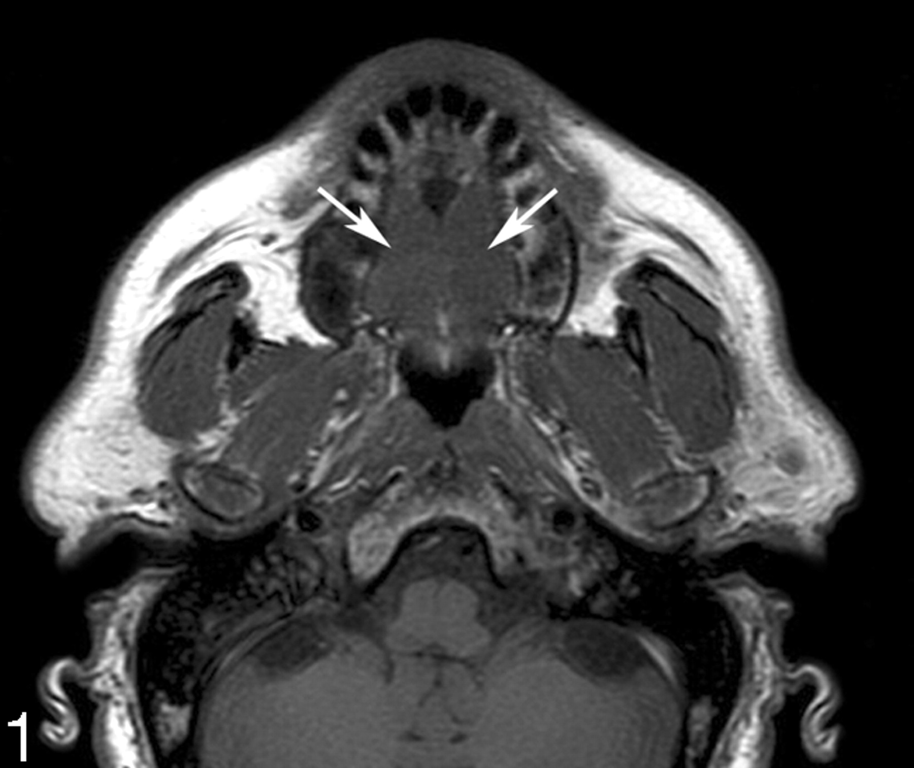
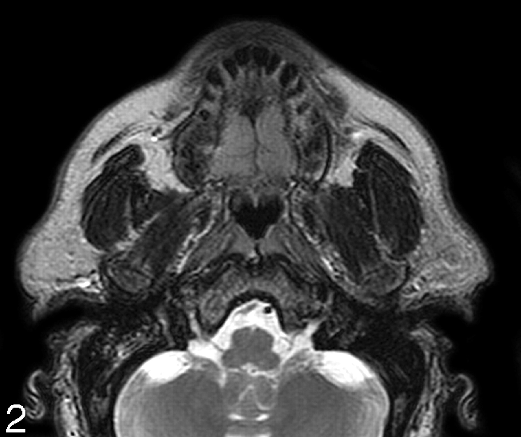
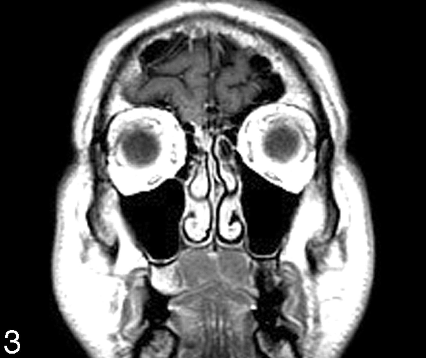
A 62-year-old man presented with a 3-week history of bilateral neck fullness and a mass in his palate. He had no pertinent prior medical history. Physical examination showed bilateral multiple nontender lymphadenopathy and a large firm palatal mass with a midline ridge. Blood work revealed leukocytosis and he was diagnosed as having chronic lymphocytic leukemia, stage 1. MR imaging showed smoothly lobulated soft tissue masses confined to the oral surface of the hard palate on either side of the midline. Compared with the skeletal muscle, the masses had overall intermediate T1-weighted (Fig. 1) and moderately high T2-weighted (Fig 2) signal intensity and enhanced uniformly (Fig 3). The patient also had diffuse homogeneous discrete lymphadenopathy. At this time, he is being clinically watched for any progression of disease, which would signal the initiation of appropriate therapy.

Fig 1.
  • Download figure
  • Open in new tab
  • Download powerpoint
Fig 1.

Axial T1-weighted MR image shows nodular soft tissue masses (arrows) of low-intermediate signal intensity along the oral surface at the level of hard palate.

Fig 2.
  • Download figure
  • Open in new tab
  • Download powerpoint
Fig 2.

Axial T2-weighted MR image shows that the masses have an intermediate signal intensity that is higher than in Fig. 1. The smoothly lobulated nature of the masses with a midline cleft can be well appreciated.

Fig 3.
  • Download figure
  • Open in new tab
  • Download powerpoint
Fig 3.

Coronal contrast enhanced T1-weighted MR image shows uniform enhancement of the masses confined to the oral surface of the hard palate with an incomplete midline furrow.

Discussion

The leukemias are divided into acute and chronic forms based on their clinical course, and the cell line involved further classifies them into myelocytic and lymphocytic types. The immunologically incompetent small neoplastic lymphocytes replace the bone marrow and infiltrate lymph nodes and various organs. The marrow replacement leads to a gradual loss of other cell lines, which manifests clinically as malaise (anemia), hemorrhage (thrombocytopenia), and infection (loss of competent white blood cells). The recirculating neoplastic lymphocytes, which make up more than 90% of the peripheral blood white cell count, infiltrate and enlarge the lymph nodes and visceral organs.4

The most common oral manifestations in leukemia, in order of frequency, include gingival hemorrhage, mucosal ulcers, infections, and gingival hyperplasia in tooth-bearing areas.6 Tooth loosening can occur from loss of the periodontal membrane. Leukemic infiltration of the tonsils and salivary glands can lead to their enlargement.4 In the head and neck, CLL most commonly presents as diffuse lymphadenopathy, often with quite large nodes. The oral manifestations of the chronic leukemias are less common than those associated with the acute leukemias.3, 5 In turn, the oral manifestations associated with CLL are the least common of all the chronic leukemias.

Palatal enlargement secondary to leukemic infiltration is rare. To our knowledge, there are only 3 prior, similar cases in the English language literature; none had CT or MR imaging studies.3–5 In these 3 prior patients, as with our patient, the palatal swelling was a painless, soft to firm, lobular mass with an overlying intact mucosa and an incomplete midline furrow. The relatively symmetric configuration of this process makes other palatal masses such as squamous carcinoma and minor salivary gland tumors quite unlikely.

Benign follicular lymphoid hyperplasia (FLH) affecting the oral surface of the hard palate is morphologically identical and, therefore, confusing in appearance and presentation. FLH in this location may present as a slowly growing, smoothly lobulated submucosal mass. Associated multicentric oral lesions and lymphadenopathy may also be present. Because isolated palatal enlargement can be the presenting feature of both CLL and FLH, and because FLH can recur after excision, it is important to distinguish these entities through pathologic examination.4, 7 Unlike CLL, which is a monoclonal leukemic infiltrate, FLH is characterized by an attenuated follicular lymphoid infiltrate within the lamina propria, replete with germinal centers.7, 8 These patients with either CLL or FLH palatal disease are typically asymptomatic for the palatal enlargement, except for ill-fitting upper dentures.4, 8,9

Other diagnostic considerations should include lymphomas of all grades, whether primary nodal or extranodal.4, 7, 9 Even though palatal involvement by lymphomas can present as soft, nontender, bilateral palatal swellings, in many cases, the palatal disease is unilateral, hard, painful, and accompanied by bone destruction.7 The final diagnostic differentiation between CLL, FHL, and the lymphomas can only be accomplished by pathologic examination.

Although a variety of other lesions (such as minor salivary gland neoplasms, torus palatinus, and infections and abscesses) can affect the hard palate, they are easily distinguished clinically and histologically from CLL.7

No prior description of the imaging findings of palatal involvement in CLL is available in literature. In our case, the MR imaging showed smoothly lobulated, uniformly enhancing soft tissue masses on either side of the midline confined to the oral cavity surface of the hard palate. There was overall intermediate T1-weighted and moderately high T2-weighted signal intensity (compared with skeletal muscle). These signal intensities are nonspecific with regard to the cell type that composes the mass. However, they suggest that this is not an inflammation-related process, because these latter conditions usually have higher T2-weighted signal intensities. In addition, the patient had the typical diffuse lymphadenopathy associated with CLL.10 The imaging findings correlated well with the clinical description of this lesion found in the literature.3–5

Summary

Palatal involvement in CLL is rare and has only been reported 3 previous times in the nonradiology literature. To our knowledge this is the first imaging description of this entity. Based on our experience, when smoothly lobulated, homogeneous masses are identified on the oral surface of the hard palate, especially in a patient who has a diffuse lymphadenopathy, the diagnosis of CLL should be considered.

References

  1. ↵
    Arber DA, Medeiros LJ. Hematopoietic lesions. In: Diagnostic Surgical Pathology of the Head and Neck. Gnepp DR, ed. Philadelphia: WB Saunders;2001 :725–76
  2. ↵
    Ioachim HL. Chronic lymphocytic leukemia. In: Lymph Node Pathology. 2nd ed. Philadelphia: JB Lippincott;1994 :531–538
  3. ↵
    Chaudhry AP, Sabes WR, Gorlin RJ. Unusual oral manifestations of chronic lymphatic leukemia. Report of a case. Oral Surg Oral Med Oral Pathol 1962;15:446–49
    PubMed
  4. ↵
    Henefer EP, Nelson JF, Beaupre EM. Palatal enlargement in chronic lymphocytic leukemia: report of case. J Oral Surg 1970;28:371–75
    PubMed
  5. ↵
    Porter SR, Matthews RW, Scully C. Chronic lymphocytic leukemia with gingival and palatal deposits. J Clin Periodontol 1994;21:559–61
    PubMed
  6. ↵
    Lynch MA, Ship II. Oral manifestations of leukemia: a postdiagnostic study. J Am Dent Assoc 1967;75:1139–44
    PubMed
  7. ↵
    Harsany DL, Ross J, Fee WE. Follicular lymphoid hyperplasia of the hard palate simulating lymphoma. Otolaryngol Head Neck Surg 1980;88:349–56
    PubMed
  8. ↵
    Menasce LP, Shanks JH, Banerjee SS, et al. Follicular lymphoid hyperplasia of the hard palate and oral mucosa: report of three cases and a review of the literature. Histopathology 2001;39:353–58
    PubMed
  9. ↵
    Nelson MA Jr, Stiff RH. An unusual manifestation of lymphoma. Oral Surg Oral Med Oral Pathol 1967;23:351–53
    PubMed
  10. ↵
    Som PM, Brandwein MS. Lymph nodes. In: Som PM, Curtin HD, eds. Head and Neck Imaging. 4th ed. St. Louis: Mosby;2003 :1865–934
  • Received April 14, 2005.
  • Accepted after revision September 30, 2005.
  • Copyright © American Society of Neuroradiology
View Abstract
PreviousNext
Back to top

In this issue

American Journal of Neuroradiology: 27 (8)
American Journal of Neuroradiology
Vol. 27, Issue 8
September 2006
  • Table of Contents
  • Index by author
Advertisement
Print
Download PDF
Email Article

Thank you for your interest in spreading the word on American Journal of Neuroradiology.

NOTE: We only request your email address so that the person you are recommending the page to knows that you wanted them to see it, and that it is not junk mail. We do not capture any email address.

Enter multiple addresses on separate lines or separate them with commas.
Palatal Enlargement in Chronic Lymphocytic Leukemia
(Your Name) has sent you a message from American Journal of Neuroradiology
(Your Name) thought you would like to see the American Journal of Neuroradiology web site.
CAPTCHA
This question is for testing whether or not you are a human visitor and to prevent automated spam submissions.
Cite this article
P. Vibhute, E. Carneiro, E. Genden, P.M. Som
Palatal Enlargement in Chronic Lymphocytic Leukemia
American Journal of Neuroradiology Sep 2006, 27 (8) 1649-1650;

Citation Manager Formats

  • BibTeX
  • Bookends
  • EasyBib
  • EndNote (tagged)
  • EndNote 8 (xml)
  • Medlars
  • Mendeley
  • Papers
  • RefWorks Tagged
  • Ref Manager
  • RIS
  • Zotero
0 Responses
Respond to this article
Share
Bookmark this article
Palatal Enlargement in Chronic Lymphocytic Leukemia
P. Vibhute, E. Carneiro, E. Genden, P.M. Som
American Journal of Neuroradiology Sep 2006, 27 (8) 1649-1650;
del.icio.us logo Twitter logo Facebook logo Mendeley logo
  • Tweet Widget
  • Facebook Like
  • Google Plus One

Jump to section

  • Article
    • Abstract
    • Case Report
    • Discussion
    • Summary
    • References
  • Figures & Data
  • Info & Metrics
  • Responses
  • References
  • PDF

Related Articles

  • No related articles found.
  • PubMed
  • Google Scholar

Cited By...

  • No citing articles found.
  • Crossref
  • Google Scholar

This article has not yet been cited by articles in journals that are participating in Crossref Cited-by Linking.

More in this TOC Section

  • FDG Uptake and Thyroid Cancer Post-BRAF Therapy
  • Normal Facial Nerve Enhancement on VIBE Sequence
  • Comprehensive Review of Inner Ear Anatomy on Photon-Counting CT
Show more Head and Neck Imaging

Similar Articles

Advertisement

Indexed Content

  • Current Issue
  • Accepted Manuscripts
  • Article Preview
  • Past Issues
  • Editorials
  • Editor's Choice
  • Fellows' Journal Club
  • Letters to the Editor
  • Video Articles

Cases

  • Case Collection
  • Archive - Case of the Week
  • Archive - Case of the Month
  • Archive - Classic Case

More from AJNR

  • Trainee Corner
  • Imaging Protocols
  • MRI Safety Corner

Multimedia

  • AJNR Podcasts
  • AJNR Scantastics

Resources

  • Turnaround Time
  • Submit a Manuscript
  • Submit a Video Article
  • Submit an eLetter to the Editor/Response
  • Manuscript Submission Guidelines
  • Statistical Tips
  • Fast Publishing of Accepted Manuscripts
  • Graphical Abstract Preparation
  • Imaging Protocol Submission
  • Evidence-Based Medicine Level Guide
  • Publishing Checklists
  • Author Policies
  • Become a Reviewer/Academy of Reviewers
  • News and Updates

About Us

  • About AJNR
  • Editorial Board
  • Editorial Board Alumni
  • Alerts
  • Permissions
  • Not an AJNR Subscriber? Join Now
  • Advertise with Us
  • Librarian Resources
  • Feedback
  • Terms and Conditions
  • AJNR Editorial Board Alumni

American Society of Neuroradiology

  • Not an ASNR Member? Join Now

© 2025 by the American Society of Neuroradiology All rights, including for text and data mining, AI training, and similar technologies, are reserved.
Print ISSN: 0195-6108 Online ISSN: 1936-959X

Powered by HighWire