Advertisement

Main menu

  • Home
  • Content
    • Current Issue
    • Accepted Manuscripts
    • Article Preview
    • Past Issue Archive
    • Video Articles
    • AJNR Case Collection
    • Case of the Week Archive
    • Case of the Month Archive
    • Classic Case Archive
  • Special Collections
    • Advancing NeuroMRI with High-Relaxivity Contrast Agents
    • AJNR Awards
    • Low-Field MRI
    • Alzheimer Disease
    • ASNR Foundation Special Collection
    • Photon-Counting CT
    • View All
  • Multimedia
    • AJNR Podcasts
    • AJNR SCANtastic
    • Trainee Corner
    • MRI Safety Corner
    • Imaging Protocols
  • For Authors
    • Submit a Manuscript
    • Submit a Video Article
    • Submit an eLetter to the Editor/Response
    • Manuscript Submission Guidelines
    • Statistical Tips
    • Fast Publishing of Accepted Manuscripts
    • Graphical Abstract Preparation
    • Imaging Protocol Submission
    • Author Policies
  • About Us
    • About AJNR
    • Editorial Board
    • Editorial Board Alumni
  • More
    • Become a Reviewer/Academy of Reviewers
    • Subscribers
    • Permissions
    • Alerts
    • Feedback
    • Advertisers
    • ASNR Home

User menu

  • Alerts
  • Log in

Search

  • Advanced search
American Journal of Neuroradiology

American Journal of Neuroradiology

ASHNR American Society of Functional Neuroradiology ASHNR American Society of Pediatric Neuroradiology ASSR
  • Alerts
  • Log in

Advanced Search

  • Home
  • Content
    • Current Issue
    • Accepted Manuscripts
    • Article Preview
    • Past Issue Archive
    • Video Articles
    • AJNR Case Collection
    • Case of the Week Archive
    • Case of the Month Archive
    • Classic Case Archive
  • Special Collections
    • Advancing NeuroMRI with High-Relaxivity Contrast Agents
    • AJNR Awards
    • Low-Field MRI
    • Alzheimer Disease
    • ASNR Foundation Special Collection
    • Photon-Counting CT
    • View All
  • Multimedia
    • AJNR Podcasts
    • AJNR SCANtastic
    • Trainee Corner
    • MRI Safety Corner
    • Imaging Protocols
  • For Authors
    • Submit a Manuscript
    • Submit a Video Article
    • Submit an eLetter to the Editor/Response
    • Manuscript Submission Guidelines
    • Statistical Tips
    • Fast Publishing of Accepted Manuscripts
    • Graphical Abstract Preparation
    • Imaging Protocol Submission
    • Author Policies
  • About Us
    • About AJNR
    • Editorial Board
    • Editorial Board Alumni
  • More
    • Become a Reviewer/Academy of Reviewers
    • Subscribers
    • Permissions
    • Alerts
    • Feedback
    • Advertisers
    • ASNR Home
  • Follow AJNR on Twitter
  • Visit AJNR on Facebook
  • Follow AJNR on Instagram
  • Join AJNR on LinkedIn
  • RSS Feeds

AJNR Awards, New Junior Editors, and more. Read the latest AJNR updates


Improved Turnaround Times | Median time to first decision: 12 days

OtherHead and Neck Imaging

Pituicytoma: Diagnostic Features on Selective Carotid Angiography and MR Imaging

W.N. Gibbs, E.S. Monuki, M.E. Linskey and A.N. Hasso
American Journal of Neuroradiology September 2006, 27 (8) 1639-1642;
W.N. Gibbs
  • Find this author on Google Scholar
  • Find this author on PubMed
  • Search for this author on this site
E.S. Monuki
  • Find this author on Google Scholar
  • Find this author on PubMed
  • Search for this author on this site
M.E. Linskey
  • Find this author on Google Scholar
  • Find this author on PubMed
  • Search for this author on this site
A.N. Hasso
  • Find this author on Google Scholar
  • Find this author on PubMed
  • Search for this author on this site
  • Article
  • Figures & Data
  • Info & Metrics
  • Responses
  • References
  • PDF
Loading

Article Figures & Data

Figures

  • Fig 1.
    • Download figure
    • Open in new tab
    • Download powerpoint
    Fig 1.

    A, Sagittal MR image shows a rounded mass in the suprasellar region, extending to the third ventricle. B, Coronal enhanced scan shows intense enhancement of the mass lesion, which appears to originate in the suprasellar region with fairly well-defined margins. The pituitary stalk, however, is not seen as a structure separate from the mass.

  • Fig 2.
    • Download figure
    • Open in new tab
    • Download powerpoint
    Fig 2.

    Selective bilateral internal carotid artery (ICA) angiograms. A, Early arterial lateral, (B) late arterial anteroposterior (AP), and (C) venous lateral magnified views of the left ICA injection. There are numerous vascular pedicles arising from the supraclinoid portion of the ICA that represent the various inferior and superior hypophyseal branches (arrows, A and B) that supply the neurohypophysis and hypothalamus. The meningohypophyseal trunk, which supplies the inferior hypophyseal artery as well as the dorsal meningeal artery, is also visible, arising from the posterior genu of the cavernous ICA (arrowheads). During the venous phase (C), the tumor stain is apparent, extending from the suprasellar region upwards in a “dumbbell” or “mushroom” pattern. There is a prominent portal vein draining into the dural venous sinus (arrow). D, Late venous phase of the lateral injection of the right ICA. The shape of the tumor stain is well seen with a caudad extension along the enlarged pituitary stalk and cephalad extension into the hypothalamus. The delayed tumor stain and prominence of the meningohypophyseal trunk at first glance suggest a meningioma of the diaphragma sella and suprasellar region.
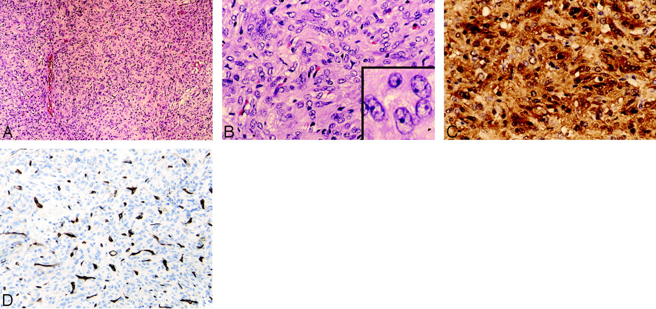
  • Fig 3.
    • Download figure
    • Open in new tab
    • Download powerpoint
    Fig 3.

    Tumor histopathology.

    A, The solid and cellular neoplasm is composed of rounded-to-spindled cells growing in a storiform pattern. No necrosis, nuclear palisading (Schwannoma), whorls (meningioma), or collagen bands (solitary fibrous tumor) are detected. Occasional large-caliber vessels are present, but the rich capillary network of the tumor is difficult to discern at low power (H&E, original magnification ×10).

    B, The tumor cells have abundant eosinophilic cytoplasm that lacks granularity. Tumor cell nuclei are large and have open chromatin, and many have a small distinct nucleolus. The rich capillary network is now visible. No mitotic figures, Rosenthal fibers, or eosinophilic granular bodies (pilocytic astrocytoma) are seen (H&E, original magnification ×20).

    C, The tumor shows strong and diffuse staining for S-100 in both the cytoplasm and nuclei of individual tumor cells (S-100, H&E, original magnification ×20, ×40).

    D, The endothelial cell marker CD34 highlights the rich vascular/capillary network of the tumor, whereas tumor cells are negative for this marker (CD34 stain, original magnification ×20).
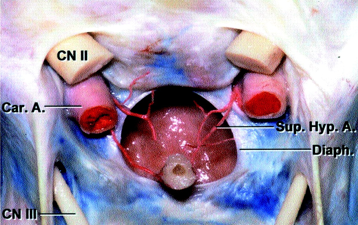
  • Fig 4.
    • Download figure
    • Open in new tab
    • Download powerpoint
    Fig 4.

    Color anatomic dissection of the sellar region. Frontal view of the optic chiasm and dissected diaphragma sella. The superior hypophyseal arteries are seen to branch into the pituitary stalk (Pit. Stalk) and extend cephalad toward the optic chiasm. (Reprinted with permission from Rhoton AL Jr. The sellar region. Neurosurgery 2002;51(suppl 1):335–74, [8.1])

PreviousNext
Back to top

In this issue

American Journal of Neuroradiology: 27 (8)
American Journal of Neuroradiology
Vol. 27, Issue 8
September 2006
  • Table of Contents
  • Index by author
Advertisement
Print
Download PDF
Email Article

Thank you for your interest in spreading the word on American Journal of Neuroradiology.

NOTE: We only request your email address so that the person you are recommending the page to knows that you wanted them to see it, and that it is not junk mail. We do not capture any email address.

Enter multiple addresses on separate lines or separate them with commas.
Pituicytoma: Diagnostic Features on Selective Carotid Angiography and MR Imaging
(Your Name) has sent you a message from American Journal of Neuroradiology
(Your Name) thought you would like to see the American Journal of Neuroradiology web site.
CAPTCHA
This question is for testing whether or not you are a human visitor and to prevent automated spam submissions.
Cite this article
W.N. Gibbs, E.S. Monuki, M.E. Linskey, A.N. Hasso
Pituicytoma: Diagnostic Features on Selective Carotid Angiography and MR Imaging
American Journal of Neuroradiology Sep 2006, 27 (8) 1639-1642;

Citation Manager Formats

  • BibTeX
  • Bookends
  • EasyBib
  • EndNote (tagged)
  • EndNote 8 (xml)
  • Medlars
  • Mendeley
  • Papers
  • RefWorks Tagged
  • Ref Manager
  • RIS
  • Zotero
0 Responses
Respond to this article
Share
Bookmark this article
Pituicytoma: Diagnostic Features on Selective Carotid Angiography and MR Imaging
W.N. Gibbs, E.S. Monuki, M.E. Linskey, A.N. Hasso
American Journal of Neuroradiology Sep 2006, 27 (8) 1639-1642;
del.icio.us logo Twitter logo Facebook logo Mendeley logo
  • Tweet Widget
  • Facebook Like
  • Google Plus One

Jump to section

  • Article
    • Abstract
    • Case Report
    • Discussion
    • References
  • Figures & Data
  • Info & Metrics
  • Responses
  • References
  • PDF

Related Articles

  • No related articles found.
  • PubMed
  • Google Scholar

Cited By...

  • Pituicytoma, Spindle Cell Oncocytoma, and Granular Cell Tumor: Clarification and Meta-Analysis of the World Literature since 1893
  • Pituicytoma: case report and literature review
  • Crossref
  • Google Scholar

This article has not yet been cited by articles in journals that are participating in Crossref Cited-by Linking.

More in this TOC Section

  • Diagnostic Performance of US in Neck Node NIRADS 2
  • Hydrops Herniation into the Semicircular Canals
  • ASL Sensitivity for Head and Neck Paraganglioma
Show more HEAD AND NECK IMAGING

Similar Articles

Advertisement

Indexed Content

  • Current Issue
  • Accepted Manuscripts
  • Article Preview
  • Past Issues
  • Editorials
  • Editor's Choice
  • Fellows' Journal Club
  • Letters to the Editor
  • Video Articles

Cases

  • Case Collection
  • Archive - Case of the Week
  • Archive - Case of the Month
  • Archive - Classic Case

More from AJNR

  • Trainee Corner
  • Imaging Protocols
  • MRI Safety Corner
  • Book Reviews

Multimedia

  • AJNR Podcasts
  • AJNR Scantastics

Resources

  • Turnaround Time
  • Submit a Manuscript
  • Submit a Video Article
  • Submit an eLetter to the Editor/Response
  • Manuscript Submission Guidelines
  • Statistical Tips
  • Fast Publishing of Accepted Manuscripts
  • Graphical Abstract Preparation
  • Imaging Protocol Submission
  • Evidence-Based Medicine Level Guide
  • Publishing Checklists
  • Author Policies
  • Become a Reviewer/Academy of Reviewers
  • News and Updates

About Us

  • About AJNR
  • Editorial Board
  • Editorial Board Alumni
  • Alerts
  • Permissions
  • Not an AJNR Subscriber? Join Now
  • Advertise with Us
  • Librarian Resources
  • Feedback
  • Terms and Conditions
  • AJNR Editorial Board Alumni

American Society of Neuroradiology

  • Not an ASNR Member? Join Now

© 2025 by the American Society of Neuroradiology All rights, including for text and data mining, AI training, and similar technologies, are reserved.
Print ISSN: 0195-6108 Online ISSN: 1936-959X

Powered by HighWire