
Article Figures & Data
Figures
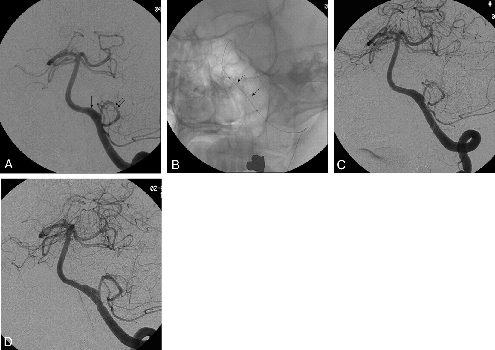
- Fig. 1.
Images from the case of a 50-year-old woman (patient 7) who had a fusiform aneurysm of the distal intracranial left vertebral artery.
A, Anteroposterior projection angiogram of the left vertebral artery disclosed a fusiform aneurysm of the distal intracranial portion (arrow) that is proximal to the vertebrobasilar junction and distal to the left posterior inferior cerebellar artery (PICA, double arrows).
B, Unsubtracted image demonstrates the deployed stent (arrows) across the aneurysm.
C, Anteroposterior projection angiogram obtained immediately after stent placement demonstrates no change in the fusiform aneurysm.
D, Angiogram obtained 19 months after stent deployment reveals no occlusion of the aneurysm.
- Fig. 2.
Angiograms in a 37-year-old man (patient 5) with dissecting aneurysm of the distal intracranial left vertebral artery treated with double stent method.
A, Anteroposterior projection shows a dissecting aneurysm of the distal intracranial left vertebral artery (arrow).
B, Angiogram obtained immediately after double stent placement demonstrates partial resolution of the aneurysm.
C, Angiogram obtained 6 months after double stent placement demonstrates complete healing of the aneurysm with restoration of the normal lumen.
- Fig. 3.
Angiograms in a 52-year-old man (patient 11) with dissecting aneurysm of the distal intracranial right vertebral artery treated with stent-assisted coiling.
A, The stent was placed across the aneurysm neck.
B, The aneurysm is occluded incompletely with coils.
C, Digital subtraction angiogram shows nearly completely occluded aneurysm with preservation of the parent artery.
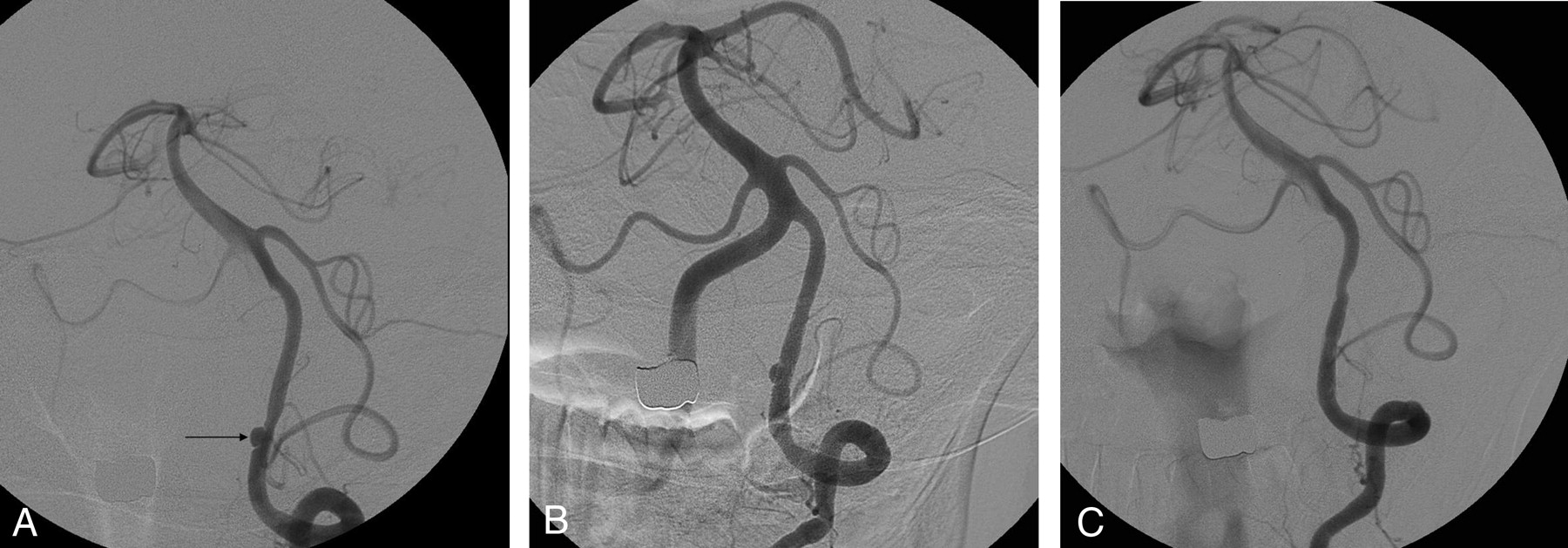
- Fig. 4.
Images from the case of a 55-year-old woman (patient 9) who had suffered SAH from a wide-necked aneurysm of the distal intracranial right vertebral artery.
A, Anteroposterior projection angiogram of the right vertebral artery disclosed an aneurysm of the distal intracranial portion that is proximal to the vertebrobasilar junction and distal to the right posterior inferior cerebellar artery.
B, The aneurysm was occluded near completely with coils.
C, Angiogram obtained 3 months after stent-assisted coiling demonstrates recanalization of the neck.


Tables
- Table 1:
Summary of the clinical characteristics in 16 patients with vertebrobasilar dissections treated with stents and/or coils
Patient No./Age (y)/Sex Clinical Presentations MRS on Admission Angiographic Sign Location 1/52/F Vertebrobasilar insufficiency 1 Fusiform dilation Left VA proximial to PICA 2/33/M Infarction, medullary 2 String sign Left VA across PICA 3/55/F Infarction, pontine 1 Pearl and string sign Right VA distal to PICA 4/54/M Infarction, cerebellar 1 Pearl and string sign Left VA across PICA 5/37/M Headache 1 Pearl and string sign Left VA distal to PICA 6/46/M Headache 1 Pearl and string sign Right VA proximal to PICA 7/50/F Headache 1 Fusiform dilation Left Va distal to PICA 8/67/M Infarction, medullary 2 Pearl and string sign Right Va proximal to PICA 9/55/F Subarachnoid hemorrhage 3 Fusiform dilation Right VA distal to PICA 10/64/M Headache 1 Pearl and string sign Left VA across PICA 11/52/M Vertebrobasilar insufficiency 1 Fusiform dilation Right VA across PICA 12/48/F Headache 1 Fusiform dilation Left Va proximal to PICA 13/53/M Subarachnoid hemorrhage 3 Pearl and string sign Left VA across PiCA 14/48/F Infarction, pontine 1 Pearl and string sign Right VA proximal to PICA Note:—MRS indicates modified Rankin Scale; VA, vertebral artery; PICA, posterior inferior cerebellar artery.
- Table 2:
Summary of treatment options, complication related to treatment, and angiographic and clinical outcomes
Patient No. Treatment Stent Type and Size (mm) Complications Immediate AO Follow-up AO at 6–12 Months Follow-up MRS 1 SPA AVE S670 4.0 × 24 None Incomplete Incomplete (stable) 1 2 SPA AVE S670 3.5 × 30 None Complete Complete 1 3 SAC AVE S670 3.5 × 24 None Complete Complete 0 4 SPA* AVE S670 3.0 × 9, 3.0 × 12 None Incomplete Complete 1 5 SPA* AVE S670 3.0 × 12, 3.5 × 12 None Incomplete Complete 0 6 SPA AVE S670 4.0 × 30 None Incomplete Incomplete (stable) 1 7 SPA AVE S670 3.0 × 24 None Incomplete Incomplete (stable) 0 8 SPA AVE Microdrive 3.5 × 24 None Incomplete Incomplete (stable) 1 9 SAC BS Neuroform 3.5 × 15 None Incomplete Incomplete (unstable) 2 10 SAC BS Neuroform 4.0 × 20 None Near complete Complete 1 11 SAC BS Neuroform 4.0 × 20 None Incomplete Complete 0 12 SPA* AVE Microdrive 3.0 × 12, 3.5 × 12 None Incomplete Complete 1 13 SPA* JOSTENT Flexmaster 2.75 × 19AVE Microdrive 2.75 × 24 Temporary vasospasm Incomplete Incomplete (stable) 1 14 SPA AVE Microdrive 3.5 × 24 None Near complete ND 1 Note:—AO indicates angiographic outcome; MRS, modified Rankin Scale; SPA, stent placement alone; SAC, stent-assisted coiling; AVE, Arterial Vascular Engineering; BS, Boston Scientific; ND, not done.
* Double stent method.


