Advertisement

Main menu

  • Home
  • Content
    • Current Issue
    • Accepted Manuscripts
    • Article Preview
    • Past Issue Archive
    • Video Articles
    • AJNR Case Collection
    • Case of the Week Archive
    • Case of the Month Archive
    • Classic Case Archive
  • Special Collections
    • AJNR Awards
    • Low-Field MRI
    • Alzheimer Disease
    • ASNR Foundation Special Collection
    • Photon-Counting CT
    • View All
  • Multimedia
    • AJNR Podcasts
    • AJNR SCANtastic
    • Trainee Corner
    • MRI Safety Corner
    • Imaging Protocols
  • For Authors
    • Submit a Manuscript
    • Submit a Video Article
    • Submit an eLetter to the Editor/Response
    • Manuscript Submission Guidelines
    • Statistical Tips
    • Fast Publishing of Accepted Manuscripts
    • Graphical Abstract Preparation
    • Imaging Protocol Submission
    • Author Policies
  • About Us
    • About AJNR
    • Editorial Board
    • Editorial Board Alumni
  • More
    • Become a Reviewer/Academy of Reviewers
    • Subscribers
    • Permissions
    • Alerts
    • Feedback
    • Advertisers
    • ASNR Home

User menu

  • Alerts
  • Log in

Search

  • Advanced search
American Journal of Neuroradiology

American Journal of Neuroradiology

ASHNR American Society of Functional Neuroradiology ASHNR American Society of Pediatric Neuroradiology ASSR
  • Alerts
  • Log in

Advanced Search

  • Home
  • Content
    • Current Issue
    • Accepted Manuscripts
    • Article Preview
    • Past Issue Archive
    • Video Articles
    • AJNR Case Collection
    • Case of the Week Archive
    • Case of the Month Archive
    • Classic Case Archive
  • Special Collections
    • AJNR Awards
    • Low-Field MRI
    • Alzheimer Disease
    • ASNR Foundation Special Collection
    • Photon-Counting CT
    • View All
  • Multimedia
    • AJNR Podcasts
    • AJNR SCANtastic
    • Trainee Corner
    • MRI Safety Corner
    • Imaging Protocols
  • For Authors
    • Submit a Manuscript
    • Submit a Video Article
    • Submit an eLetter to the Editor/Response
    • Manuscript Submission Guidelines
    • Statistical Tips
    • Fast Publishing of Accepted Manuscripts
    • Graphical Abstract Preparation
    • Imaging Protocol Submission
    • Author Policies
  • About Us
    • About AJNR
    • Editorial Board
    • Editorial Board Alumni
  • More
    • Become a Reviewer/Academy of Reviewers
    • Subscribers
    • Permissions
    • Alerts
    • Feedback
    • Advertisers
    • ASNR Home
  • Follow AJNR on Twitter
  • Visit AJNR on Facebook
  • Follow AJNR on Instagram
  • Join AJNR on LinkedIn
  • RSS Feeds

AJNR Awards, New Junior Editors, and more. Read the latest AJNR updates

Research ArticleFUNCTIONAL

A Functional MRI Study: Cerebral Laterality for Lexical-Semantic Processing and Human Voice Perception

M. Koeda, H. Takahashi, N. Yahata, K. Asai, Y. Okubo and H. Tanaka
American Journal of Neuroradiology August 2006, 27 (7) 1472-1479;
M. Koeda
  • Find this author on Google Scholar
  • Find this author on PubMed
  • Search for this author on this site
H. Takahashi
  • Find this author on Google Scholar
  • Find this author on PubMed
  • Search for this author on this site
N. Yahata
  • Find this author on Google Scholar
  • Find this author on PubMed
  • Search for this author on this site
K. Asai
  • Find this author on Google Scholar
  • Find this author on PubMed
  • Search for this author on this site
Y. Okubo
  • Find this author on Google Scholar
  • Find this author on PubMed
  • Search for this author on this site
H. Tanaka
  • Find this author on Google Scholar
  • Find this author on PubMed
  • Search for this author on this site
  • Article
  • Figures & Data
  • Info & Metrics
  • Responses
  • References
  • PDF
Loading

Article Figures & Data

Figures

  • Tables
  • Fig. 1.
    • Download figure
    • Open in new tab
    • Download powerpoint
    Fig. 1.

    A, reverse sentences (rSEN); B, identifiable nonvocal sounds (SND). C, sentences (SEN).
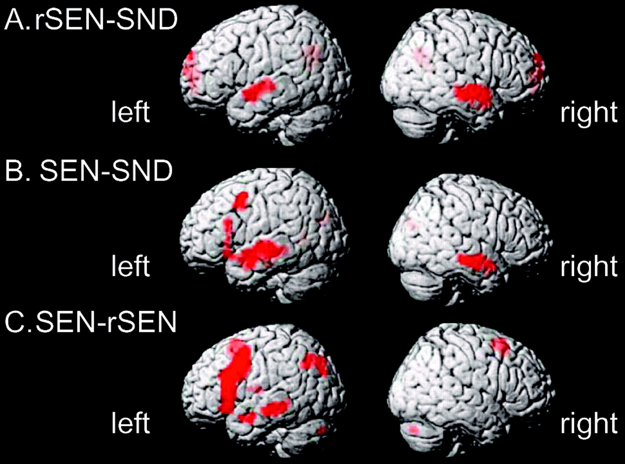
  • Fig. 2.
    • Download figure
    • Open in new tab
    • Download powerpoint
    Fig. 2.

    Activated areas revealed by the contrasts. rSEN-SND (A), SEN-SND (B), and SEN-rSEN (C) with a statistical threshold of P < .00005 (random effect model uncorrected; extent threshold at 10 voxels). SEN, sentences; rSEN, reverse sentences; SND, identifiable nonvocal sounds.
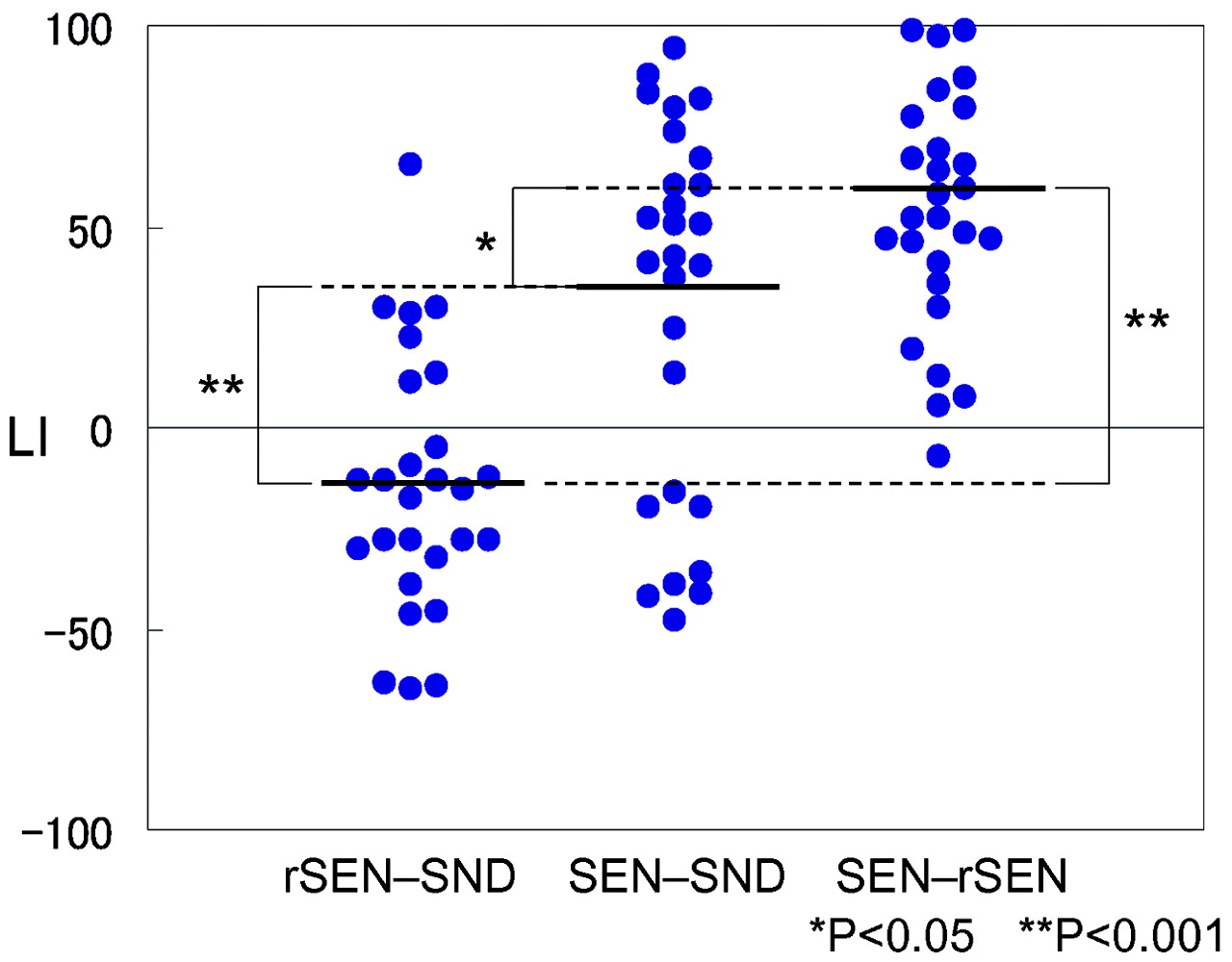
  • Fig. 3.
    • Download figure
    • Open in new tab
    • Download powerpoint
    Fig. 3.

    LI distribution of the temporal activation under rSEN-SND, SEN-SND, and SEN-rSEN contrasts: The bold line shows the mean of LI under each contrast. One-way ANOVA and multiple comparison by Bonferroni test for individual LI in temporal activation was significantly different among the 3 contrasts (ANOVA: F (2, 78) = 26.28, P < .001, Bonferroni: P < .05). *, P < .05; **, P < .001.

  • Fig. 4.
    • Download figure
    • Open in new tab
    • Download powerpoint
    Fig. 4.

    Individual variability of LI of the temporal cortices: symmetrical or right-lateralized activation was observed in 22 of 27 subjects (81.4%) under the rSEN-SND contrast in the temporal cortices. Although 9 of 27 subjects (33.3%) showed symmetrical or right-lateralized activation under the SEN-SND contrast in the temporal cortices, all subjects showed left hemisphere dominance under the SEN-rSEN contrast. *, P < .05; **, P < .001.

Tables

  • Figures
    • View popup
    Table 1

    Cognitive systems involved in listening to SEN, rSEN, and SND

    SNDrSENSENrSEN-SNDSEN-SNDSEN-rSEN
    Lexical-semantic processing−−+−++
    Human voice perception−++++−
    Tonal fluctuation+++−−−
    Attention+++−−−
    Primary auditory processing+++−−−
    • Note:—SND indicates nonvocal sounds; rSEN, reverse sentences; SEN, sentences. SND, rSEN, and SEN are in the context of subjects scanned by functional MR imaging while listening to these sounds.

    • View popup
    Table 2

    Peak coordinates (x, y, z) and their z values of cerebral activation under three contrasts (rSEN-SND, SEN-SND, SEN-rSEN)

    Brain RegionsContrast
    rSEN-SNDSEN-SNDSEN-rSEN
    LeftRightLeftRightLeftRight
    xyzz Valuexyzz Valuexyzz Valuexyzz Valuexyzz Valuexyzz Value
    Frontal cortices
        MFG BA6−459456.29−459458.29
        IFG
            Operculum BA9/46−5421214.54−5421278.38
            Triangular BA45−542104.38−542436.45
        SFG BA6−315606.61
    Temporal cortices
        Anterior
            STS BA22−51−3−66.28513−36.16−570−96.68
            MTG BA21−510−124.93510−96.83−57−3−186.6463−12−186.42−540−215.90
        Central
            STS BA22−51−9−36.1451−6−36.34−54−1204.86
            MTG BA2154−12−96.83−57−15−154.8361−21−94.82−57−27−126.69
        Heschl’s gyrus BA4154−21124.16
        Posterior
            STS BA22−48−2134.68
            MTG BA2145−18−94.68−54−33−94.90−54−33−66.38
    Parietal cortices
        Precuneus BA19−33−78365.75
    Cerebellum9−78−336.16
    • Note:—SND indicates identifiable nonvocal sounds; rSEN, reverse sentences; SEN, sentences. SND, rSEN, and SEN are in the context of subjects scanned by functional MR imaging while listening to these sounds. MFG indicates middle frontal gyrus; IFG, inferior frontal gyrus; SFG, superior frontal gyrus; STS, superior temporal sulcus; MTG, middle temporal gyrus. Activation differences were considered significant at height threshold (P < .00005, random effect model uncorrected) and extent threshold (10 voxels).

PreviousNext
Back to top

In this issue

American Journal of Neuroradiology: 27 (7)
American Journal of Neuroradiology
Vol. 27, Issue 7
August 2006
  • Table of Contents
  • Index by author
Advertisement
Print
Download PDF
Email Article

Thank you for your interest in spreading the word on American Journal of Neuroradiology.

NOTE: We only request your email address so that the person you are recommending the page to knows that you wanted them to see it, and that it is not junk mail. We do not capture any email address.

Enter multiple addresses on separate lines or separate them with commas.
A Functional MRI Study: Cerebral Laterality for Lexical-Semantic Processing and Human Voice Perception
(Your Name) has sent you a message from American Journal of Neuroradiology
(Your Name) thought you would like to see the American Journal of Neuroradiology web site.
CAPTCHA
This question is for testing whether or not you are a human visitor and to prevent automated spam submissions.
Cite this article
M. Koeda, H. Takahashi, N. Yahata, K. Asai, Y. Okubo, H. Tanaka
A Functional MRI Study: Cerebral Laterality for Lexical-Semantic Processing and Human Voice Perception
American Journal of Neuroradiology Aug 2006, 27 (7) 1472-1479;

Citation Manager Formats

  • BibTeX
  • Bookends
  • EasyBib
  • EndNote (tagged)
  • EndNote 8 (xml)
  • Medlars
  • Mendeley
  • Papers
  • RefWorks Tagged
  • Ref Manager
  • RIS
  • Zotero
0 Responses
Respond to this article
Share
Bookmark this article
A Functional MRI Study: Cerebral Laterality for Lexical-Semantic Processing and Human Voice Perception
M. Koeda, H. Takahashi, N. Yahata, K. Asai, Y. Okubo, H. Tanaka
American Journal of Neuroradiology Aug 2006, 27 (7) 1472-1479;
del.icio.us logo Twitter logo Facebook logo Mendeley logo
  • Tweet Widget
  • Facebook Like
  • Google Plus One

Jump to section

  • Article
    • Abstract
    • Materials and Methods
    • Results
    • Discussion
    • Conclusion
    • Appendix 1: Contents of the Sentences
    • Appendix 2: Contents of the Questions in the Pilot Study Questionnaire
    • Acknowledgments
    • Footnotes
    • References
  • Figures & Data
  • Info & Metrics
  • Responses
  • References
  • PDF

Related Articles

  • No related articles found.
  • PubMed
  • Google Scholar

Cited By...

  • Cerebral laterality of writing in right- and left- handers: A functional transcranial Doppler ultrasound study
  • Crossref
  • Google Scholar

This article has not yet been cited by articles in journals that are participating in Crossref Cited-by Linking.

More in this TOC Section

  • Kurtosis and Epileptogenic Tubers: A Pilot Study
  • Glutaric Aciduria Type 1: DK vs. Conventional MRI
  • Multiparametric MRI in PEDS Pontine Glioma
Show more FUNCTIONAL

Similar Articles

Advertisement

Indexed Content

  • Current Issue
  • Accepted Manuscripts
  • Article Preview
  • Past Issues
  • Editorials
  • Editor's Choice
  • Fellows' Journal Club
  • Letters to the Editor
  • Video Articles

Cases

  • Case Collection
  • Archive - Case of the Week
  • Archive - Case of the Month
  • Archive - Classic Case

More from AJNR

  • Trainee Corner
  • Imaging Protocols
  • MRI Safety Corner
  • Book Reviews

Multimedia

  • AJNR Podcasts
  • AJNR Scantastics

Resources

  • Turnaround Time
  • Submit a Manuscript
  • Submit a Video Article
  • Submit an eLetter to the Editor/Response
  • Manuscript Submission Guidelines
  • Statistical Tips
  • Fast Publishing of Accepted Manuscripts
  • Graphical Abstract Preparation
  • Imaging Protocol Submission
  • Evidence-Based Medicine Level Guide
  • Publishing Checklists
  • Author Policies
  • Become a Reviewer/Academy of Reviewers
  • News and Updates

About Us

  • About AJNR
  • Editorial Board
  • Editorial Board Alumni
  • Alerts
  • Permissions
  • Not an AJNR Subscriber? Join Now
  • Advertise with Us
  • Librarian Resources
  • Feedback
  • Terms and Conditions
  • AJNR Editorial Board Alumni

American Society of Neuroradiology

  • Not an ASNR Member? Join Now

© 2025 by the American Society of Neuroradiology All rights, including for text and data mining, AI training, and similar technologies, are reserved.
Print ISSN: 0195-6108 Online ISSN: 1936-959X

Powered by HighWire