Advertisement

Main menu

  • Home
  • Content
    • Current Issue
    • Accepted Manuscripts
    • Article Preview
    • Past Issue Archive
    • Video Articles
    • AJNR Case Collection
    • Case of the Week Archive
    • Case of the Month Archive
    • Classic Case Archive
  • Special Collections
    • AJNR Awards
    • Low-Field MRI
    • Alzheimer Disease
    • ASNR Foundation Special Collection
    • Photon-Counting CT
    • View All
  • Multimedia
    • AJNR Podcasts
    • AJNR SCANtastic
    • Trainee Corner
    • MRI Safety Corner
    • Imaging Protocols
  • For Authors
    • Submit a Manuscript
    • Submit a Video Article
    • Submit an eLetter to the Editor/Response
    • Manuscript Submission Guidelines
    • Statistical Tips
    • Fast Publishing of Accepted Manuscripts
    • Graphical Abstract Preparation
    • Imaging Protocol Submission
    • Author Policies
  • About Us
    • About AJNR
    • Editorial Board
    • Editorial Board Alumni
  • More
    • Become a Reviewer/Academy of Reviewers
    • Subscribers
    • Permissions
    • Alerts
    • Feedback
    • Advertisers
    • ASNR Home

User menu

  • Alerts
  • Log in

Search

  • Advanced search
American Journal of Neuroradiology

American Journal of Neuroradiology

ASHNR American Society of Functional Neuroradiology ASHNR American Society of Pediatric Neuroradiology ASSR
  • Alerts
  • Log in

Advanced Search

  • Home
  • Content
    • Current Issue
    • Accepted Manuscripts
    • Article Preview
    • Past Issue Archive
    • Video Articles
    • AJNR Case Collection
    • Case of the Week Archive
    • Case of the Month Archive
    • Classic Case Archive
  • Special Collections
    • AJNR Awards
    • Low-Field MRI
    • Alzheimer Disease
    • ASNR Foundation Special Collection
    • Photon-Counting CT
    • View All
  • Multimedia
    • AJNR Podcasts
    • AJNR SCANtastic
    • Trainee Corner
    • MRI Safety Corner
    • Imaging Protocols
  • For Authors
    • Submit a Manuscript
    • Submit a Video Article
    • Submit an eLetter to the Editor/Response
    • Manuscript Submission Guidelines
    • Statistical Tips
    • Fast Publishing of Accepted Manuscripts
    • Graphical Abstract Preparation
    • Imaging Protocol Submission
    • Author Policies
  • About Us
    • About AJNR
    • Editorial Board
    • Editorial Board Alumni
  • More
    • Become a Reviewer/Academy of Reviewers
    • Subscribers
    • Permissions
    • Alerts
    • Feedback
    • Advertisers
    • ASNR Home
  • Follow AJNR on Twitter
  • Visit AJNR on Facebook
  • Follow AJNR on Instagram
  • Join AJNR on LinkedIn
  • RSS Feeds

AJNR Awards, New Junior Editors, and more. Read the latest AJNR updates

OtherBRAIN

MR Spectroscopy-Aided Differentiation: “Giant” Extra-Axial Tuberculoma Masquerading as Meningioma

P.C. Khanna, S. Godinho, D.P. Patkar, S.A. Pungavkar and M.A. Lawande
American Journal of Neuroradiology August 2006, 27 (7) 1438-1440;
P.C. Khanna
  • Find this author on Google Scholar
  • Find this author on PubMed
  • Search for this author on this site
S. Godinho
  • Find this author on Google Scholar
  • Find this author on PubMed
  • Search for this author on this site
D.P. Patkar
  • Find this author on Google Scholar
  • Find this author on PubMed
  • Search for this author on this site
S.A. Pungavkar
  • Find this author on Google Scholar
  • Find this author on PubMed
  • Search for this author on this site
M.A. Lawande
  • Find this author on Google Scholar
  • Find this author on PubMed
  • Search for this author on this site
  • Article
  • Figures & Data
  • Info & Metrics
  • Responses
  • References
  • PDF
Loading

Abstract

SUMMARY: Tuberculosis is common in the developing world and in developed nations secondary to increasing immunocompromise in the population. It commonly causes meningitis and parenchymal tuberculomas. We present a case of an unusual masslike “giant” extra-axial tuberculoma during pregnancy. Unusual morphology and size at imaging made meningioma a close differential. MR spectroscopy served to complement MR imaging, providing diagnostic confirmation and depicted findings characteristic of a tuberculoma.

Tuberculosis, an important public health problem compounded by the upsurge of the human immunodeficiency virus usually results from Mycobacterium tuberculosis infection, though Mycobacterium bovis, a frequent pathogen of the past, and Mycobacterium avium complex (M avium and M intracellulare) are infrequently encountered. Central nervous system (CNS) tuberculomas typically result from hematogenous dissemination and, upon histologic examination, are granulomas with central caseous necrosis.

Although intra-axial tuberculous granulomata are the more common variety, extra-axial lesions are rarely encountered1–6 and may present diagnostic dilemmas that need to be resolved by imaging and allied techniques, so as to circumvent unnecessary chemotherapy, biopsies, and surgery. MR spectroscopy is one such technique that we used.

Case Report

A 25-year-old pregnant patient (gravida 1, para 0) presented at 27 weeks’ gestation with severe headaches and focal right-sided convulsions of 3 months’ duration without fever, papilledema, or elevated blood pressure. Elevated erythrocyte sedimentation rate (ESR) was noted. Postpartum lumbar puncture and CSF analysis after imaging studies revealed mild lymphocytic predominance and positive polymerase chain reaction (PCR) for tuberculosis. Antituberculous therapy was begun after suspending breast-feeding.

Two MR examinations (1.5T EchoSpeed; GE Medical Systems, Milwaukee, Wis) obtained at 27 weeks and postpartum revealed an unchanged 3.8 × 2.75 × 3.0-cm left frontal extraparenchymal mass lesion that appeared hypointense on T1-weighted images (T1WI; Fig 1A), with a thick, isohypointense rim. On T2-weighted images (T2WI, Fig 1B) and fluid-attenuated inversion recovery (FLAIR), it appeared predominantly hypointense with central hyperintensity, consistent with necrosis. The lesion appeared dark on diffusion-weighted images (DWI; Fig 2A). Contrast (gadolinium-diethylene-triaminepentaacetic acid [Gd-DTPA]) enhanced MR (Fig 2B) depicted prominent heterogeneous central enhancement with a thick, peripheral nonenhancing wall and peripheral rim-enhancement. Adjoining meningeal enhancement (“dural tail”), adjacent CSF cleft, mass-effect, and vasogenic edema were noted. MR angiography and venography revealed no abnormal vascularity or occlusion. MR perfusion revealed no changes in cerebral blood volume or flow. A differential diagnosis of inflammatory granuloma (with leptomeningitis and cerebritis) versus meningioma was considered.

Fig 1.
  • Download figure
  • Open in new tab
  • Download powerpoint
Fig 1.

Axial T1WI (A; fast spin echo; TR/TE = 2015ms/9.7ms) and T2WI (B; fast spin echo; TR/TE = 4360ms/83ms) depicting a lobulated, dura-based lesion in the left frontal convexity that is predominantly hypointense on T1WI with a thick, iso-hypointense rim. On T2WI, it is of mixed intensity but predominantly hypointense with a hyperintense center. Displacement of the underlying parenchyma and a CSF cleft are identified (arrows). A large component of vasogenic edema is visualized.

Fig 2.
  • Download figure
  • Open in new tab
  • Download powerpoint
Fig 2.

Axial DWI (A; TR/TE = 3000ms/11ms) depicts a dark-appearing lesion, with a hyperintense rim (arrows). Axial contrast-enhanced T1WI (B; Gd-DTPA enhanced fast spin echo; TR/TE = 2015ms/9.7ms) depicts prominent central enhancement, a thick peripheral nonenhancing component and rim-enhancement beyond this. There is thick enhancement of adjoining meninges (“dural-tail” sign, arrow) with mild enhancement of meninges in the anterior interhemispheric region and the right cerebral convexity (not shown).

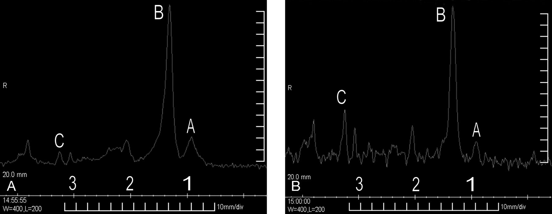
A volume of interest of 8.0 mL was selected from the center of the lesion on T2WI and image-guided, single-voxel, point-resolved MR spectroscopy (PRESS; repetition time [TR]/echo time [TE] = 1500ms /35ms) was used, because with this technique, lipid contamination in volumes abutting fat structures is negligible. Multivoxel MR spectroscopy was obtained as well. Findings were consistent with necrotic inflammatory granuloma/tuberculoma, revealing elevated lipid-lactate and choline (Cho) peaks with barely detectable N-acetylaspartate (NAA) and creatine (Cr). No abnormal peak was detected corresponding to alanine. Follow-up MR spectroscopy (Fig 3A,B) remained unchanged. Long TE (144 ms; Fig 3B) MR spectroscopy sequence obtained from an identical location did not suppress the 1.33-ppm peak, confirming predominance of lipid. Findings were more conspicuous with multivoxel spectroscopy.

Fig 3.
  • Download figure
  • Open in new tab
  • Download powerpoint
Fig 3.

Single-voxel PRESS MR spectroscopy (A; TR/TE = 1500ms/35ms). Adequate water suppression has been achieved. Reading from right to left, peaks A and B (tall and broad) at 0.9 and 1.33 ppm, respectively, represent typical long-chain lipids (lipid/lactate). NAA and Cr are barely detectable. A small Cho peak, C, is seen to resonate at 3.2 ppm. Long TE MR spectroscopy (B; TR/TE = 1500ms/144ms) depicts persistence of predominant lipid peak at 1.33 ppm.

Based on imaging, spectroscopic, and clinical features, a diagnosis of giant extra-axial tuberculoma was made. Surgical excision biopsy confirmed the diagnosis.

Discussion

Although intraparenchymal tuberculomas are common, solid extra-axial tuberculous masses are extremely uncommon.1 We believe our case is unique in that our patient was pregnant and immunocompetent, MR findings mimicked those of a similarly located isolated meningioma, and we were able to successfully use spectroscopy to confirm our diagnosis.

Extra-axial tuberculomas simulating meningiomas may be located in the frontoparietal areas2 and pontine, pericallosal,4 and suprasellar cisterns. Cystic1 and en-plaque meningioma5 mimics and those caused by M avium (noted in patients with systemic lupus erythematosus3) can be encountered. Most appear hypointense to isointense to gray matter1–6 on T1WI and T2WI and variable on DWI, often with a hyperintense rim; those with hyperintense centers on T2WI are usually hyperintense on DWI.7 Meningiomas typically appear isointense to gray matter on T1WI and T2WI and DWI, unless atypical or aggressive.8 Signal intensity is a function of intralesional lipids, macrophages, fibrosis, and cellular infiltrates.9

In vivo proton MR spectroscopy has been studied extensively in this context. Tuberculomas are characterized by a prominent decrease in NAA/Cr and slight decrease in NAA/Cho.10 Lipid-lactate peaks are usually elevated (86% of tuberculomas11). Paradoxically, lipid/Cr may occasionally be decreased relative to normal cerebral parenchyma, probably resulting from small dimensions of most tuberculomas relative to voxel volume. 3D multivoxel proton spectroscopy with 2D chemical shift imaging interrogates relatively small voxel volumes and overcomes this paradox.12 Meningiomas invariably have elevated alanine. A high lipid/Cr ratio may also be noted.10 “Finger-printing” of M tuberculosis cell-wall biochemicals in tuberculomas is now possible, facilitating their detection.13 T1-weighted magnetization transfer MR imaging is a useful adjunct to MR spectroscopy and shows promise in tissue characterization of CNS tuberculomas.13

Conclusions

When diagnostic dilemmas present themselves, MR spectroscopy considered in perspective with MR imaging and clinicopathologic features can be useful in certain situations. Rare as they are, extra-axial tuberculomas may masquerade as meningiomas. To our knowledge, ours is the first report of an extra-axial “giant” tuberculoma that bore a striking resemblance to meningioma and in which diagnostic confirmation was obtained using proton MR spectroscopy that was later corroborated by surgical biopsy and histopathology.

References

  1. ↵
    Shindo A, Honda C, Baba Y. A case of an intracranial tuberculoma, mimicking meningioma that developed during treatment with anti-tuberculous agents. No Shinkei Geka 1999;27:837–41
    PubMed
  2. ↵
    Isenmann S, Zimmermann DR, Wichmann W, et al. Tuberculoma mimicking meningioma of the falx cerebri. PCR diagnosis of mycobacterial DNA from formalin-fixed tissue. Clin Neuropathol 1996;15:155–58
    PubMed
  3. ↵
    Di Patre PL, Radziszewski W, Martin NA, et al. A meningioma-mimicking tumor caused by Mycobacterium avium complex in an immunocompromised patient. Am J Surg Pathol 2000;24:136–39
    CrossRefPubMed
  4. ↵
    Adachi K, Yoshida K, Tomita H, et al. Tuberculoma mimicking falx meningioma–case report. Neurol Med Chir (Tokyo) 2004;44:489–92
    PubMed
  5. ↵
    Bauer J, Johnson RF, Levy JM, et al. Tuberculoma presenting as an en plaque meningioma. Case report. J Neurosurg 1996;85:685–88
    PubMed
  6. ↵
    Lindner A, Schneider C, Hofmann E, et al. Isolated meningeal tuberculoma mimicking meningioma: case report. Surg Neurol 1995;43:81–84
    CrossRefPubMed
  7. ↵
    Batra A, Tripathi RP. Diffusion-weighted magnetic resonance imaging and magnetic resonance spectroscopy in the evaluation of focal cerebral tubercular lesions. Acta Radiol 2004;45:679–88
    PubMed
  8. ↵
    Filippi CG, Edgar MA, Ulug AM, et al. Appearance of meningiomas on diffusion-weighted images: correlating diffusion constants with histopathologic findings. AJNR Am J Neuroradiol 2001;22:65–72
    Abstract/FREE Full Text
  9. ↵
    Gupta RK, Pandey R, Khan EM, et al. Intracranial tuberculomas: MRI signal intensity correlation with histopathology and localised proton spectroscopy. Magn Reson Imaging 1993;11:443–49
    CrossRefPubMed
  10. ↵
    Bulakbasi N, Kocaoglu M, Örs F, et al. Combination of single-voxel proton MR spectroscopy and apparent diffusion coefficient calculation in the evaluation of common brain tumors. AJNR Am J Neuroradiol 2003;24:225–33
    Abstract/FREE Full Text
  11. ↵
    Pretell EJ, Martinot C Jr., Garcia HH, et al. Cysticercosis working group in Peru. Differential diagnosis between cerebral tuberculosis and neurocysticercosis by magnetic resonance spectroscopy. J Comput Assist Tomogr 2005;29:112–14
    CrossRefPubMed
  12. ↵
    Gonen O, Murdoch JB, Stoyanova R, et al. 3D multivoxel proton spectroscopy of human brain using a hybrid of 8th-order Hadamard encoding with 2D chemical shift imaging. Magn Reson Med 1998;39:34–40
    PubMed
  13. ↵
    Gupta RK, Husain M, Vatsal DK, et al. Comparative evaluation of magnetization transfer MR imaging and in-vivo proton MR spectroscopy in brain tuberculomas. Magn Reson Imaging 2002;20:375–81
    CrossRefPubMed
  • Received July 11, 2005.
  • Accepted after revision August 31, 2005.
  • Copyright © American Society of Neuroradiology
View Abstract
PreviousNext
Back to top

In this issue

American Journal of Neuroradiology: 27 (7)
American Journal of Neuroradiology
Vol. 27, Issue 7
August 2006
  • Table of Contents
  • Index by author
Advertisement
Print
Download PDF
Email Article

Thank you for your interest in spreading the word on American Journal of Neuroradiology.

NOTE: We only request your email address so that the person you are recommending the page to knows that you wanted them to see it, and that it is not junk mail. We do not capture any email address.

Enter multiple addresses on separate lines or separate them with commas.
MR Spectroscopy-Aided Differentiation: “Giant” Extra-Axial Tuberculoma Masquerading as Meningioma
(Your Name) has sent you a message from American Journal of Neuroradiology
(Your Name) thought you would like to see the American Journal of Neuroradiology web site.
CAPTCHA
This question is for testing whether or not you are a human visitor and to prevent automated spam submissions.
Cite this article
P.C. Khanna, S. Godinho, D.P. Patkar, S.A. Pungavkar, M.A. Lawande
MR Spectroscopy-Aided Differentiation: “Giant” Extra-Axial Tuberculoma Masquerading as Meningioma
American Journal of Neuroradiology Aug 2006, 27 (7) 1438-1440;

Citation Manager Formats

  • BibTeX
  • Bookends
  • EasyBib
  • EndNote (tagged)
  • EndNote 8 (xml)
  • Medlars
  • Mendeley
  • Papers
  • RefWorks Tagged
  • Ref Manager
  • RIS
  • Zotero
0 Responses
Respond to this article
Share
Bookmark this article
MR Spectroscopy-Aided Differentiation: “Giant” Extra-Axial Tuberculoma Masquerading as Meningioma
P.C. Khanna, S. Godinho, D.P. Patkar, S.A. Pungavkar, M.A. Lawande
American Journal of Neuroradiology Aug 2006, 27 (7) 1438-1440;
del.icio.us logo Twitter logo Facebook logo Mendeley logo
  • Tweet Widget
  • Facebook Like
  • Google Plus One

Jump to section

  • Article
    • Abstract
    • Case Report
    • Discussion
    • Conclusions
    • References
  • Figures & Data
  • Info & Metrics
  • Responses
  • References
  • PDF

Related Articles

  • No related articles found.
  • PubMed
  • Google Scholar

Cited By...

  • No citing articles found.
  • Crossref
  • Google Scholar

This article has not yet been cited by articles in journals that are participating in Crossref Cited-by Linking.

More in this TOC Section

  • Multimodal CT Provides Improved Performance for Lacunar Infarct Detection
  • Optimal MRI Sequence for Identifying Occlusion Location in Acute Stroke: Which Value of Time-Resolved Contrast-Enhanced MRA?
  • Evaluating the Effects of White Matter Multiple Sclerosis Lesions on the Volume Estimation of 6 Brain Tissue Segmentation Methods
Show more BRAIN

Similar Articles

Advertisement

Indexed Content

  • Current Issue
  • Accepted Manuscripts
  • Article Preview
  • Past Issues
  • Editorials
  • Editor's Choice
  • Fellows' Journal Club
  • Letters to the Editor
  • Video Articles

Cases

  • Case Collection
  • Archive - Case of the Week
  • Archive - Case of the Month
  • Archive - Classic Case

More from AJNR

  • Trainee Corner
  • Imaging Protocols
  • MRI Safety Corner
  • Book Reviews

Multimedia

  • AJNR Podcasts
  • AJNR Scantastics

Resources

  • Turnaround Time
  • Submit a Manuscript
  • Submit a Video Article
  • Submit an eLetter to the Editor/Response
  • Manuscript Submission Guidelines
  • Statistical Tips
  • Fast Publishing of Accepted Manuscripts
  • Graphical Abstract Preparation
  • Imaging Protocol Submission
  • Evidence-Based Medicine Level Guide
  • Publishing Checklists
  • Author Policies
  • Become a Reviewer/Academy of Reviewers
  • News and Updates

About Us

  • About AJNR
  • Editorial Board
  • Editorial Board Alumni
  • Alerts
  • Permissions
  • Not an AJNR Subscriber? Join Now
  • Advertise with Us
  • Librarian Resources
  • Feedback
  • Terms and Conditions
  • AJNR Editorial Board Alumni

American Society of Neuroradiology

  • Not an ASNR Member? Join Now

© 2025 by the American Society of Neuroradiology All rights, including for text and data mining, AI training, and similar technologies, are reserved.
Print ISSN: 0195-6108 Online ISSN: 1936-959X

Powered by HighWire