
Article Figures & Data
Figures
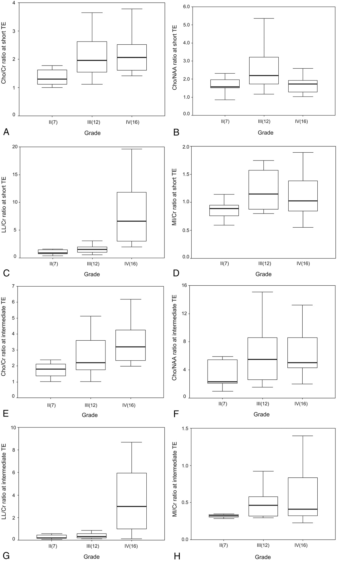
- Fig 1.
Box plots of the ratios of the metabolites at both TE sequences in the patients with cerebral gliomas. The horizontal thick line is the median, the upper and lower ends of the boxes are the 3rd and 1st quartiles, and the vertical lines show the full range of values in the data. The extremes and outliers were omitted.
A, Cho/Cr at short TE; B, Cho/NAA at short TE; C, LL/Cr at short TE; D, mIns/Cr at short TE; E, Cho/Cr at intermediate TE; F, Cho/NAA at intermediate TE; G, LL/Cr at intermediate TE; H, mIns/Cr at intermediate TE.
- Fig 2.
A 35-year-old man with low-grade (grade II) astrocytoma.
A, Axial T2-weighted image reveals diffuse infiltrating glioma involving left basal ganglia and frontotemporal area with a square voxel (6.2 cm3).
B, 1H-MR spectrum obtained at short TE (2000/35) shows increased Cho/Cr ratio (1.9:1), increased Cho/NAA ratio (2.3:1) and increased lipid-lactate (LL)/Cr ratio (1.6:1).
C, 1H-MR spectrum obtained at intermediate TE (1500/144) shows more increased Cho/Cr ratio (2.4:1), more increased Cho/NAA ratio (4.9:1) and hardly discernible peak of lipid-lactate between 0.9 and 1.3 ppm (LL/Cr ratio: 0.3:1). The baseline of the spectrum is noisier than B.
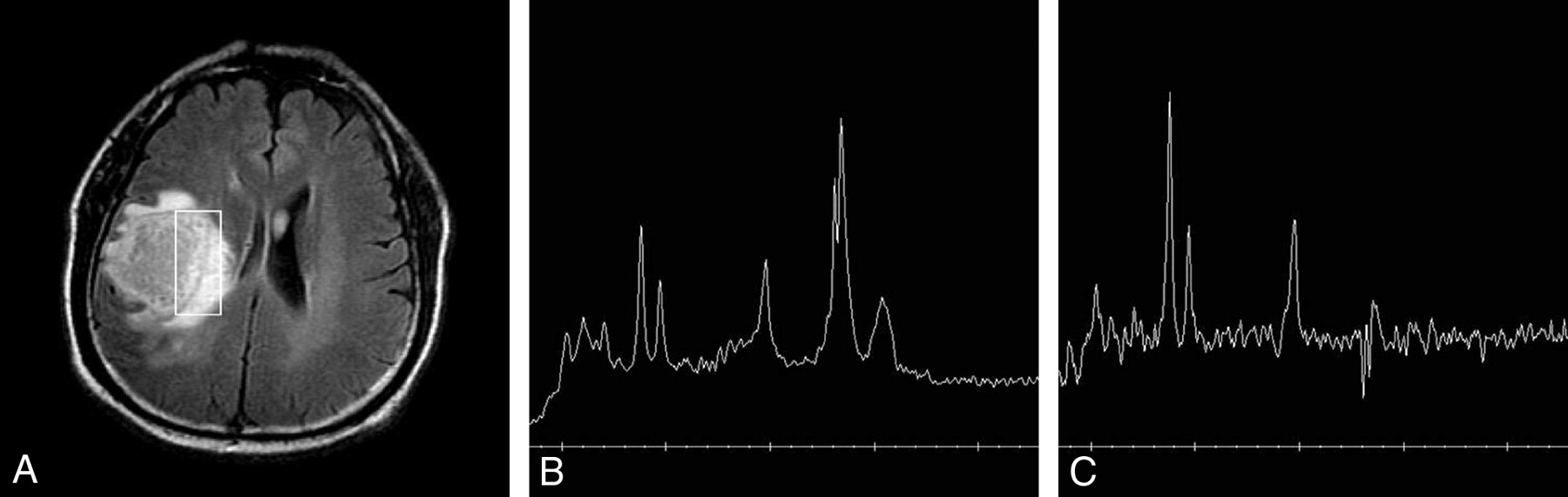
- Fig 3.
A 38-year-old man with anaplastic (grade III) oligodendroglioma.
A, Axial FLAIR image reveals a infiltrating tumor in the right parietal lobe with a square voxel (4.2 cm3).
B, 1H-MR spectrum obtained at short TE (2000/35) shows increased Cho/Cr ratio (2.4:1), increased Cho/NAA ratio (4.1:1), and increased lipid-lactate (LL)/Cr ratio (2.1:1).
C, 1H-MR spectrum obtained at intermediate TE (1500/144) shows more increased Cho/Cr ratio (2.9:1), more increased Cho/NAA ratio (5.7:1), and much smaller peaks of lipid-lactate at 1.3 ppm (LL/Cr: 0.8:1). The baseline of the spectrum is noisier than B.
- Fig 4.
A 61-year-old man with glioblastoma (grade IV).
A, Axial FLAIR image reveals a large necrotic tumor in the right frontal lobe with a rectangular voxel (8.0 cm3).
B, 1H-MR spectrum obtained at short TE (2000/35) shows increased Cho/Cr ratio (1.5:1), increased Cho/NAA ratio (1.2:1), and markedly increased LL/Cr ratio (3.8:1) at 1.3 ppm.
C, 1H-MR spectrum obtained at intermediate TE (1500/144) shows more increased Cho/Cr ratio of (2.2:1), more increased Cho/NAA ratio (2.1:1), and a smaller inverted lactate peak with smaller lipid peaks at 1.2–1.4 and 0.9 ppm (LL/Cr: 0.9:1). The baseline of the spectrum is noisier than B.
- Fig 5.
Graph shows 4 ROC curves of Cho/Cr and LL/Cr ratios at both TEs for differentiation of high-grade glioma from low-grade glioma. Az value (area under the ROC curve) is the highest in LL/Cr ratio at short TE and lowest in Cho/Cr ratio at intermediate TE. But there are no significant differences among all the Az values.


Tables
- Table 1:
Metabolite ratios of 3 grades of cerebral gliomas at both echo time (TE) sequences
Short TE Intermediate TE Cho/Cr Cho/NAA LL/Cr mIns/Cr Cho/Cr Cho/NAA LL/Cr mIns/Cr Grade II (n = 7) 1.52 ± 0.66 1.88 ± 0.96 1.07 ± 0.46 0.86 ± 0.19 1.92 ± 0.89 4.57 ± 4.35 0.30 ± 0.21 0.36 ± 0.11 Grade III (n = 12) 2.09 ± 0.82 2.51 ± 1.20 1.62 ± 0.80 1.23 ± 0.37 2.64 ± 1.39 8.14 ± 9.52 0.38 ± 0.23 0.49 ± 0.19 Grade IV (n = 16) 2.21 ± 0.79 1.66 ± 0.46 10.31 ± 10.22 1.15 ± 0.52 4.25 ± 3.83 6.31 ± 3.54 4.20 ± 4.95 0.74 ± 0.75 Note:—Cho indicates choline, Cr, creatine; NAA, N-acetylaspartate; LL, lipid and lactate; MI, myo-inositol.
- Table 2:
Metabolite ratios of low-grade (II) and high-grade (III + IV) cerebral gliomas at both echo time (TE) sequences
Short TE Intermediate TE Cho/Cr Cho/NAA LL/Cr mIns/Cr Cho/Cr Cho/NAA LL/Cr mIns/Cr Low grade (II) (n = 7) 1.52 ± 0.66 1.88 ± 0.96 1.07 ± 0.46 0.86 ± 0.19 1.92 ± 0.89 4.57 ± 4.35 0.30 ± 0.21 0.36 ± 0.11 High grade (III + IV) (n = 28) 2.17 ± 0.79 2.02 ± 0.94 6.17 ± 8.33 1.18 ± 0.45 3.56 ± 3.10 7.09 ± 6.69 2.56 ± 4.17 0.63 ± 0.59 P value .0152 NS <.001 NS .0111 NS .0152 NS Note:—Cho indicates choline, Cr, creatine; NAA, N-acetylaspartate; LL, lipid and lactate; mIns, myo-inositol; NS, not significant.
- Table 3:
Diagnostic accuracies of Cho/Cr and/or LL/Cr ratios at short echo time (TE), intermediate TE, and both TEs in differentiation between low-grade (II) and high-grade (III + IV) gliomas using the cutoff (threshold) values obtained with minimum C1 error
Accuracy (%) Sensitivity (%) Specificity (%) PPV (%) NPV (%) Grading with Cho/Cr and/or LL/Cr at short TE 85.7 92.9 57.1 89.7 66.7 Grading with Cho/Cr and/or LL/Cr at intermediate TE 82.9 89.3 57.1 89.3 57.1 Grading with Cho/Cr and/or LL/Cr at both TEs 85.7 96.4 42.9 87.1 75.0 Note:—Cho indicates choline, Cr, creatine; NAA, N-acetylaspartate; LL, lipid and lactate; mIns, myo-inositol; PPV, positive predictive value; NPV, negative predictive value.


