Advertisement

Main menu

  • Home
  • Content
    • Current Issue
    • Accepted Manuscripts
    • Article Preview
    • Past Issue Archive
    • Video Articles
    • AJNR Case Collection
    • Case of the Week Archive
    • Case of the Month Archive
    • Classic Case Archive
  • Special Collections
    • AJNR Awards
    • Low-Field MRI
    • Alzheimer Disease
    • ASNR Foundation Special Collection
    • Photon-Counting CT
    • View All
  • Multimedia
    • AJNR Podcasts
    • AJNR SCANtastic
    • Trainee Corner
    • MRI Safety Corner
    • Imaging Protocols
  • For Authors
    • Submit a Manuscript
    • Submit a Video Article
    • Submit an eLetter to the Editor/Response
    • Manuscript Submission Guidelines
    • Statistical Tips
    • Fast Publishing of Accepted Manuscripts
    • Graphical Abstract Preparation
    • Imaging Protocol Submission
    • Author Policies
  • About Us
    • About AJNR
    • Editorial Board
    • Editorial Board Alumni
  • More
    • Become a Reviewer/Academy of Reviewers
    • Subscribers
    • Permissions
    • Alerts
    • Feedback
    • Advertisers
    • ASNR Home

User menu

  • Alerts
  • Log in

Search

  • Advanced search
American Journal of Neuroradiology

American Journal of Neuroradiology

ASHNR American Society of Functional Neuroradiology ASHNR American Society of Pediatric Neuroradiology ASSR
  • Alerts
  • Log in

Advanced Search

  • Home
  • Content
    • Current Issue
    • Accepted Manuscripts
    • Article Preview
    • Past Issue Archive
    • Video Articles
    • AJNR Case Collection
    • Case of the Week Archive
    • Case of the Month Archive
    • Classic Case Archive
  • Special Collections
    • AJNR Awards
    • Low-Field MRI
    • Alzheimer Disease
    • ASNR Foundation Special Collection
    • Photon-Counting CT
    • View All
  • Multimedia
    • AJNR Podcasts
    • AJNR SCANtastic
    • Trainee Corner
    • MRI Safety Corner
    • Imaging Protocols
  • For Authors
    • Submit a Manuscript
    • Submit a Video Article
    • Submit an eLetter to the Editor/Response
    • Manuscript Submission Guidelines
    • Statistical Tips
    • Fast Publishing of Accepted Manuscripts
    • Graphical Abstract Preparation
    • Imaging Protocol Submission
    • Author Policies
  • About Us
    • About AJNR
    • Editorial Board
    • Editorial Board Alumni
  • More
    • Become a Reviewer/Academy of Reviewers
    • Subscribers
    • Permissions
    • Alerts
    • Feedback
    • Advertisers
    • ASNR Home
  • Follow AJNR on Twitter
  • Visit AJNR on Facebook
  • Follow AJNR on Instagram
  • Join AJNR on LinkedIn
  • RSS Feeds

AJNR Awards, New Junior Editors, and more. Read the latest AJNR updates

OtherSpine Imaging and Spine Image-Guided Interventions

Oligodendroglioma Metastatic to Bone Marrow

Firas Al-Ali, Aaron J. Hendon, Marcia K. Liepman, Jennifer L. Wisniewski, Mark J. Krinock and Kevin Beckman
American Journal of Neuroradiology October 2005, 26 (9) 2410-2414;
Firas Al-Ali
  • Find this author on Google Scholar
  • Find this author on PubMed
  • Search for this author on this site
Aaron J. Hendon
  • Find this author on Google Scholar
  • Find this author on PubMed
  • Search for this author on this site
Marcia K. Liepman
  • Find this author on Google Scholar
  • Find this author on PubMed
  • Search for this author on this site
Jennifer L. Wisniewski
  • Find this author on Google Scholar
  • Find this author on PubMed
  • Search for this author on this site
Mark J. Krinock
  • Find this author on Google Scholar
  • Find this author on PubMed
  • Search for this author on this site
Kevin Beckman
  • Find this author on Google Scholar
  • Find this author on PubMed
  • Search for this author on this site
  • Article
  • Figures & Data
  • Info & Metrics
  • Responses
  • References
  • PDF
Loading

Article Figures & Data

Figures

  • Fig 1.
    • Download figure
    • Open in new tab
    • Download powerpoint
    Fig 1.

    The figures show a small well-delineated nonenhancing right temporal lobe mass with predominately low signal intensity in T1-weighted (A) and predominately increased signal intensity in T2-weighted (B) images, involving white and gray marrow consistent with oligodendroglioma.

  • Fig 2.
    • Download figure
    • Open in new tab
    • Download powerpoint
    Fig 2.

    The neoplasm is of moderate cellularity and composed of relatively uniform cells with round nuclei. Mitotic figures or pleomorphism is not identified.

  • Fig 3.
    • Download figure
    • Open in new tab
    • Download powerpoint
    Fig 3.

    Four years later, interval tumor recurrence is seen in the posterior aspect of the surgical cavity with a homogeneous increased signal intensity seen in the T2-weighted (A) image and low signal intensity in the T1-weighted (B) image without enhancement.
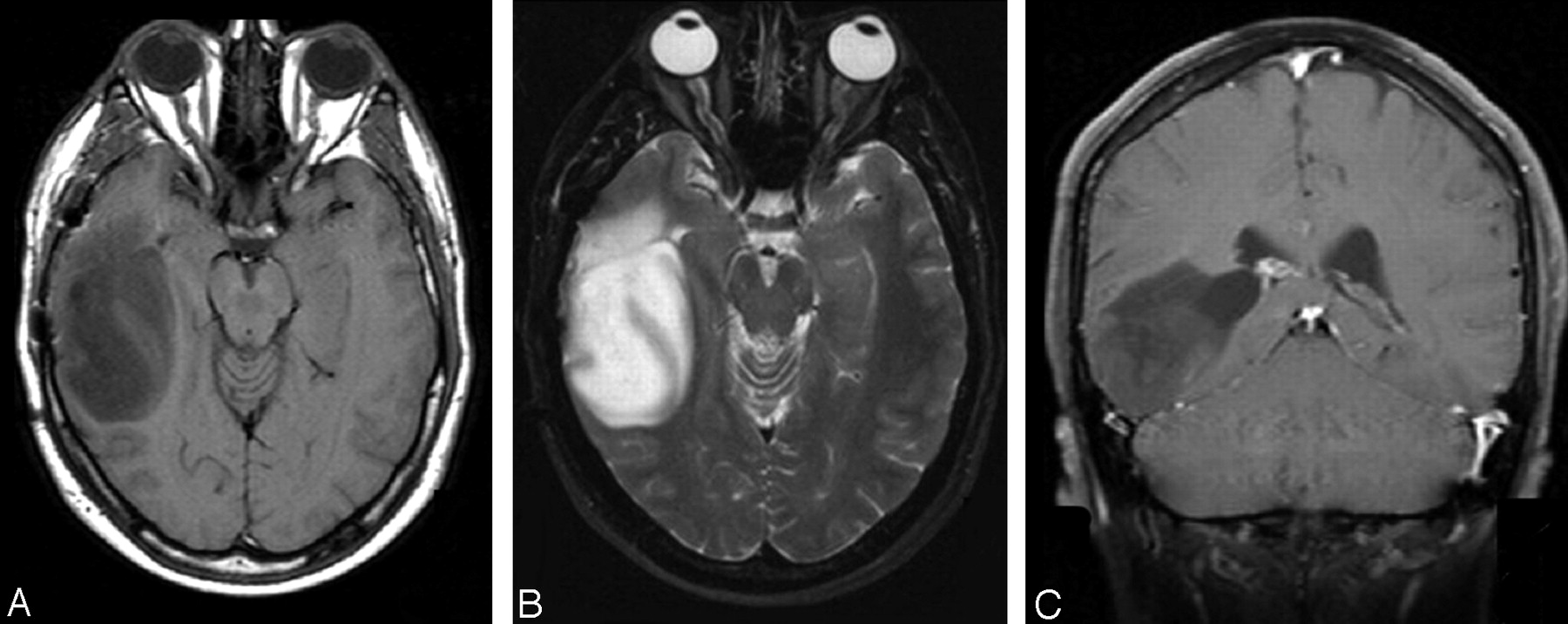
  • Fig 4.
    • Download figure
    • Open in new tab
    • Download powerpoint
    Fig 4.

    A second tumor recurrence is seen 8 years after initial diagnosis. A large homogenous mass is present, abutting the right lateral ventricle with low signal intensity on T1-weighted (A) and increased signal intensity on T2-weighted (B) images. No evidence of abnormal enhancement and of either subependymal or leptomeningeal spread is seen on the coronal T1-weighted postgadolinium (C) image.
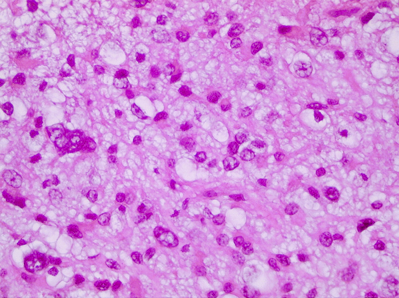
  • Fig 5.
    • Download figure
    • Open in new tab
    • Download powerpoint
    Fig 5.

    Histologic image shows anaplastic oligodendroglioma with increased cellularity, with mitotic activity and increased nuclear pleomorphism.
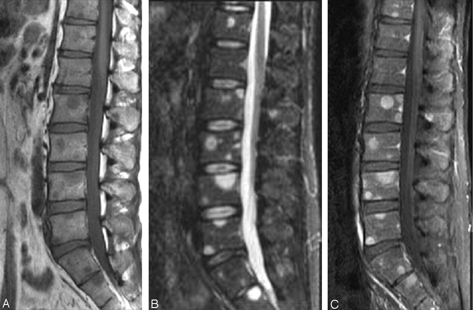
  • Fig 6.
    • Download figure
    • Open in new tab
    • Download powerpoint
    Fig 6.

    There are numerous areas of signal-intensity abnormality throughout the vertebral body with abnormal low signal intensity in T1-weighted (A) images corresponding to increased signal intensity on T2-weighted (B) images and diffuse abnormal enhancement after gadolinium administration (C). Note the absence of a compression deformity despite the presence of diffuse bony metastases.
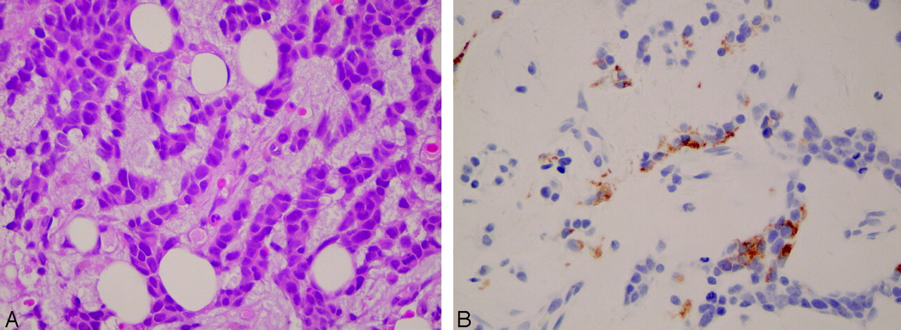
  • Fig 7.
    • Download figure
    • Open in new tab
    • Download powerpoint
    Fig 7.

    Metastatic poorly differentiated neoplasm in the bone marrow with scant cytoplasm and large hyperchromatic nuclei (A). The same specimen shows cytoplasmic staining for glial fibrillary acidic protein in tumor cells in the bone marrow (B).

  • Fig 8.
    • Download figure
    • Open in new tab
    • Download powerpoint
    Fig 8.

    The sagittal T1-weighted (A) image demonstrates abnormal diffuse low signal intensity of the vertebral body consistent with replacement of the normal fatty marrow by tumor cells as well as interval development of a posterior epidural mass compressing the cord. Sagittal T1-weighted postgadolinium with fat saturation (B) image demonstrates intense enhancement of the epidural mass with a dural tail sign as well as diffuse enhancement of the vertebral bodies. Sagittal T2-weighted with fat saturation (C) image demonstrates the epidural mass having homogeneous increased signal intensity with subtle but diffuse increased signal intensity of the vertebral body.

PreviousNext
Back to top

In this issue

American Journal of Neuroradiology: 26 (9)
American Journal of Neuroradiology
Vol. 26, Issue 9
1 Oct 2005
  • Table of Contents
  • Index by author
Advertisement
Print
Download PDF
Email Article

Thank you for your interest in spreading the word on American Journal of Neuroradiology.

NOTE: We only request your email address so that the person you are recommending the page to knows that you wanted them to see it, and that it is not junk mail. We do not capture any email address.

Enter multiple addresses on separate lines or separate them with commas.
Oligodendroglioma Metastatic to Bone Marrow
(Your Name) has sent you a message from American Journal of Neuroradiology
(Your Name) thought you would like to see the American Journal of Neuroradiology web site.
CAPTCHA
This question is for testing whether or not you are a human visitor and to prevent automated spam submissions.
Cite this article
Firas Al-Ali, Aaron J. Hendon, Marcia K. Liepman, Jennifer L. Wisniewski, Mark J. Krinock, Kevin Beckman
Oligodendroglioma Metastatic to Bone Marrow
American Journal of Neuroradiology Oct 2005, 26 (9) 2410-2414;

Citation Manager Formats

  • BibTeX
  • Bookends
  • EasyBib
  • EndNote (tagged)
  • EndNote 8 (xml)
  • Medlars
  • Mendeley
  • Papers
  • RefWorks Tagged
  • Ref Manager
  • RIS
  • Zotero
0 Responses
Respond to this article
Share
Bookmark this article
Oligodendroglioma Metastatic to Bone Marrow
Firas Al-Ali, Aaron J. Hendon, Marcia K. Liepman, Jennifer L. Wisniewski, Mark J. Krinock, Kevin Beckman
American Journal of Neuroradiology Oct 2005, 26 (9) 2410-2414;
del.icio.us logo Twitter logo Facebook logo Mendeley logo
  • Tweet Widget
  • Facebook Like
  • Google Plus One

Jump to section

  • Article
    • Abstract
    • Case Report
    • Discussion
    • References
  • Figures & Data
  • Info & Metrics
  • Responses
  • References
  • PDF

Related Articles

  • No related articles found.
  • PubMed
  • Google Scholar

Cited By...

  • Bone Marrow Metastases from a 1p/19q Co-deleted Oligodendroglioma - A Case Report
  • Crossref
  • Google Scholar

This article has not yet been cited by articles in journals that are participating in Crossref Cited-by Linking.

More in this TOC Section

  • Management Outcomes For VO Spine Biopsy
  • Cone Beam CT Myelography
  • Post-Procedural Brachial Neuritis Features
Show more Spine Imaging and Spine Image-Guided Interventions

Similar Articles

Advertisement

Indexed Content

  • Current Issue
  • Accepted Manuscripts
  • Article Preview
  • Past Issues
  • Editorials
  • Editor's Choice
  • Fellows' Journal Club
  • Letters to the Editor
  • Video Articles

Cases

  • Case Collection
  • Archive - Case of the Week
  • Archive - Case of the Month
  • Archive - Classic Case

More from AJNR

  • Trainee Corner
  • Imaging Protocols
  • MRI Safety Corner
  • Book Reviews

Multimedia

  • AJNR Podcasts
  • AJNR Scantastics

Resources

  • Turnaround Time
  • Submit a Manuscript
  • Submit a Video Article
  • Submit an eLetter to the Editor/Response
  • Manuscript Submission Guidelines
  • Statistical Tips
  • Fast Publishing of Accepted Manuscripts
  • Graphical Abstract Preparation
  • Imaging Protocol Submission
  • Evidence-Based Medicine Level Guide
  • Publishing Checklists
  • Author Policies
  • Become a Reviewer/Academy of Reviewers
  • News and Updates

About Us

  • About AJNR
  • Editorial Board
  • Editorial Board Alumni
  • Alerts
  • Permissions
  • Not an AJNR Subscriber? Join Now
  • Advertise with Us
  • Librarian Resources
  • Feedback
  • Terms and Conditions
  • AJNR Editorial Board Alumni

American Society of Neuroradiology

  • Not an ASNR Member? Join Now

© 2025 by the American Society of Neuroradiology All rights, including for text and data mining, AI training, and similar technologies, are reserved.
Print ISSN: 0195-6108 Online ISSN: 1936-959X

Powered by HighWire