
Article Figures & Data
Figures
- Fig 1.
Schematic diagram of the lateral and anteroposterior views show the origin of the proatlantal artery from the external carotid artery at the second vertebral body level. Occipital artery origin from proatlantal artery is seen.
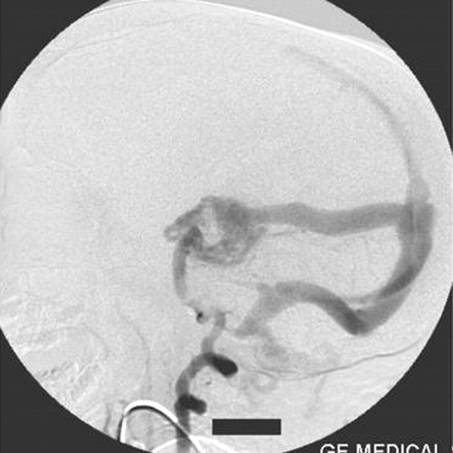
- Fig 2.
Right vertebral artery injection lateral view angiogram shows filling of the Galen’s vein fistula and the falcine sinus.
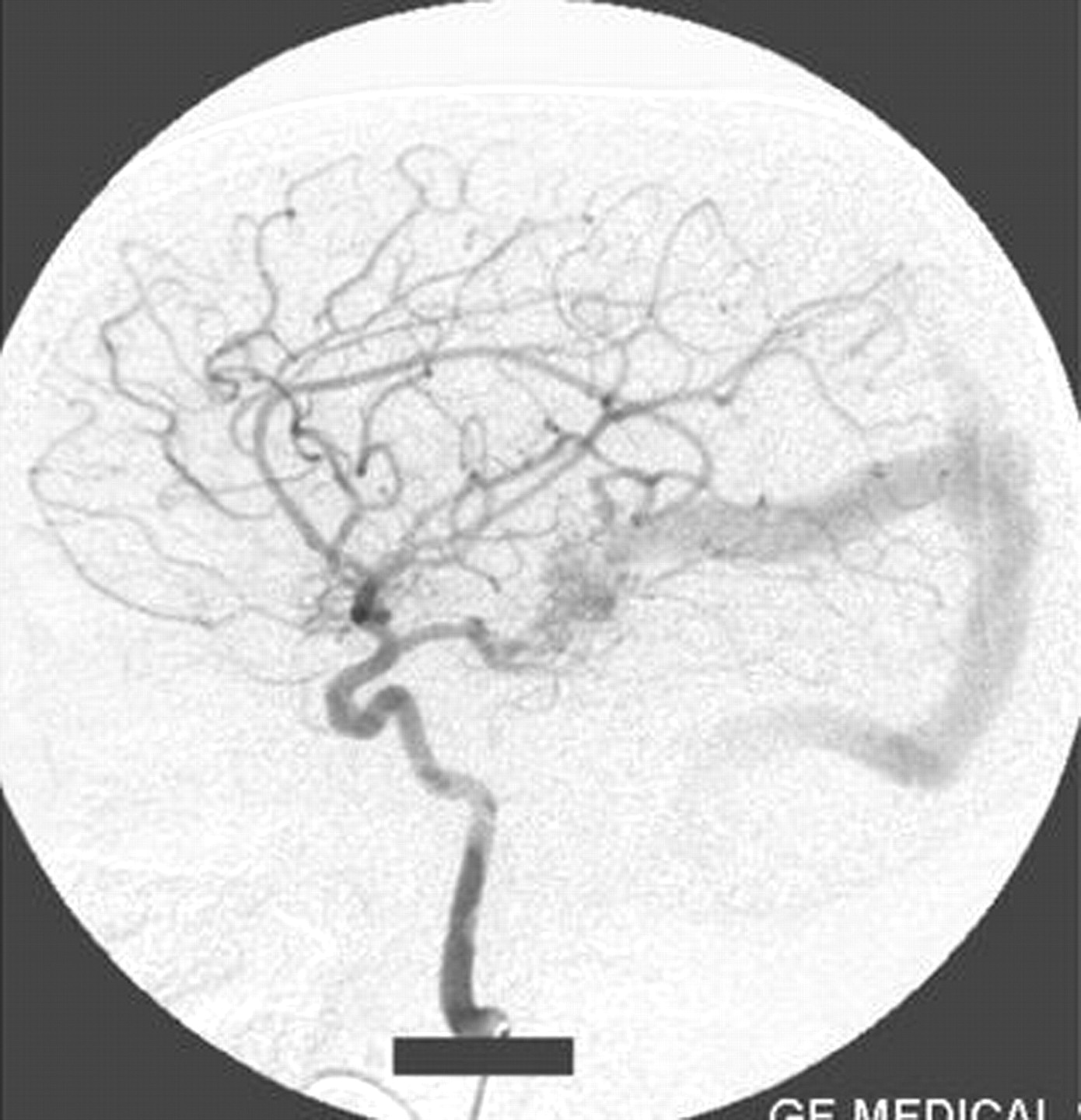
- Fig 3.
Right internal carotid artery injection lateral view angiogram shows the filling of the fistula through the posterior communicating artery.
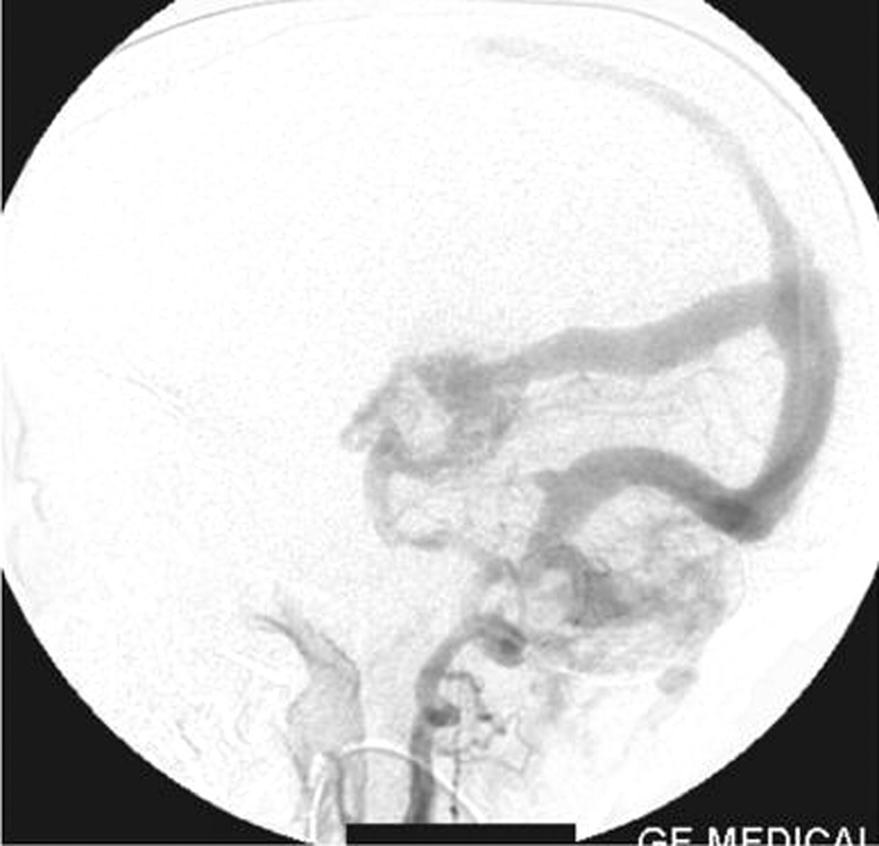
- Fig 4.
Left internal carotid artery injection lateral view angiogram shows the filling of the fistula through the posterior communicating artery.
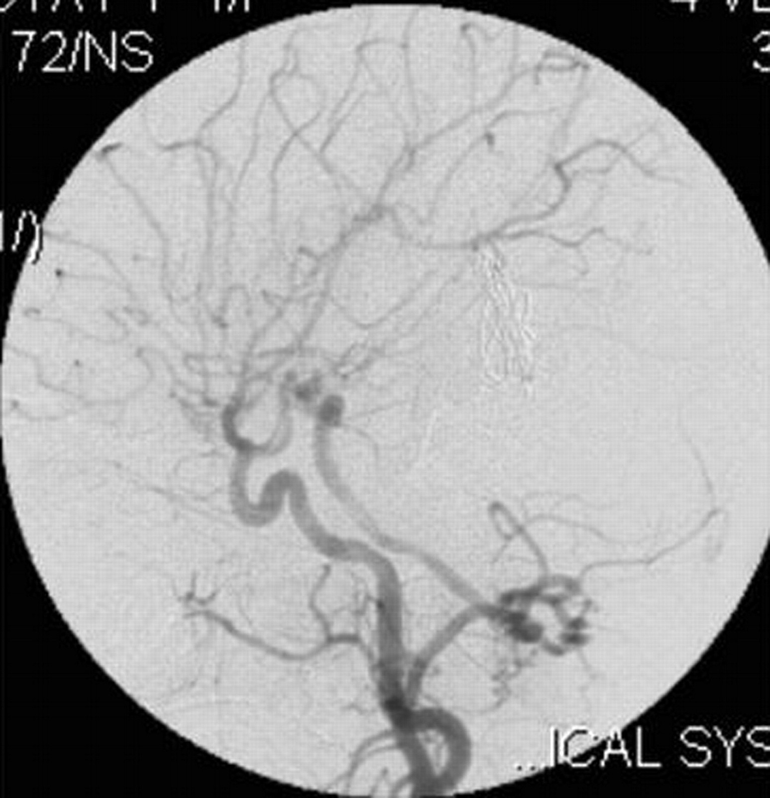
- Fig 5.
Left vertebral artery injection lateral view angiogram shows the fistula filling from the thalamoperforator arteries.
- Fig 6.
Right external carotid artery injection lateral view angiogram shows the proatlantal artery.
- Fig 7.
Right external artery injection anteroposterior view angiogram shows the opacification of the proatlantal artery of the right side.
- Fig 8.
Left external carotid artery injection lateral view angiogram shows the filling of the vertebral artery via the left proatlantal artery.
- Fig 9.
Left external carotid artery injection anteroposterior view angiogram shows the proatlantal artery and the opacification of the vertebral artery.
- Fig 10.
Right common carotid artery injection angiogram shows the origin of the proatlantal artery and the filling of the vertebral artery (case 2).


In this issue


Jump to section
Related Articles
Cited By...
- No citing articles found.