Advertisement

Main menu

  • Home
  • Content
    • Current Issue
    • Accepted Manuscripts
    • Article Preview
    • Past Issue Archive
    • Video Articles
    • AJNR Case Collection
    • Case of the Week Archive
    • Case of the Month Archive
    • Classic Case Archive
  • Special Collections
    • AJNR Awards
    • Low-Field MRI
    • Alzheimer Disease
    • ASNR Foundation Special Collection
    • Photon-Counting CT
    • View All
  • Multimedia
    • AJNR Podcasts
    • AJNR SCANtastic
    • Trainee Corner
    • MRI Safety Corner
    • Imaging Protocols
  • For Authors
    • Submit a Manuscript
    • Submit a Video Article
    • Submit an eLetter to the Editor/Response
    • Manuscript Submission Guidelines
    • Statistical Tips
    • Fast Publishing of Accepted Manuscripts
    • Graphical Abstract Preparation
    • Imaging Protocol Submission
    • Author Policies
  • About Us
    • About AJNR
    • Editorial Board
    • Editorial Board Alumni
  • More
    • Become a Reviewer/Academy of Reviewers
    • Subscribers
    • Permissions
    • Alerts
    • Feedback
    • Advertisers
    • ASNR Home

User menu

  • Alerts
  • Log in

Search

  • Advanced search
American Journal of Neuroradiology

American Journal of Neuroradiology

ASHNR American Society of Functional Neuroradiology ASHNR American Society of Pediatric Neuroradiology ASSR
  • Alerts
  • Log in

Advanced Search

  • Home
  • Content
    • Current Issue
    • Accepted Manuscripts
    • Article Preview
    • Past Issue Archive
    • Video Articles
    • AJNR Case Collection
    • Case of the Week Archive
    • Case of the Month Archive
    • Classic Case Archive
  • Special Collections
    • AJNR Awards
    • Low-Field MRI
    • Alzheimer Disease
    • ASNR Foundation Special Collection
    • Photon-Counting CT
    • View All
  • Multimedia
    • AJNR Podcasts
    • AJNR SCANtastic
    • Trainee Corner
    • MRI Safety Corner
    • Imaging Protocols
  • For Authors
    • Submit a Manuscript
    • Submit a Video Article
    • Submit an eLetter to the Editor/Response
    • Manuscript Submission Guidelines
    • Statistical Tips
    • Fast Publishing of Accepted Manuscripts
    • Graphical Abstract Preparation
    • Imaging Protocol Submission
    • Author Policies
  • About Us
    • About AJNR
    • Editorial Board
    • Editorial Board Alumni
  • More
    • Become a Reviewer/Academy of Reviewers
    • Subscribers
    • Permissions
    • Alerts
    • Feedback
    • Advertisers
    • ASNR Home
  • Follow AJNR on Twitter
  • Visit AJNR on Facebook
  • Follow AJNR on Instagram
  • Join AJNR on LinkedIn
  • RSS Feeds

AJNR Awards, New Junior Editors, and more. Read the latest AJNR updates

OtherBRAIN

Bilateral Cerebral Venous Angioma Associated with Varices: A Case Report and Review of the Literature

Surjith Vattoth, Sukalyan Purkayastha, E.R. Jayadevan and Arun Kumar Gupta
American Journal of Neuroradiology October 2005, 26 (9) 2320-2322;
Surjith Vattoth
  • Find this author on Google Scholar
  • Find this author on PubMed
  • Search for this author on this site
Sukalyan Purkayastha
  • Find this author on Google Scholar
  • Find this author on PubMed
  • Search for this author on this site
E.R. Jayadevan
  • Find this author on Google Scholar
  • Find this author on PubMed
  • Search for this author on this site
Arun Kumar Gupta
  • Find this author on Google Scholar
  • Find this author on PubMed
  • Search for this author on this site
  • Article
  • Figures & Data
  • Info & Metrics
  • Responses
  • References
  • PDF
Loading

Abstract

Summary: A varix occurring with a cerebral venous angioma is a rare combination of cerebral vascular malformations. The most common vascular anomaly associated with a venous angioma is a cavernous angioma. We report a case of bilateral supratentorial venous angioma with 2 varices involving the angioma situated on the right frontotemporal region. Multiple subependymal veins were seen in the right supratentorial region, which joined to form multiple collector veins, which, in turn, unified into a tortuous channel in the region of the sylvian fissure and continued into venous varices.

Cerebral varices in combination with a venous angioma are very rare. Only 8 cases were reported through 1995 (1). After that, to the best of our knowledge, no case has been reported in the literature. Also, the occurrence of multiple venous angiomas is very rare. In our patient, CT demonstrated a venous varix associated with a right-sided supratentorial venous angioma. Digital subtraction angiography further delineated the presence of 2 varices associated with this venous angioma. CT and digital subtraction angiography also demonstrated a left supratentorial venous angioma in our patient. To the best of our knowledge, this is the first reported case of double supratentorial venous angiomas with the larger one associated with 2 varices.

Case Report

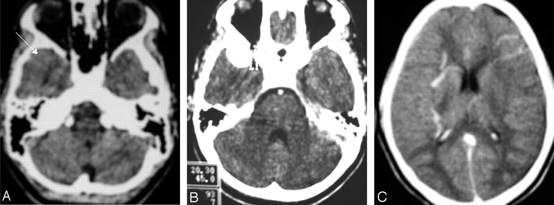
A 14-year-old girl presented with 2 episodes of generalized seizures within a week. She had no history of seizure or any other significant medical illness. Findings of her routine blood investigations, neurologic examination, and other systemic examinations were normal. Plain CT of the head showed an isoattenuated extraaxial well-defined oval lesion anterior to the right temporal pole (Fig 1A). On contrast administration, this lesion was seen to be intensely enhancing and measured 2 × 1.6 cm, which was suggestive of a vascular structure (Fig 1B). Multiple subependymal veins were seen in the right supratentorial region, which joined to form multiple collector veins, which in turn unified into a tortuous channel in the region of the sylvian fissure and continued to the region of this enhancing vascular structure, probably a venous varix. Abnormal enhancing vascular channels were also noted in the left frontal region, extending to the periphery from a subependymal location (Fig 1C). Therefore, the possibility of an arteriovenous malformation with an aneurysm or venous ectasia with dural feeders was considered. Because multiple vascular channels drained into collector veins, the possibility of a venous angioma with venous ectasia was also considered.

Fig 1.
  • Download figure
  • Open in new tab
  • Download powerpoint
Fig 1.

A, Noncontrast axial CT scan shows an isoattenuated extraaxial lesion anterior to the right temporal pole. B, Contrast-enhanced axial CT scan shows intense enhancement of the lesion. C, Contrast-enhanced axial CT scan shows multiple subependymal veins joining to form collector veins unifying into a tortuous channel in the region of the right sylvian fissure and continuing to the region of the anterior temporal enhancing extraaxial lesion. Abnormal enhancing vessels are also seen in the left frontal region.

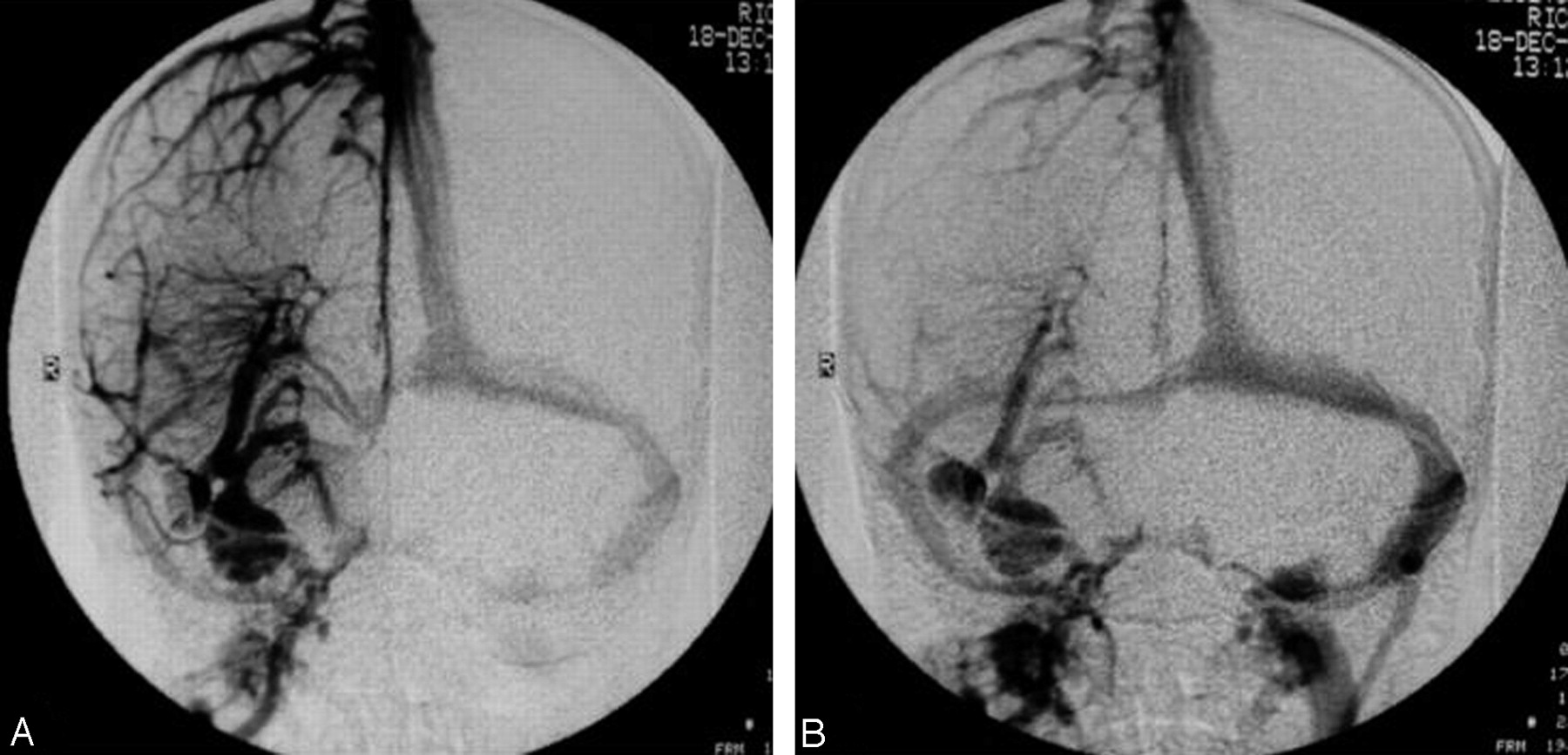
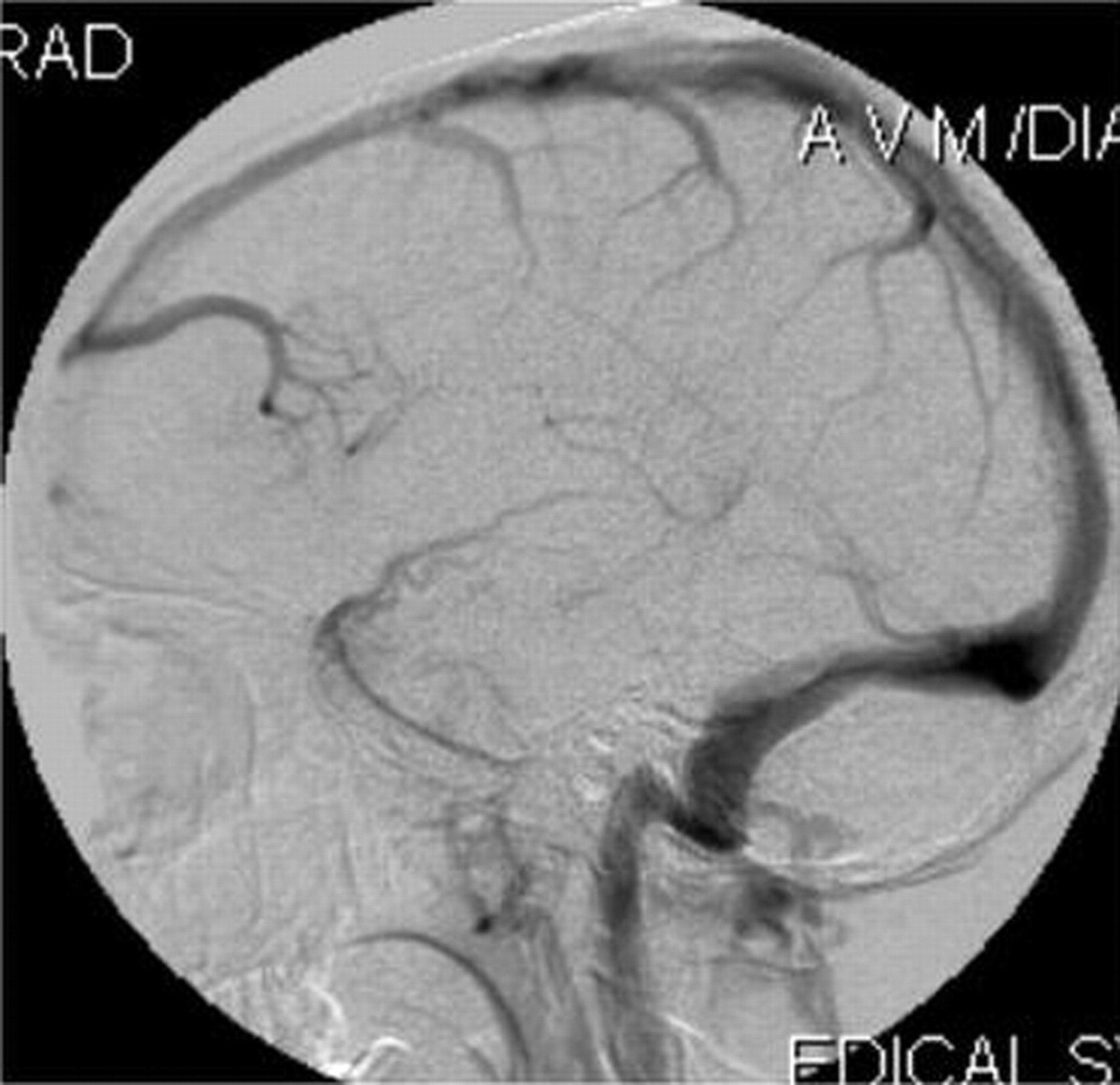
Digital subtraction angiography showed a normal arterial phase of the right internal carotid artery (Fig 2), whereas the venous phase in anteroposterior and lateral views showed multiple subependymal veins joining to form 4 collector veins, which, in turn, coursed anteroinferiorly to drain into 2 large venous varices in the region of the superficial middle cerebral vein (Fig 3, 4). These features were suggestive of a venous angioma with varices. A left internal carotid artery injection showed another supratentorial venous angioma in the frontal region, without any venous varix, that drained into the superior sagittal sinus (Fig 5). Findings in the arterial phase were normal on the left side. The posterior circulation was normal.

Fig 2.
  • Download figure
  • Open in new tab
  • Download powerpoint
Fig 2.

A, -B, Right and left internal carotid artery angiograms show a normal arterial phase.

Fig 3.
  • Download figure
  • Open in new tab
  • Download powerpoint
Fig 3.

A, -B, Anteroposterior view of early and late venous phases of the right internal carotid artery angiograms shows the presence of 2 varices and the venous angioma.

Fig 4.
  • Download figure
  • Open in new tab
  • Download powerpoint
Fig 4.

Lateral view of the venous phase of the right internal carotid artery angiogram shows a classic “Medusa head” appearance with a draining vein varix in the region of the right superficial middle cerebral vein.

Fig 5.
  • Download figure
  • Open in new tab
  • Download powerpoint
Fig 5.

Lateral view of the venous phase of the left internal carotid artery angiogram shows a classic “Medusa head” appearance draining into the superior sagittal sinus.

Discussion

Giant venous varices are a common association with an arteriovenous malformation, but a venous varix occurring in a venous angioma is very rare and only 8 cases have been reported in the literature (1). The most common vascular anomaly associated with a venous angioma is a cavernous angioma (2). Varices associated with a venous angioma occur in the draining veins of the angioma, and some case reports have shown that these varices can also bleed (3, 4).

Double supratentorial venous angiomas are rare. In 40 patients with radiologically diagnosed venous angioma, Fujii et al (5) found only one case with a double venous angioma. Garner et al (6) encountered no patient with a double lesion among his 100 patients having cerebral venous angioma. In a review of the literature of varices occurring with a cerebral venous angioma by Uchino et al (1), all 8 cases were supratentorial with varices involving the draining veins. They proposed that the increased flow in the draining vein might have increased the venous blood pressure causing a portion with a weaker wall to dilate.

Our case demonstrated 2 venous varices in the larger venous angioma involving the draining vein. Kazumata et al (7) described multiple varicose venous dilations that involved unilateral superficial cerebral veins accompanied by a varix of the internal jugular vein. Also, bilaterality of the supratentorial venous angioma associated with varices makes the present case highly unusual. Surgery is contraindicated in these patients because these venous structures drain the normal parts of the brain and surgery can result in extensive venous infarcts. There is a risk of hemorrhage especially if the patient develops focal stenosis of the terminal segment of the collector vein (8). Another potential complication is venous infarction, which may occur if the draining vein becomes thrombosed (9).

McCormick (10) categorized a pure intracranial varix as a venous malformation. Knowledge of the association of venous varices with venous angioma is essential to diagnose this combination correctly on CT or MR imaging and not to mistake it for the more common association of venous varices with an arteriovenous malformation. Digital subtraction angiography can confirm the diagnosis.

References

  1. ↵
    Uchino A, Hasuo K, Matsumoto S, Ikezaki K, Masuda.K. Varix occurring with cerebral venous angioma: a case report and review of the literature. Neuroradiology 1995;37:29–31
    PubMed
  2. ↵
    Wilms G, Bleus E, Demaerel P, et al. Simultaneous occurrence of developmental venous anomalies and cavernous angiomas. AJNR Am J Neuroradiol 1994;15:1247–1254
    Abstract/FREE Full Text
  3. ↵
    Maiuri F, Gangemi M, Iaconetta G. Giant intracranial varix associated with venous angioma and intracerebral hemorrhage. Acta Neurol (Napoli) 1990;12:231–236
  4. ↵
    Roda JM, Bencosme J, Isla A, et al. Intraventricular varix causing hemorrhage: case report. J Neurosurg 1988;68:472–473
    PubMed
  5. ↵
    Fujii K, Matsushima T, Inamura T, Fukui M. Natural history and choice of treatment in 40 patients with medullary venous malformation. Neurosurg Rev 1992;15:13–20
    CrossRefPubMed
  6. ↵
    Garner TB, Curling OD Jr, Kelly DL Jr, Laster DW. The natural history of intracranial venous angiomas. J Neurosurg 1991;75:715–722
    PubMed
  7. ↵
    Kazumata K, Fujimoto S, Idosaka H, Kuroda S, Nunomura M, Houkin K. Multiple varices in the unilateral cerebral venous system. AJNR Am J Neuroradiol 1999;20:1243–1244
    Abstract/FREE Full Text
  8. ↵
    Osborn AG. Diagnostic neuroradiology. St. Louis, MO: Mosby-Year Book;1994 :314–320
  9. ↵
    Lai PH, Chen PC, Pan HB, Yang CF. Venous infarction from a venous angioma occurring after thrombosis of a drainage vein. AJR Am J Roentgenol 1999;172:1698–1699
    PubMed
  10. ↵
    McCormick WF. The pathology of angiomas. In: Fein JM, Flamm ES, eds. Cerebrovascular surgery,vol. 6 . New York: Springer-Verlag;1985 :1075–1095
  • Received December 26, 2004.
  • Accepted after revision January 5, 2005.
  • Copyright © American Society of Neuroradiology
View Abstract
PreviousNext
Back to top

In this issue

American Journal of Neuroradiology: 26 (9)
American Journal of Neuroradiology
Vol. 26, Issue 9
1 Oct 2005
  • Table of Contents
  • Index by author
Advertisement
Print
Download PDF
Email Article

Thank you for your interest in spreading the word on American Journal of Neuroradiology.

NOTE: We only request your email address so that the person you are recommending the page to knows that you wanted them to see it, and that it is not junk mail. We do not capture any email address.

Enter multiple addresses on separate lines or separate them with commas.
Bilateral Cerebral Venous Angioma Associated with Varices: A Case Report and Review of the Literature
(Your Name) has sent you a message from American Journal of Neuroradiology
(Your Name) thought you would like to see the American Journal of Neuroradiology web site.
CAPTCHA
This question is for testing whether or not you are a human visitor and to prevent automated spam submissions.
Cite this article
Surjith Vattoth, Sukalyan Purkayastha, E.R. Jayadevan, Arun Kumar Gupta
Bilateral Cerebral Venous Angioma Associated with Varices: A Case Report and Review of the Literature
American Journal of Neuroradiology Oct 2005, 26 (9) 2320-2322;

Citation Manager Formats

  • BibTeX
  • Bookends
  • EasyBib
  • EndNote (tagged)
  • EndNote 8 (xml)
  • Medlars
  • Mendeley
  • Papers
  • RefWorks Tagged
  • Ref Manager
  • RIS
  • Zotero
0 Responses
Respond to this article
Share
Bookmark this article
Bilateral Cerebral Venous Angioma Associated with Varices: A Case Report and Review of the Literature
Surjith Vattoth, Sukalyan Purkayastha, E.R. Jayadevan, Arun Kumar Gupta
American Journal of Neuroradiology Oct 2005, 26 (9) 2320-2322;
del.icio.us logo Twitter logo Facebook logo Mendeley logo
  • Tweet Widget
  • Facebook Like
  • Google Plus One

Jump to section

  • Article
    • Abstract
    • Case Report
    • Discussion
    • References
  • Figures & Data
  • Info & Metrics
  • Responses
  • References
  • PDF

Related Articles

  • No related articles found.
  • PubMed
  • Google Scholar

Cited By...

  • Association of venous varix and developmental venous anomaly: report of a case and review of literature
  • A benchmark approach to hemorrhage risk management of cavernous malformations
  • Crossref
  • Google Scholar

This article has not yet been cited by articles in journals that are participating in Crossref Cited-by Linking.

More in this TOC Section

  • Progression of Microstructural Damage in Spinocerebellar Ataxia Type 2: A Longitudinal DTI Study
  • SWI or T2*: Which MRI Sequence to Use in the Detection of Cerebral Microbleeds? The Karolinska Imaging Dementia Study
  • Statin Therapy Does Not Affect the Radiographic and Clinical Profile of Patients with TIA and Minor Stroke
Show more BRAIN

Similar Articles

Advertisement

Indexed Content

  • Current Issue
  • Accepted Manuscripts
  • Article Preview
  • Past Issues
  • Editorials
  • Editor's Choice
  • Fellows' Journal Club
  • Letters to the Editor
  • Video Articles

Cases

  • Case Collection
  • Archive - Case of the Week
  • Archive - Case of the Month
  • Archive - Classic Case

More from AJNR

  • Trainee Corner
  • Imaging Protocols
  • MRI Safety Corner

Multimedia

  • AJNR Podcasts
  • AJNR Scantastics

Resources

  • Turnaround Time
  • Submit a Manuscript
  • Submit a Video Article
  • Submit an eLetter to the Editor/Response
  • Manuscript Submission Guidelines
  • Statistical Tips
  • Fast Publishing of Accepted Manuscripts
  • Graphical Abstract Preparation
  • Imaging Protocol Submission
  • Evidence-Based Medicine Level Guide
  • Publishing Checklists
  • Author Policies
  • Become a Reviewer/Academy of Reviewers
  • News and Updates

About Us

  • About AJNR
  • Editorial Board
  • Editorial Board Alumni
  • Alerts
  • Permissions
  • Not an AJNR Subscriber? Join Now
  • Advertise with Us
  • Librarian Resources
  • Feedback
  • Terms and Conditions
  • AJNR Editorial Board Alumni

American Society of Neuroradiology

  • Not an ASNR Member? Join Now

© 2025 by the American Society of Neuroradiology All rights, including for text and data mining, AI training, and similar technologies, are reserved.
Print ISSN: 0195-6108 Online ISSN: 1936-959X

Powered by HighWire