
Article Figures & Data
Figures
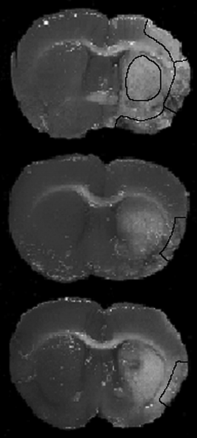
- Fig 1.
TTC-stained brain sections on the level of the optic chiasm. The white or grayish tissue is unstained and is equivalent to necrotic tissue. The upper section demonstrates a complete infarction of the MCA-supplied territory. Borders of the parietal, temporal, and piriform cortex are outlined as well as the basal ganglia. The middle section demonstrates partial necrosis of the temporal cortex, whereas the lower sections demonstrate complete necrosis.
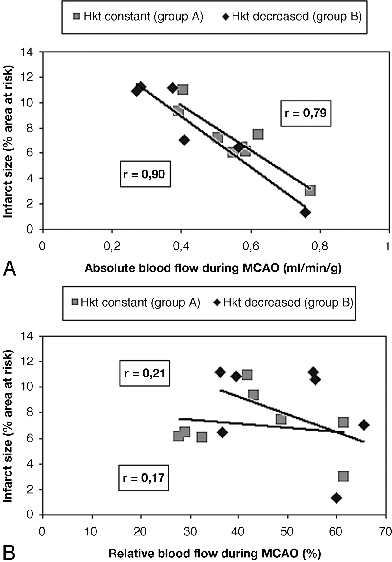
- Fig 2.
Relationships between absolute CBF and relative CBFC at 30 minutes post-MCAO and infarct size in groups A (A) and B (B) (expressed as percentage of total brain size). Hkt indicates hematocrit.


Tables
Parameter Baseline 30 min Post-MCA Occlusion 30 min Post-reperfusion AOPmax (mmHg) 126 ± 19 106 ± 22* 101 ± 19* AOPmin (mmHg) 84 ± 21 75 ± 20* 68 ± 9* Heart rate (beats/min) 211 ± 27 245 ± 39* 246 ± 48* Blood pH 7.35 ± 0.03 7.39 ± 0.04 7.37 ± 0.03 Pco2 (mmHg) 38.9 ± 3.8 33.0 ± 6.4 34.4 ± 7.6 Po2 (mmHg) 98.0 ± 19.2 93.6 ± 23.9 92.7 ± 15.0 HCO3− (mM/L) 21.1 ± 2.6 20.9 ± 2.9 21.2 ± 3.3 SO2 (%) 94.2 ± 3.4 92.6 ± 3.7 93.2 ± 2.7 Hematocrit (%) 41.3 ± 3.5 41.7 ± 5.1 42.0 ± 6.7 K+ (mM/L) 3.9 ± 0.2 4.9 ± 0.4* Na+ (mM/L) 138 ± 1.6 136 ± 1.8 Ca++ (mM/L) 1.2 ± 0.08 1.2 ± 0.07 Note.—MCA indicates middle cerebral artery; AOPmax, maximal aortic pressure; AOPmin, minimal aortic pressure.
* P < .05 compared to baseline.
Parameter Baseline 30 min Post-MCA Occlusion 30 min Post-reperfusion AOPmax (mmHg) 124 ± 10.5 112 ± 12* 110 ± 10* AOPmin (mmHg) 89 ± 11 78 ± 7* 78 ± 14* Heart rate (beats/min) 210 ± 24 214 ± 23* 222 ± 19* Blood pH 7.37 ± 0.05 7.40 ± 0.03 7.38 ± 0.01 Pco2 (mmHg) 36.3 ± 1.4 42.0 ± 3.5 35.4 ± 1.5 Po2 (mmHg) 105.8 ± 19.2 113.1 ± 28.3 107.8 ± 16.4 HCO3− (mM/L) 22.0 ± 2.4 23.6 ± 4.8 24.8 ± 3.8 SO2 (%) 96.0 ± 1.3 94.4 ± 1.3 95.2 ± 2.4 Hematocrit (%) 40.2 ± 4.6 34.8 ± 5.9* 27.1 ± 7.5* K+ (mM/L) 3.6 ± 0.5 3.8 ± 0.3* Na+ (mM/L) 141 ± 2.9 137 ± 0.8 Ca++ (mM/L) 1.1 ± 0.08 1.2 ± 0.1 Note.—MCA indicates middle cerebral artery; AOPmax, maximal aortic pressure; AOPmin, minimal aortic pressure.
* P < .05 compared to baseline.
- TABLE 3:
Cerebral blood flow alterations in animals of group A (MCA occlusion, blood replaced)
Brain Areas Absolute CBF (ml/g/min) Baseline 30 min Post-MCA Occlusion 30 min Post-reperfusion PCi 0.88 ± 0.26 0.51 ± 0.21* 0.42 ± 0.28* (98 ± 25%) (46 ± 30%; 58 ± 29%)* (65 ± 39%; 48 ± 23%)* TCi 0.74 ± 0.24 0.20 ± 0.15* 0.23 ± 0.21* (94 ± 23%) (20 ± 18%; 27 ± 20%)* (30 ± 28%; 31 ± 25%)* PiCi 0.41 ± 0.21 0.33 ± 0.18 0.28 ± 0.20 (94 ± 28%) (63 ± 33%; 81 ± 30%)* (54 ± 27%; 68 ± 27%)* BGi 0.62 ± 0.19 0.53 ± 0.22 0.48 ± 0.18 (82 ± 30%) 62 ± 31%; 77 ± 30%)* (63 ± 30%; 61 ± 30%)* PCni 0.90 ± 0.37 1.11 ± 0.72 0.64 ± 0.47* TCni 0.79 ± 0.35 0.97 ± 0.43* 0.77 ± 0.27 PiCni 0.44 ± 0.19 0.53 ± 0.19 0.53 ± 0.19 BGni 0.85 ± 0.35 0.86 ± 0.34 0.66 ± 0.31* Note.—MCA indicates middle cerebral artery; CBF, cerebral blood flow; PCi, infarcted parietal cortex (PCni, noninfarcted); TCi, infarcted temporal cortex (TCni, noninfarcted); PiCi, infarcted piriform cortex (PiCini, noninfarcted); BGi, infarcted basal ganglia (BGni, noninfarcted).
Values in parentheses show relative CBF in areas of the infarcted hemisphere corresponding to contralateral noninfarcted areas (CBFC) or its baseline blood flow (CBFB), respectively.
* P < .05 compared to baseline.
- TABLE 4:
Cerebral blood flow alterations in animals of group B (MCA occlusion, no blood donation)
Brain Areas Baseline 30 min Post-MCA Occlusion 30 min Post-reperfusion PCi 1.02 ± 0.33 0.54 ± 0.30* 0.44 ± 0.31* (98 ± 22%) (46 ± 27%; 53 ± 32%)* (51 ± 26%; 43 ± 19%)* TCi 0.86 ± 0.43 0.27 ± 0.19* 0.29 ± 0.20* (97 ± 26%) (30 ± 15%; 31 ± 19%)* (32 ± 23%; 34 ± 26%)* PiCi 0.60 ± 0.28 0.40 ± 0.20 0.33 ± 0.17* (95 ± 22%) (66 ± 30%; 68 ± 26%)* (52 ± 31%; 55 ± 29%)* BGi 0.60 ± 0.27 0.55 ± 0.23 0.49 ± 0.22 (98 ± 28%) (55 ± 27%; 69 ± 34%)* (72 ± 36%; 61 ± 27%)* PCni 1.04 ± 0.34 1.18 ± 0.50 0.87 ± 0.39* TCni 0.88 ± 0.43 0.88 ± 0.49* 0.90 ± 0.48 PiCni 0.63 ± 0.26 0.60 ± 0.26 0.62 ± 0.27 BGni 0.82 ± 0.40 1.00 ± 0.48 0.67 ± 0.34* Note.—MCA indicates middle cerebral artery; CBF, cerebral blood flow; PCi, infarcted parietal cortex (PCni, noninfarcted); TCi, infarcted temporal cortex (TCni, noninfarcted); PiCi, infarcted piriform cortex (PiCini, noninfarcted); BGi, infarcted basal ganglia (BGni, noninfarcted).
Values in parentheses show relative CBF in areas of the infarcted hemisphere corresponding to contralateral noninfarcted areas (CBFC) or its baseline blood flow (CBFB), respectively.
* P < .05 compared to baseline.
In this issue


Jump to section
Related Articles
Cited By...
- Predicting experimental success: a retrospective case-control study using the rat intraluminal thread model of stroke
- Congenic Fine-Mapping Identifies a Major Causal Locus for Variation in the Native Collateral Circulation and Ischemic Injury in Brain and Lower Extremity
- MicroRNA Expression in the Blood and Brain of Rats Subjected to Transient Focal Ischemia by Middle Cerebral Artery Occlusion