Advertisement

Main menu

  • Home
  • Content
    • Current Issue
    • Accepted Manuscripts
    • Article Preview
    • Past Issue Archive
    • Video Articles
    • AJNR Case Collection
    • Case of the Week Archive
    • Case of the Month Archive
    • Classic Case Archive
  • Special Collections
    • AJNR Awards
    • Low-Field MRI
    • Alzheimer Disease
    • ASNR Foundation Special Collection
    • Photon-Counting CT
    • View All
  • Multimedia
    • AJNR Podcasts
    • AJNR SCANtastic
    • Trainee Corner
    • MRI Safety Corner
    • Imaging Protocols
  • For Authors
    • Submit a Manuscript
    • Submit a Video Article
    • Submit an eLetter to the Editor/Response
    • Manuscript Submission Guidelines
    • Statistical Tips
    • Fast Publishing of Accepted Manuscripts
    • Graphical Abstract Preparation
    • Imaging Protocol Submission
    • Author Policies
  • About Us
    • About AJNR
    • Editorial Board
    • Editorial Board Alumni
  • More
    • Become a Reviewer/Academy of Reviewers
    • Subscribers
    • Permissions
    • Alerts
    • Feedback
    • Advertisers
    • ASNR Home

User menu

  • Alerts
  • Log in

Search

  • Advanced search
American Journal of Neuroradiology

American Journal of Neuroradiology

ASHNR American Society of Functional Neuroradiology ASHNR American Society of Pediatric Neuroradiology ASSR
  • Alerts
  • Log in

Advanced Search

  • Home
  • Content
    • Current Issue
    • Accepted Manuscripts
    • Article Preview
    • Past Issue Archive
    • Video Articles
    • AJNR Case Collection
    • Case of the Week Archive
    • Case of the Month Archive
    • Classic Case Archive
  • Special Collections
    • AJNR Awards
    • Low-Field MRI
    • Alzheimer Disease
    • ASNR Foundation Special Collection
    • Photon-Counting CT
    • View All
  • Multimedia
    • AJNR Podcasts
    • AJNR SCANtastic
    • Trainee Corner
    • MRI Safety Corner
    • Imaging Protocols
  • For Authors
    • Submit a Manuscript
    • Submit a Video Article
    • Submit an eLetter to the Editor/Response
    • Manuscript Submission Guidelines
    • Statistical Tips
    • Fast Publishing of Accepted Manuscripts
    • Graphical Abstract Preparation
    • Imaging Protocol Submission
    • Author Policies
  • About Us
    • About AJNR
    • Editorial Board
    • Editorial Board Alumni
  • More
    • Become a Reviewer/Academy of Reviewers
    • Subscribers
    • Permissions
    • Alerts
    • Feedback
    • Advertisers
    • ASNR Home
  • Follow AJNR on Twitter
  • Visit AJNR on Facebook
  • Follow AJNR on Instagram
  • Join AJNR on LinkedIn
  • RSS Feeds

AJNR Awards, New Junior Editors, and more. Read the latest AJNR updates

OtherPEDIATRICS

Glioblastoma Multiforme of the Conus Medullaris in a Child: Description of a Case and Literature Review

Alessandro Stecco, Claudia Quirico, Amelia Giampietro, Giuseppe Sessa, Renzo Boldorini and Alessandro Carriero
American Journal of Neuroradiology September 2005, 26 (8) 2157-2160;
Alessandro Stecco
  • Find this author on Google Scholar
  • Find this author on PubMed
  • Search for this author on this site
Claudia Quirico
  • Find this author on Google Scholar
  • Find this author on PubMed
  • Search for this author on this site
Amelia Giampietro
  • Find this author on Google Scholar
  • Find this author on PubMed
  • Search for this author on this site
Giuseppe Sessa
  • Find this author on Google Scholar
  • Find this author on PubMed
  • Search for this author on this site
Renzo Boldorini
  • Find this author on Google Scholar
  • Find this author on PubMed
  • Search for this author on this site
Alessandro Carriero
  • Find this author on Google Scholar
  • Find this author on PubMed
  • Search for this author on this site
  • Article
  • Figures & Data
  • Info & Metrics
  • Responses
  • References
  • PDF
Loading

Article Figures & Data

Figures

  • Fig 1.
    • Download figure
    • Open in new tab
    • Download powerpoint
    Fig 1.

    A–C, Fast spin-echo T2-weighted sagittal acquisition (A) shows a hyperintense and homogeneous mass at the T12 level, involving the conus and the epicomus, with a diffuse aspect and without a clear cleavability plane. Spin-echo T1-weighted sagittal (B) and coronal (postgadolinium) (C) acquisitions show a hypointense lesion with a mild patchy nodular enhancement of the upper portion, associated with a faint enhancement along the pial surface.
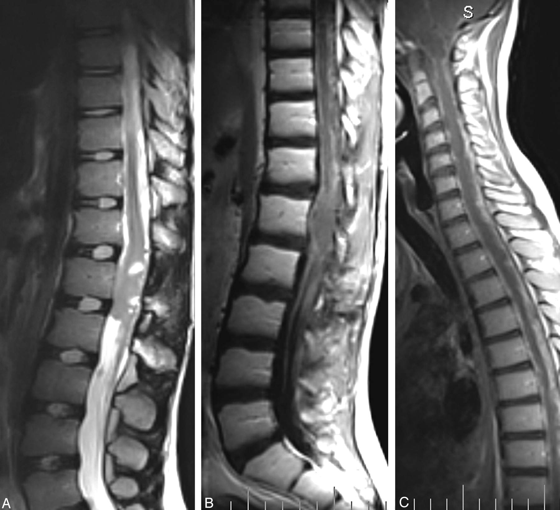
  • Fig 2.
    • Download figure
    • Open in new tab
    • Download powerpoint
    Fig 2.

    A–C, Nine-month follow-up after surgery. Fast spin-echo T2-weighted (A) and spin-echo T1-weighted (postgadolinium) (B) sagittal lumbar spine acquisitions show a local recurrence of the disease. Note the leptomeningeal spread (B, C) along the entire spinal canal, up to the posterior cerebral fossa (C).

  • Fig 3.
    • Download figure
    • Open in new tab
    • Download powerpoint
    Fig 3.

    Photomicrograph of histologic findings shows glioblastoma multiforme. Note high cellular attenuation, pleomorphism, and mitosis of neoplastic cells (hematoxylin and eosin, original magnification ×400).

  • Fig 4.
    • Download figure
    • Open in new tab
    • Download powerpoint
    Fig 4.

    Photomicrograph of histologic findings shows anti-Ki67 (MIB-one) antiserum indicating a high-proliferating index in neoplastic cells (immunoperoxidase, hematoxylin counterstaining, original magnification ×400).

PreviousNext
Back to top

In this issue

American Journal of Neuroradiology: 26 (8)
American Journal of Neuroradiology
Vol. 26, Issue 8
1 Sep 2005
  • Table of Contents
  • Index by author
Advertisement
Print
Download PDF
Email Article

Thank you for your interest in spreading the word on American Journal of Neuroradiology.

NOTE: We only request your email address so that the person you are recommending the page to knows that you wanted them to see it, and that it is not junk mail. We do not capture any email address.

Enter multiple addresses on separate lines or separate them with commas.
Glioblastoma Multiforme of the Conus Medullaris in a Child: Description of a Case and Literature Review
(Your Name) has sent you a message from American Journal of Neuroradiology
(Your Name) thought you would like to see the American Journal of Neuroradiology web site.
CAPTCHA
This question is for testing whether or not you are a human visitor and to prevent automated spam submissions.
Cite this article
Alessandro Stecco, Claudia Quirico, Amelia Giampietro, Giuseppe Sessa, Renzo Boldorini, Alessandro Carriero
Glioblastoma Multiforme of the Conus Medullaris in a Child: Description of a Case and Literature Review
American Journal of Neuroradiology Sep 2005, 26 (8) 2157-2160;

Citation Manager Formats

  • BibTeX
  • Bookends
  • EasyBib
  • EndNote (tagged)
  • EndNote 8 (xml)
  • Medlars
  • Mendeley
  • Papers
  • RefWorks Tagged
  • Ref Manager
  • RIS
  • Zotero
0 Responses
Respond to this article
Share
Bookmark this article
Glioblastoma Multiforme of the Conus Medullaris in a Child: Description of a Case and Literature Review
Alessandro Stecco, Claudia Quirico, Amelia Giampietro, Giuseppe Sessa, Renzo Boldorini, Alessandro Carriero
American Journal of Neuroradiology Sep 2005, 26 (8) 2157-2160;
del.icio.us logo Twitter logo Facebook logo Mendeley logo
  • Tweet Widget
  • Facebook Like
  • Google Plus One

Jump to section

  • Article
    • Abstract
    • Case Report
    • Discussion
    • References
  • Figures & Data
  • Info & Metrics
  • Responses
  • References
  • PDF

Related Articles

  • No related articles found.
  • PubMed
  • Google Scholar

Cited By...

  • Proliferative and Apoptotic Activity of Glioblastoma Multiforme Cells Cultured on In Ovo Model
  • Crossref
  • Google Scholar

This article has not yet been cited by articles in journals that are participating in Crossref Cited-by Linking.

More in this TOC Section

  • Early Ultrasonic Monitoring of Brain Growth and Later Neurodevelopmental Outcome in Very Preterm Infants
  • Diagnostic Value of Sylvian Fissure Hyperechogenicity in Fetal SAH
  • Feasibility and Added Value of Fetal DTI Tractography in the Evaluation of an Isolated Short Corpus Callosum: Preliminary Results
Show more Pediatrics

Similar Articles

Advertisement

Indexed Content

  • Current Issue
  • Accepted Manuscripts
  • Article Preview
  • Past Issues
  • Editorials
  • Editor's Choice
  • Fellows' Journal Club
  • Letters to the Editor
  • Video Articles

Cases

  • Case Collection
  • Archive - Case of the Week
  • Archive - Case of the Month
  • Archive - Classic Case

More from AJNR

  • Trainee Corner
  • Imaging Protocols
  • MRI Safety Corner
  • Book Reviews

Multimedia

  • AJNR Podcasts
  • AJNR Scantastics

Resources

  • Turnaround Time
  • Submit a Manuscript
  • Submit a Video Article
  • Submit an eLetter to the Editor/Response
  • Manuscript Submission Guidelines
  • Statistical Tips
  • Fast Publishing of Accepted Manuscripts
  • Graphical Abstract Preparation
  • Imaging Protocol Submission
  • Evidence-Based Medicine Level Guide
  • Publishing Checklists
  • Author Policies
  • Become a Reviewer/Academy of Reviewers
  • News and Updates

About Us

  • About AJNR
  • Editorial Board
  • Editorial Board Alumni
  • Alerts
  • Permissions
  • Not an AJNR Subscriber? Join Now
  • Advertise with Us
  • Librarian Resources
  • Feedback
  • Terms and Conditions
  • AJNR Editorial Board Alumni

American Society of Neuroradiology

  • Not an ASNR Member? Join Now

© 2025 by the American Society of Neuroradiology All rights, including for text and data mining, AI training, and similar technologies, are reserved.
Print ISSN: 0195-6108 Online ISSN: 1936-959X

Powered by HighWire