Advertisement

Main menu

  • Home
  • Content
    • Current Issue
    • Accepted Manuscripts
    • Article Preview
    • Past Issue Archive
    • Video Articles
    • AJNR Case Collection
    • Case of the Week Archive
    • Case of the Month Archive
    • Classic Case Archive
  • Special Collections
    • AJNR Awards
    • Low-Field MRI
    • Alzheimer Disease
    • ASNR Foundation Special Collection
    • Photon-Counting CT
    • View All
  • Multimedia
    • AJNR Podcasts
    • AJNR SCANtastic
    • Trainee Corner
    • MRI Safety Corner
    • Imaging Protocols
  • For Authors
    • Submit a Manuscript
    • Submit a Video Article
    • Submit an eLetter to the Editor/Response
    • Manuscript Submission Guidelines
    • Statistical Tips
    • Fast Publishing of Accepted Manuscripts
    • Graphical Abstract Preparation
    • Imaging Protocol Submission
    • Author Policies
  • About Us
    • About AJNR
    • Editorial Board
    • Editorial Board Alumni
  • More
    • Become a Reviewer/Academy of Reviewers
    • Subscribers
    • Permissions
    • Alerts
    • Feedback
    • Advertisers
    • ASNR Home

User menu

  • Alerts
  • Log in

Search

  • Advanced search
American Journal of Neuroradiology

American Journal of Neuroradiology

ASHNR American Society of Functional Neuroradiology ASHNR American Society of Pediatric Neuroradiology ASSR
  • Alerts
  • Log in

Advanced Search

  • Home
  • Content
    • Current Issue
    • Accepted Manuscripts
    • Article Preview
    • Past Issue Archive
    • Video Articles
    • AJNR Case Collection
    • Case of the Week Archive
    • Case of the Month Archive
    • Classic Case Archive
  • Special Collections
    • AJNR Awards
    • Low-Field MRI
    • Alzheimer Disease
    • ASNR Foundation Special Collection
    • Photon-Counting CT
    • View All
  • Multimedia
    • AJNR Podcasts
    • AJNR SCANtastic
    • Trainee Corner
    • MRI Safety Corner
    • Imaging Protocols
  • For Authors
    • Submit a Manuscript
    • Submit a Video Article
    • Submit an eLetter to the Editor/Response
    • Manuscript Submission Guidelines
    • Statistical Tips
    • Fast Publishing of Accepted Manuscripts
    • Graphical Abstract Preparation
    • Imaging Protocol Submission
    • Author Policies
  • About Us
    • About AJNR
    • Editorial Board
    • Editorial Board Alumni
  • More
    • Become a Reviewer/Academy of Reviewers
    • Subscribers
    • Permissions
    • Alerts
    • Feedback
    • Advertisers
    • ASNR Home
  • Follow AJNR on Twitter
  • Visit AJNR on Facebook
  • Follow AJNR on Instagram
  • Join AJNR on LinkedIn
  • RSS Feeds

AJNR Awards, New Junior Editors, and more. Read the latest AJNR updates

OtherBRAIN

Intraosseous Hemangioma of the Skull with Dural Tail Sign: Radiologic Features with Pathologic Correlation

Maria Politi, Bernd F.M. Romeike, Panagiotis Papanagiotou, Abdullah Nabhan, Tobias Struffert, Wolfgang Feiden and Wolfgang Reith
American Journal of Neuroradiology September 2005, 26 (8) 2049-2052;
Maria Politi
  • Find this author on Google Scholar
  • Find this author on PubMed
  • Search for this author on this site
Bernd F.M. Romeike
  • Find this author on Google Scholar
  • Find this author on PubMed
  • Search for this author on this site
Panagiotis Papanagiotou
  • Find this author on Google Scholar
  • Find this author on PubMed
  • Search for this author on this site
Abdullah Nabhan
  • Find this author on Google Scholar
  • Find this author on PubMed
  • Search for this author on this site
Tobias Struffert
  • Find this author on Google Scholar
  • Find this author on PubMed
  • Search for this author on this site
Wolfgang Feiden
  • Find this author on Google Scholar
  • Find this author on PubMed
  • Search for this author on this site
Wolfgang Reith
  • Find this author on Google Scholar
  • Find this author on PubMed
  • Search for this author on this site
  • Article
  • Figures & Data
  • Info & Metrics
  • Responses
  • References
  • PDF
Loading

Article Figures & Data

Figures

  • Fig 1.
    • Download figure
    • Open in new tab
    • Download powerpoint
    Fig 1.

    46-year-old woman with a gradually enlarging mass on her forehead. Noncontrast axial T1-weighted MR image (A) reveals a well-circumscribed mass, measuring 3 × 4 × 6 cm in the right frontal bone with intra- and extracranial extension. The lesion is well defined, and the signal intensity is of mixed intensity with some central hyperintensity and bone destruction. T2-weighted image (B) shows a lesion of high signal intensity with central areas of hypointensity. The lesion is well demarcated from the galea extracranially (outer uniform dark line) and the underlying dura intracranially (inner dark line). There is no surrounding edema. After gadopentetate dimeglumine administration (C), strong and homogeneous enhancement of the mass, relative enhancement of the bone marrow, and a dural tail sign are observed. The lesion extends intracranially and appears to compress the underlying brain, but without intraparenchymal extension. On the inner table in the parietal area, the small enhancing nodule corresponds to a vessel.

  • Fig 2.
    • Download figure
    • Open in new tab
    • Download powerpoint
    Fig 2.

    External carotid angiogram, lateral view, with midarterial (A) and capillary (B) phase, shows the rich vascularity of the tumor. The tumor shadow is fed by the right middle meningeal artery (black arrow) and by branches of the superficial temporal artery (white arrow).

  • Fig 3.
    • Download figure
    • Open in new tab
    • Download powerpoint
    Fig 3.

    Intraoperatively, the rich vascular network of the tumor is seen.
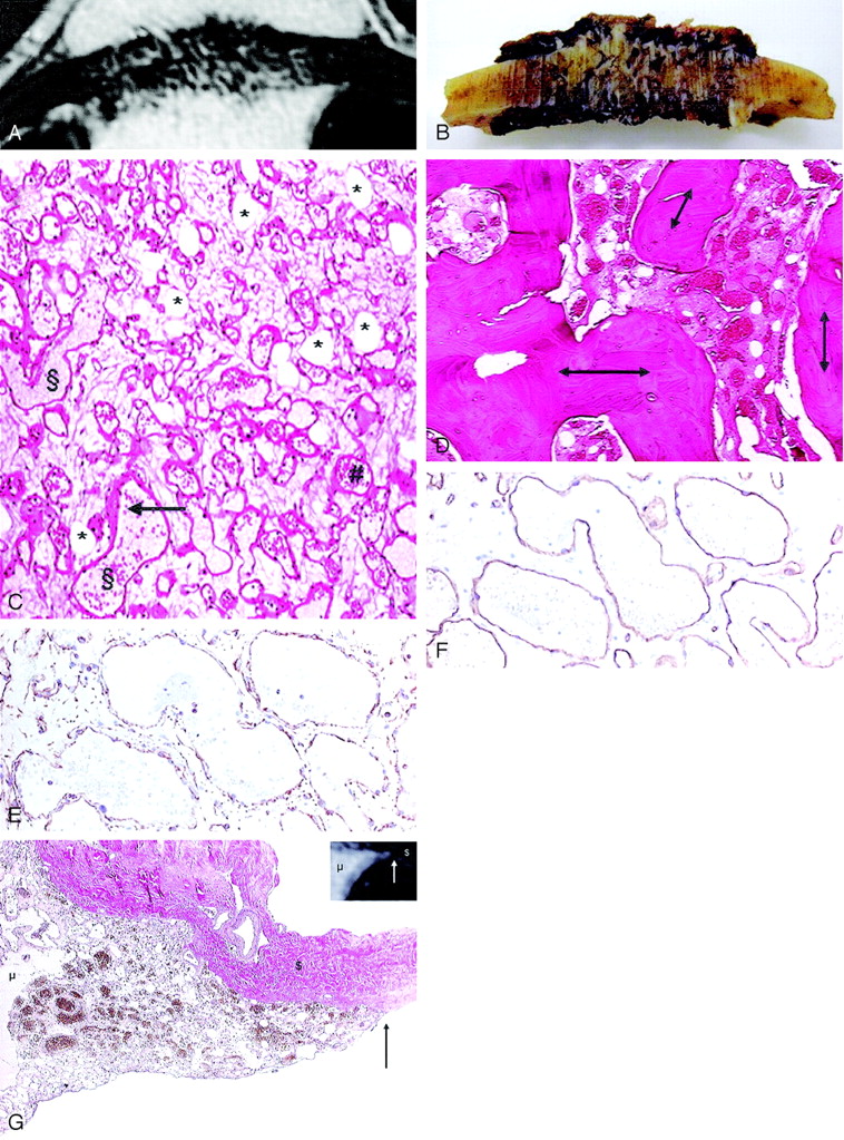
  • Fig 4.
    • Download figure
    • Open in new tab
    • Download powerpoint
    Fig 4.

    Correlation of T1-weighted contrast-enhanced MR image (A) with gross formalin-fixed specimen. Gross examination after formalin fixation shows a brown–red mass in the diploe, with radiated spicules and disruption of the outer and inner table (B); the bulk of the intra- and extracranial mass is missing as a result of the surgical excision technique. In the histomorphological representative section of the extraosseous fraction of the lesion (C), the fat cells (*) are seen as clear spaces without rimming; the vessels (§) are irregularly shaped dilated spaces lined by a single layer of inconspicuous endothelial cells (arrow) and often filled with erythrocytes (#) (hematoxylin and eosin, original magnification ×40). The vessels are arranged in a diffuse haphazard pattern. No signs of thrombosis or bleeding are seen. Intraosseous fraction (D) shows the hemangioma lying between plump bony trabeculae (double arrows) in irregularly shaped marrow spaces (hematoxylin and eosin, original magnification ×20). Immunohistochemically, the antibodies were directed against vimentin, which labels all mesenchymal cells (ie, all vessel walls and fat cells) (E), and CD34 (F), which labels endothelial cells (immunoperoxidase/diaminobenzidine method, original magnification ×100). In the correlation of the T1-weighted contrast-enhanced MR image with the histologic specimen at the site of dural tail sign (G), the dura is not invaded by the lesion; instead, the lesion grew along the inner surface of the skull. The van Gieson stain labels the cavernous hemangioma yellow–brown (mesenchymal component [μ]) and the collagenous fibers red (dura matter [$]). The arrow indicates the sharp delineation of the hemangioma (original magnification, ×20).

PreviousNext
Back to top

In this issue

American Journal of Neuroradiology: 26 (8)
American Journal of Neuroradiology
Vol. 26, Issue 8
1 Sep 2005
  • Table of Contents
  • Index by author
Advertisement
Print
Download PDF
Email Article

Thank you for your interest in spreading the word on American Journal of Neuroradiology.

NOTE: We only request your email address so that the person you are recommending the page to knows that you wanted them to see it, and that it is not junk mail. We do not capture any email address.

Enter multiple addresses on separate lines or separate them with commas.
Intraosseous Hemangioma of the Skull with Dural Tail Sign: Radiologic Features with Pathologic Correlation
(Your Name) has sent you a message from American Journal of Neuroradiology
(Your Name) thought you would like to see the American Journal of Neuroradiology web site.
CAPTCHA
This question is for testing whether or not you are a human visitor and to prevent automated spam submissions.
Cite this article
Maria Politi, Bernd F.M. Romeike, Panagiotis Papanagiotou, Abdullah Nabhan, Tobias Struffert, Wolfgang Feiden, Wolfgang Reith
Intraosseous Hemangioma of the Skull with Dural Tail Sign: Radiologic Features with Pathologic Correlation
American Journal of Neuroradiology Sep 2005, 26 (8) 2049-2052;

Citation Manager Formats

  • BibTeX
  • Bookends
  • EasyBib
  • EndNote (tagged)
  • EndNote 8 (xml)
  • Medlars
  • Mendeley
  • Papers
  • RefWorks Tagged
  • Ref Manager
  • RIS
  • Zotero
0 Responses
Respond to this article
Share
Bookmark this article
Intraosseous Hemangioma of the Skull with Dural Tail Sign: Radiologic Features with Pathologic Correlation
Maria Politi, Bernd F.M. Romeike, Panagiotis Papanagiotou, Abdullah Nabhan, Tobias Struffert, Wolfgang Feiden, Wolfgang Reith
American Journal of Neuroradiology Sep 2005, 26 (8) 2049-2052;
del.icio.us logo Twitter logo Facebook logo Mendeley logo
  • Tweet Widget
  • Facebook Like
  • Google Plus One

Jump to section

  • Article
    • Abstract
    • Case Report
    • Discussion
    • References
  • Figures & Data
  • Info & Metrics
  • Responses
  • References
  • PDF

Related Articles

  • No related articles found.
  • PubMed
  • Google Scholar

Cited By...

  • Intraosseous Venous Malformations of the Head and Neck
  • The CT Prevalence of Arrested Pneumatization of the Sphenoid Sinus in Patients with Sickle Cell Disease
  • Antemortem trauma and survival in the late Middle Pleistocene human cranium from Maba, South China
  • A rare spontaneous osteosarcoma of the calvarium in a patient with long-standing fibrous dysplasia: CT and MR findings
  • Intraosseous hemangioma in parietal bone
  • Crossref
  • Google Scholar

This article has not yet been cited by articles in journals that are participating in Crossref Cited-by Linking.

More in this TOC Section

  • Multimodal CT Provides Improved Performance for Lacunar Infarct Detection
  • Optimal MRI Sequence for Identifying Occlusion Location in Acute Stroke: Which Value of Time-Resolved Contrast-Enhanced MRA?
  • Evaluating the Effects of White Matter Multiple Sclerosis Lesions on the Volume Estimation of 6 Brain Tissue Segmentation Methods
Show more BRAIN

Similar Articles

Advertisement

Indexed Content

  • Current Issue
  • Accepted Manuscripts
  • Article Preview
  • Past Issues
  • Editorials
  • Editor's Choice
  • Fellows' Journal Club
  • Letters to the Editor
  • Video Articles

Cases

  • Case Collection
  • Archive - Case of the Week
  • Archive - Case of the Month
  • Archive - Classic Case

More from AJNR

  • Trainee Corner
  • Imaging Protocols
  • MRI Safety Corner

Multimedia

  • AJNR Podcasts
  • AJNR Scantastics

Resources

  • Turnaround Time
  • Submit a Manuscript
  • Submit a Video Article
  • Submit an eLetter to the Editor/Response
  • Manuscript Submission Guidelines
  • Statistical Tips
  • Fast Publishing of Accepted Manuscripts
  • Graphical Abstract Preparation
  • Imaging Protocol Submission
  • Evidence-Based Medicine Level Guide
  • Publishing Checklists
  • Author Policies
  • Become a Reviewer/Academy of Reviewers
  • News and Updates

About Us

  • About AJNR
  • Editorial Board
  • Editorial Board Alumni
  • Alerts
  • Permissions
  • Not an AJNR Subscriber? Join Now
  • Advertise with Us
  • Librarian Resources
  • Feedback
  • Terms and Conditions
  • AJNR Editorial Board Alumni

American Society of Neuroradiology

  • Not an ASNR Member? Join Now

© 2025 by the American Society of Neuroradiology All rights, including for text and data mining, AI training, and similar technologies, are reserved.
Print ISSN: 0195-6108 Online ISSN: 1936-959X

Powered by HighWire