
Article Figures & Data
Figures
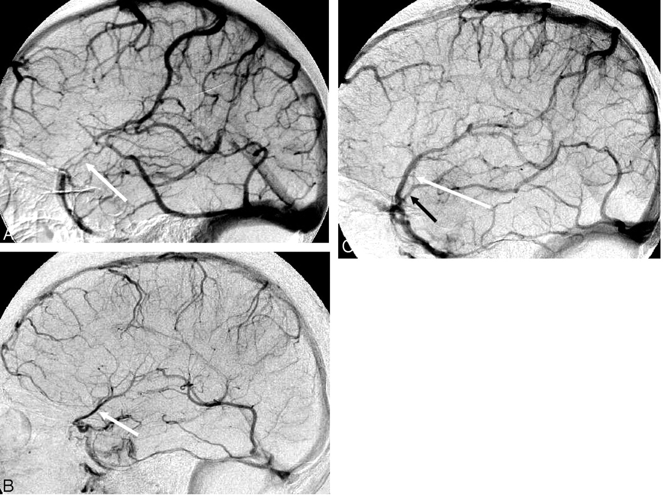
- Fig 1.
Assessment of superficial middle cerebral vein (SMCV) size. A, Two small paired SMCVs (arrow). B, Moderate-size single SMCV (arrow). C, Large SMCV (white arrow) and small temporal branch (black arrow) draining into larger SMCV.
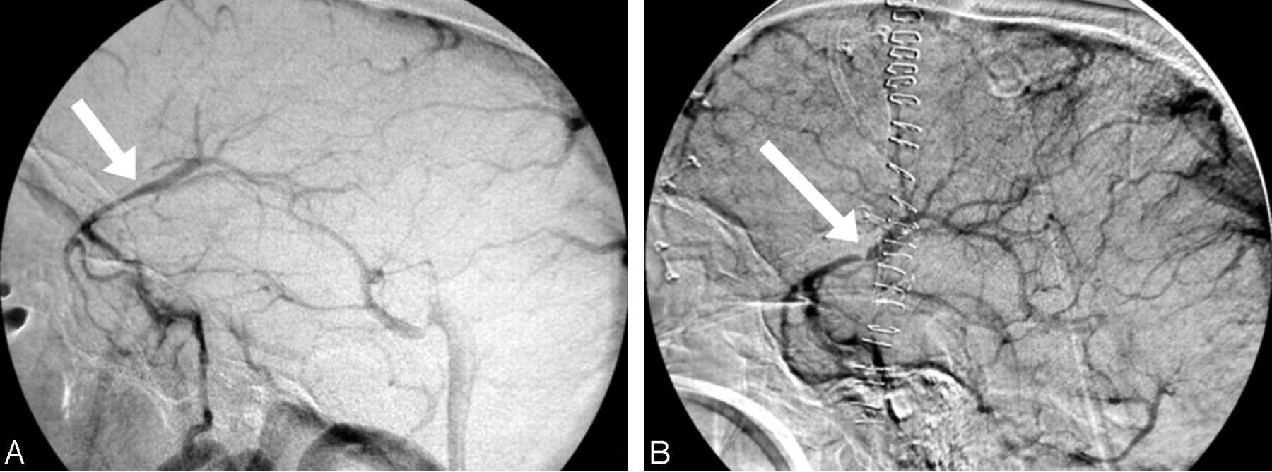
- Fig 2.
Large SMCV with distal occlusion. A, Three large branches with smaller branches join to form the SMCV. B, Mid and distal portions of the SMCV do not fill (arrow).
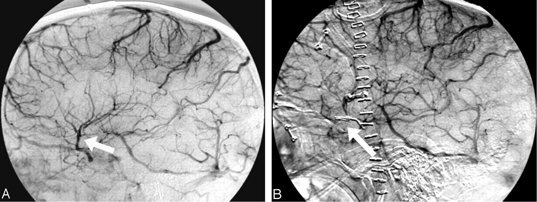
- Fig 3.
Focal attenuation of SMCV. A, Large single SMCV. B, Focal attenuation of midportion of SMCV (arrow). The entire extent of the vein fills.
- Fig 4.
Small SMCV occlusion. A, Unequal-sized and paired branches form a small SMCV. B, Almost the entire extent of the SMCV is obscured or fails to fill (arrow).
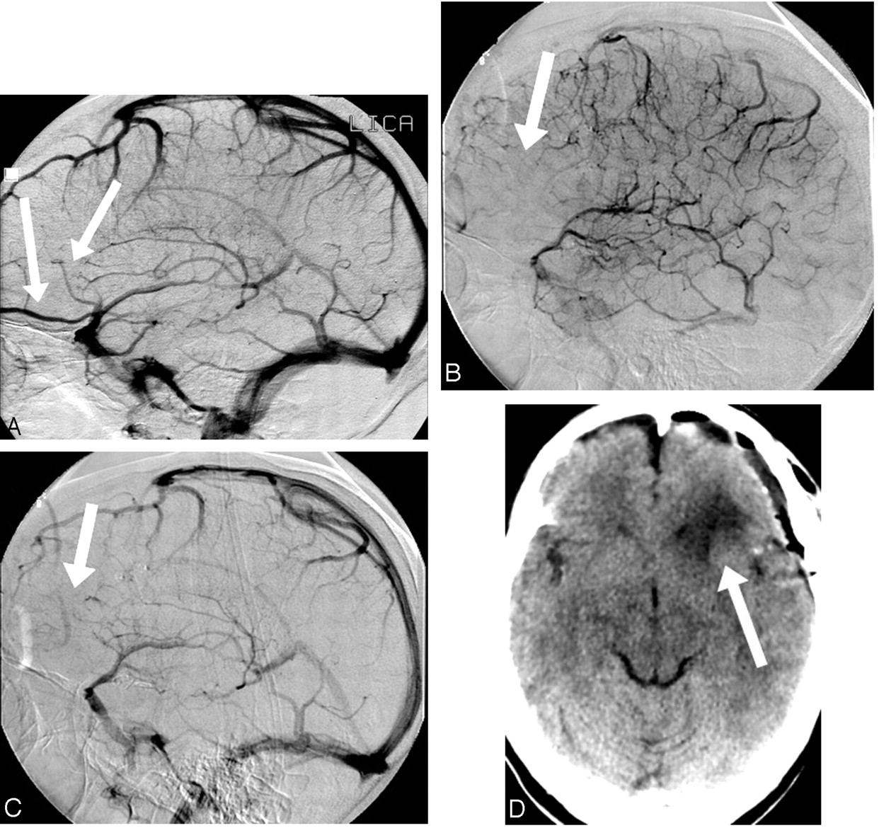
- Fig 5.
Occlusion/nonfilling of frontal lobe veins. A, Multiple large branches from the frontal (arrows) and temporal lobes coalesce to form a large SMCV trunk. B, Early venous phase shows no filling of veins and oligemic region of brain (arrow). C, During the late venous phase, the cortical veins (arrow) of the frontal lobe also fail to fill. D, CT scan shows deep frontal lobe and subcortical white matter edema. The attenuation of the cortical gray matter appears to be normal. The arterial structures are intact.
- Fig 6.
Occluded and very small SMCV and small temporal branch are associated with a small region of brain edema. A, Very small SMCV (white arrow); temporal branch (black arrow). B, Nonfilling of tiny SMCV (white arrow) and distal occlusion of temporal branch (black arrow). C, Small temporal lobe region of edema appears to correlate well with small vessel occlusions (arrows).
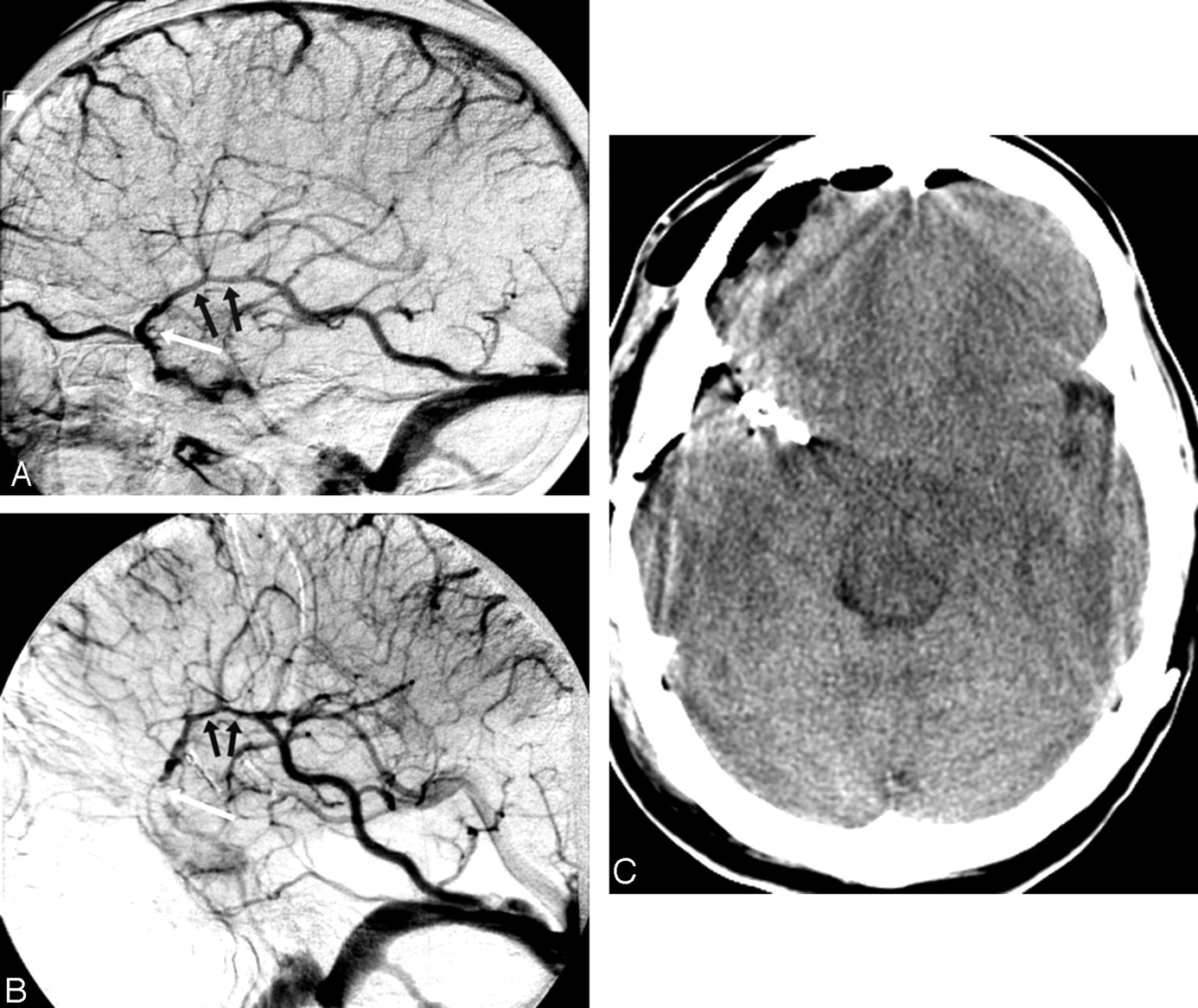
- Fig 7.
Distal occlusion of large SMCV. Brain is protected by collateral branch. A, A large collateral branch is connected to the vein of Labbé (small black arrows). Distal SMCV is depicted by white arrow. B, Occlusion of veins distally (white arrow) is protected by collaterals (black arrows). C, Postoperative CT scan is unremarkable, without significant brain edema or hemorrhage.


