
Article Figures & Data
Figures
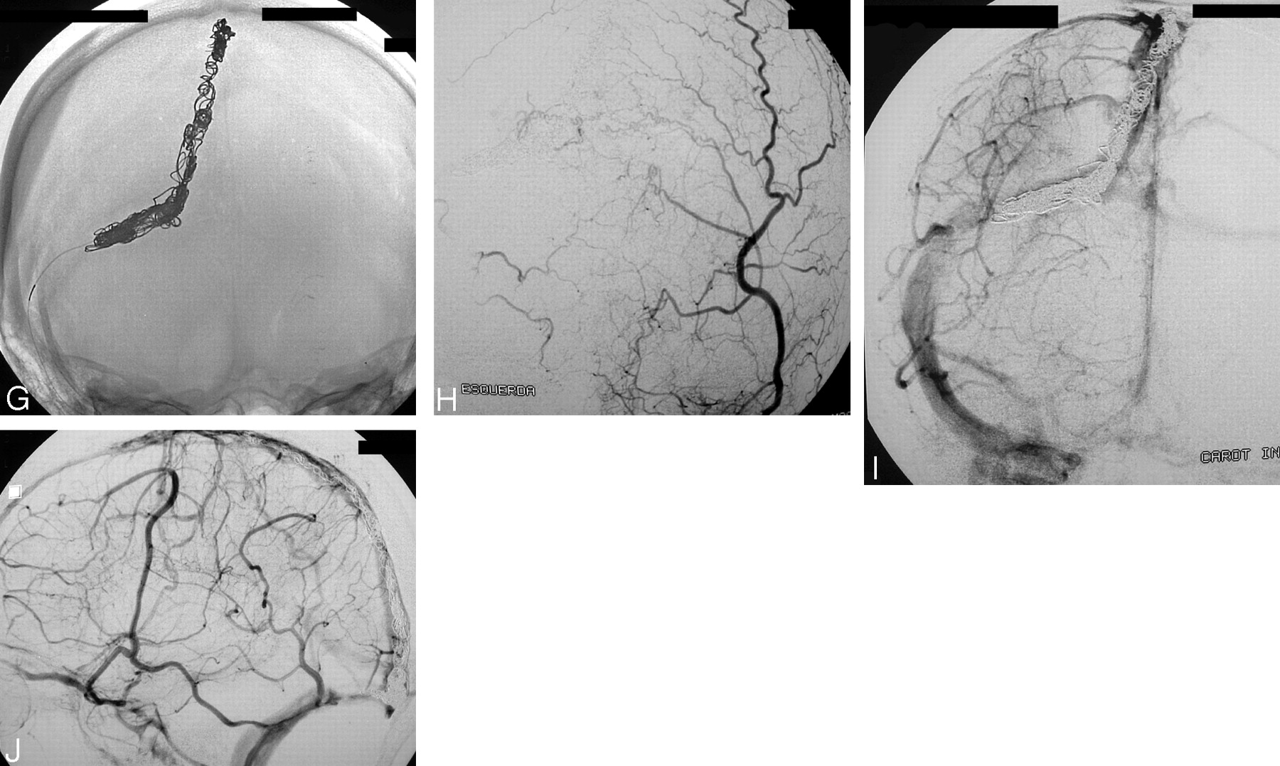
- Fig 1.
Case 4.
A and B, Right ECA and left occipital artery injections in AP view, showing DAVS in the superior sagittal sinus (SSS) and right transverse sinus (TS). Partial opacification of these sinuses is shown (arrows).
C and D, Right CCA angiograms, in arterial (C) and venous phase (D). In panel C, the arrow indicates a early partial filling of the SSS and right TS. In panel D, the arrow indicates a nonopacified area of these sinuses.
E, Superposition of the arterial and venous phases of the right CCA injection, showing in black the shunt and in white the normal venous drainage of the brain.
F, Injection into the septation (black) during the venous phase of the right ICA.
G, Conventional radiographic image, showing coils into the septation.
H, Left occipital artery injection in AP view posttreatment, showing complete occlusion of the shunt.
I and J, AP and lateral view (venous phase) of the right ICA angiogram, showing patency of the SSS and right TS.
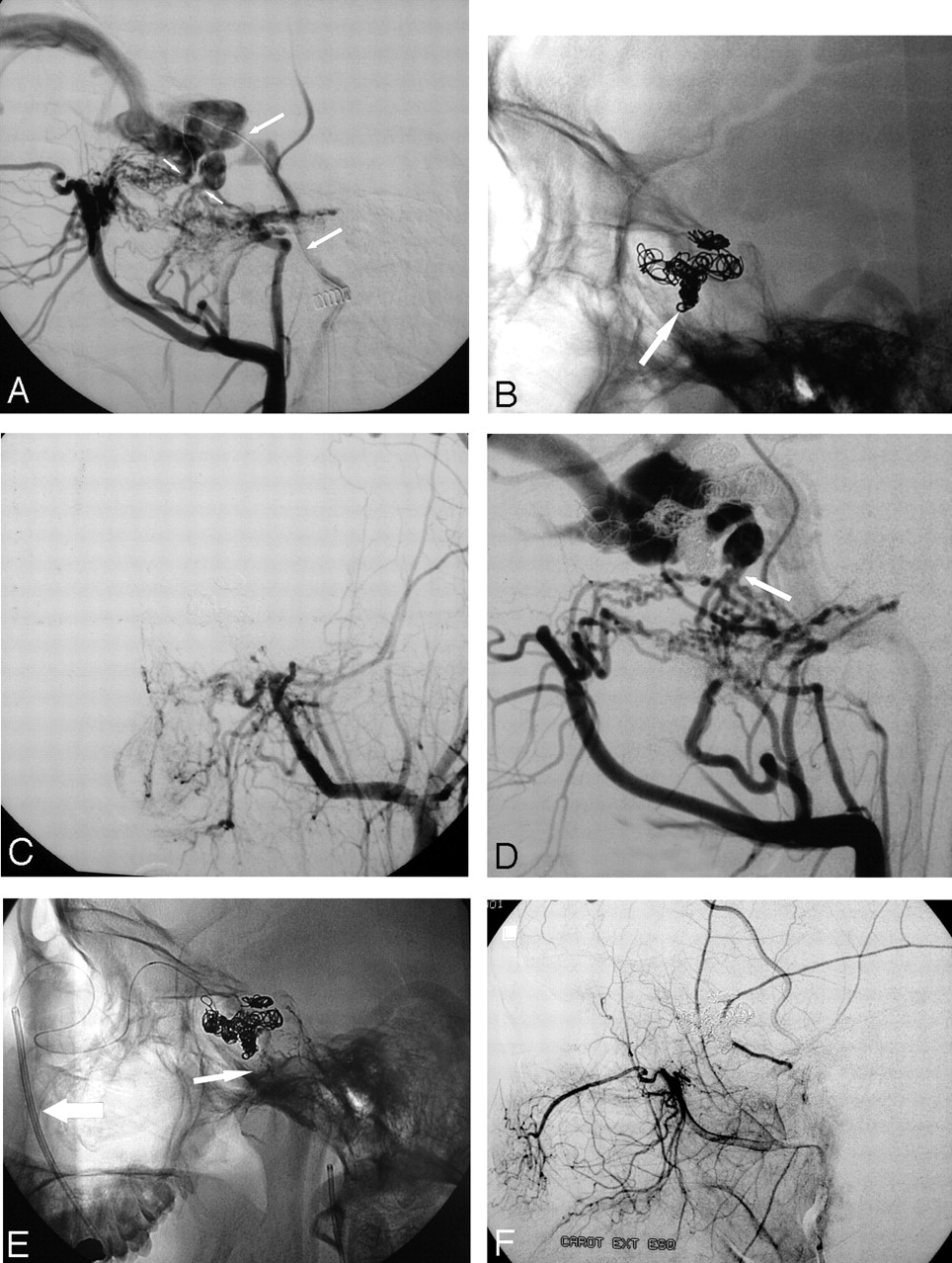
- Fig 2.
Case 2.
A, Right ECA in lateral view shows a DAVS in the distal transversal sinus. Note the convergence of the feeders toward a point.
B, Right occipital artery injection in oblique view shows the shunt is located only over an accessory sinus that has a “tubular hyperdensity” (arrow).
C and D, Superselective injection (C) and coil occlusion (D) of the dural sinus compartment.
E and F, Right occipital and right ICA angiograms, showing complete occlusion of the DAVS, while preserving patency of the lateral and sigmoid sinuses.
- Fig 3.
Case 3.
A, Left CCA angiogram shows DAVS in the SS. Arrow shows a line, which corresponds to a septation.
B, Left occipital artery angiogram after superselective transvenous dural sinus occlusion, showing complete occlusion of the shunt.
C, Venous phase of left ICA angiogram in lateral/oblique view showing coils in the septatio, and preservation of the lumen of the SS.
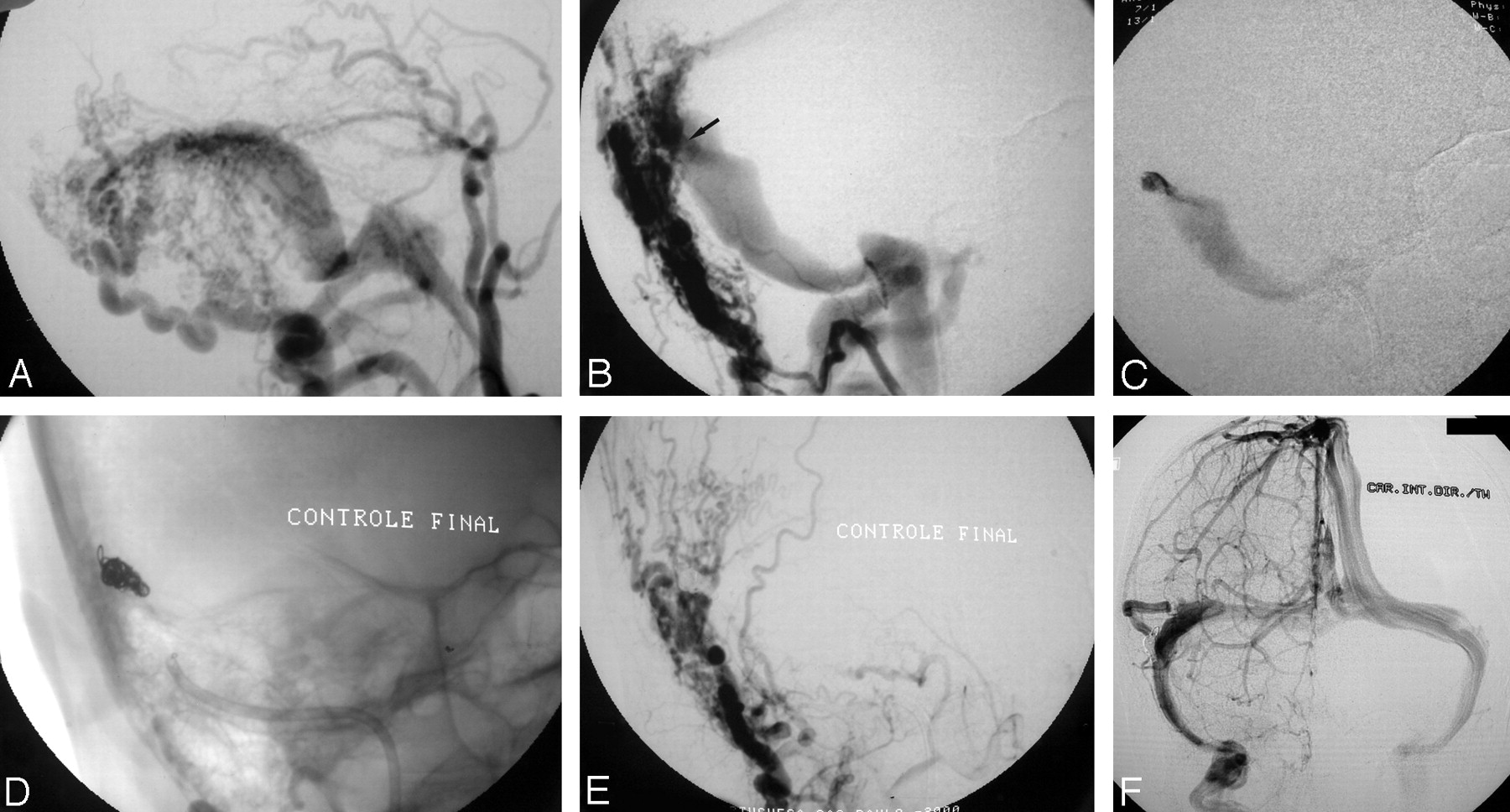
- Fig 4.
Case 1.
A, Right ascending pharyngeal artery angiogram in AP view: DAVS in the foramen magnun region, draining retrogradely toward inferior petrosal sinus and sylvian superficial vein and toward the jugular bulb. Arrow shows hyperdensity.
B, Left ascending pharyngeal artery angiogram in AP view shows that the shunt is located over an accessory sinus.
C, Left ascending pharyngeal artery angiogram in AP view after superselective transvenous dural sinus occlusion, showing complete occlusion of the shunt.
D, RICA angiogram in venous phase, in AP view, showing patency of the normal sinuses.
- Fig 5.
Case 9.
A, Left ECA angiogram in lateral view. There is convergence of feeders toward two points of the cavernous sinus (small arrows). Long arrow is showing a microcatheter into the cavernous sinus (through the inferior petrosal sinus).
B, Conventional radiograph, showing high-density cast of coils into the most anterior septation (arrow) and low-density cast in a portion of the cavernous sinus.
C, Left ECA angiogram in AP view after embolization, showing total occlusion of the DAVS.
D, Left ECA angiogram in lateral view 1 month after embolization, showing recurrence of the shunt only in the posterior septation (arrow). There is no more drainage toward the inferior petrosal sinus.
E, Conventional radiograph at the end of the second session of embolization. Large arrow show 5F guide catheter into the facial vein (transfemoral approach). Arrow shows fragments of NBCA injected in the accessory meningeal artery.
F, Left ECA injection in lateral view after embolization, showing complete occlusion of the DAVS.


Tables
Summary of patients with superselective transvenous embolization of compartment
Case Location Type of Compartment Superselective Transvenous Occlusion Total Sinus Occlusion Recurrence Results Number of Coils Follow-up Angio (mo) 1 FM ACC + − − Cure 6 3 2 TS ACC + − − Cure 6 6 3 SS SEP + − − Cure 5 4 SSS SEP + − − Cure 15 1 5 CS SEP + − − Cure 6 6 6 TS SEP + − − Cure 4 3 7 SS SEP + + − Cure 15 10 8 SS ACC + + − Cure 19 22 9 CS SEP + − +A+V Cure 6 3 10 TS SEP + − +A Cure 8 0 11 SS SEP − + − Cure 13 8 12 SS SEP + − − Cure 14 16 Note.—FM indicates foramen magnum; SS, sigmoid sinus; SSS, superior sagital sinus; CS, cavernous sinus; TS, transverse sinus; ACC, accessory sinus; SEP, septation; +, yes; −, no; A+V, arterial and venous approach; A, arterial approach.
In this issue


Jump to section
Related Articles
Cited By...
- Toward a Better Understanding of Dural Arteriovenous Fistula Angioarchitecture: Superselective Transvenous Embolization of a Sigmoid Common Arterial Collector
- Endovascular Treatment of Dural Arteriovenous Fistulas Using Transarterial Liquid Embolization in Combination with Transvenous Balloon-Assisted Protection of the Venous Sinus
- Angioarchitecture of Transverse-Sigmoid Sinus Dural Arteriovenous Fistulas: Evaluation of Shunted Pouches by Multiplanar Reformatted Images of Rotational Angiography
- ONYX versus n-BCA for embolization of cranial dural arteriovenous fistulas
- Onyx 18 embolisation of dural arteriovenous fistula via arterial and venous pathways: preliminary experience and evaluation of the short-term outcomes
- Cranial dural arteriovenous fistula: transarterial Onyx embolization experience and technical nuances
- Spontaneous Angiographic Conversion of Intracranial Dural Arteriovenous Shunt: Long-Term Follow-Up in Nontreated Patients