
Article Figures & Data
Figures
- Fig 1.
T1-, T2-, and proton density–weighted and FLAIR images (left to right) from a typical examination. LE can be seen in the white matter posterior to the ventricles.
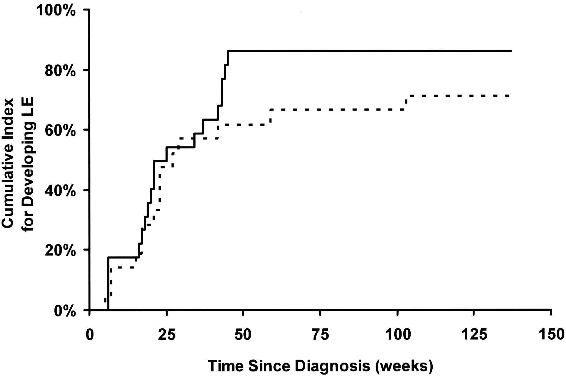
- Fig 2.
Cumulative risk for LE as a function of time since diagnosis. Solid line represents patients in the standard- and high-risk treatment arm; dashed line, those in the low-risk treatment arm.
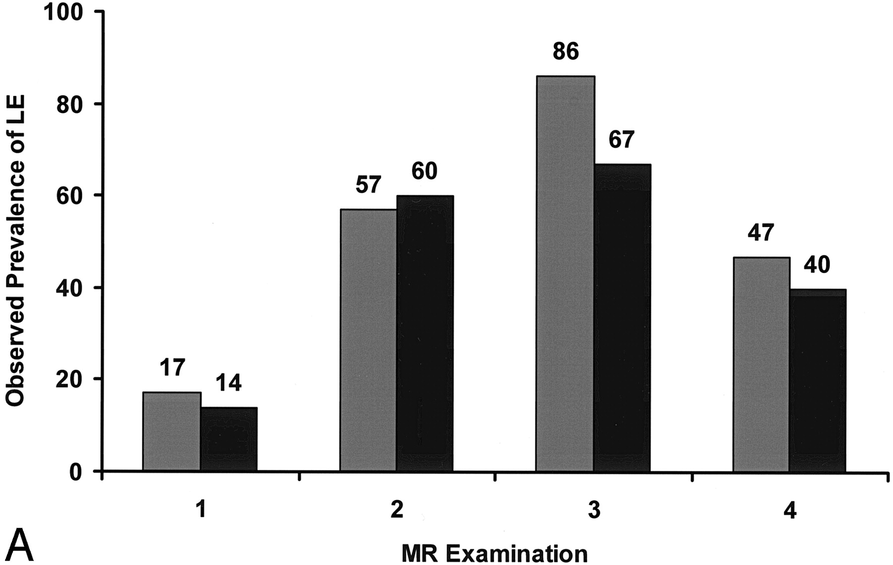
- Fig 3.
Observed prevalence of LE in patients in the standard- or high-risk arm (gray bars) and patients in the low-risk arm (black bars) of the treatment protocol.
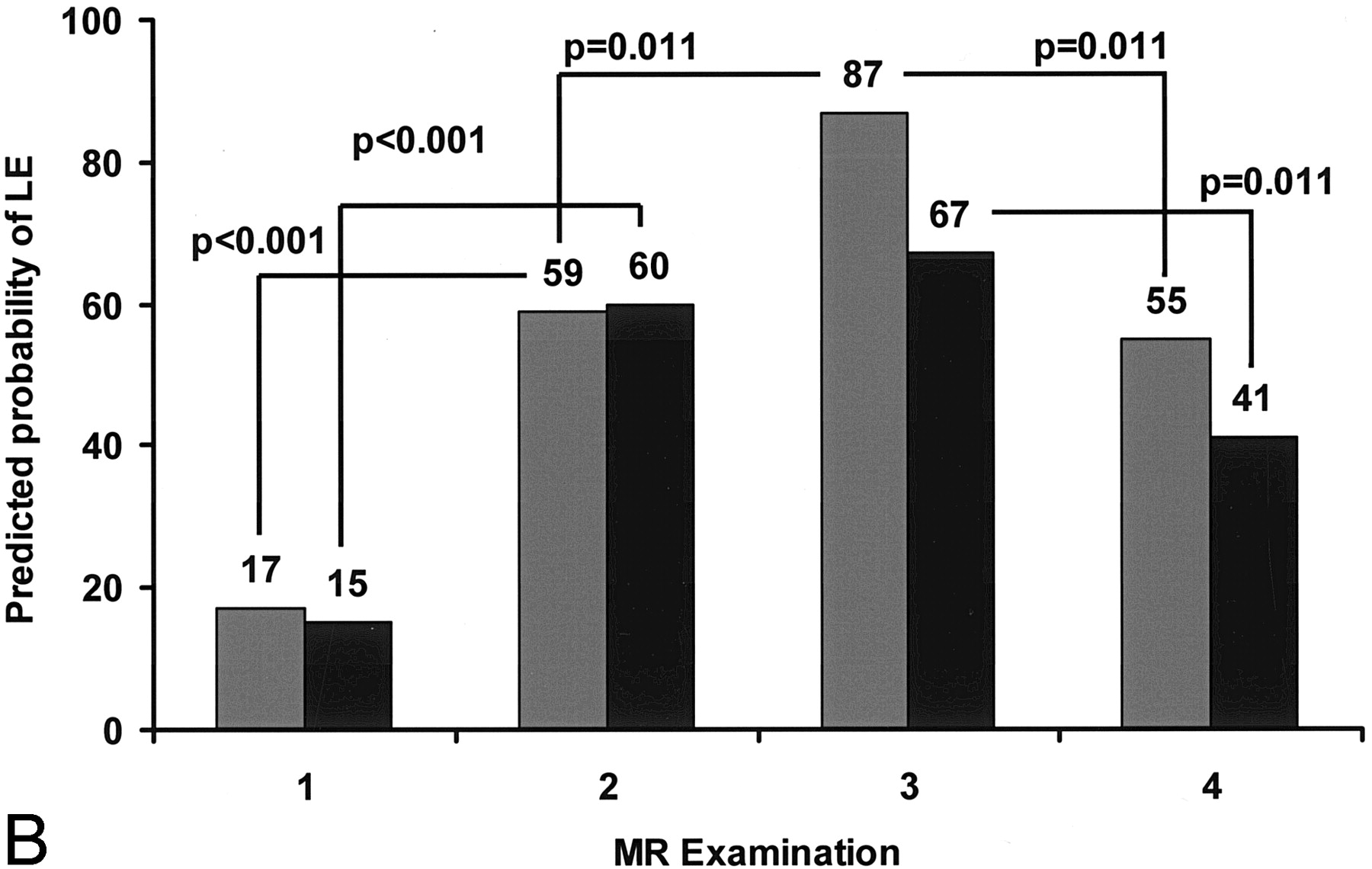
- Fig 4.
Predicted probability of LE according to the general linear model for patients in the standard- or high-risk arm (gray bars) and patients in the low-risk arm (black bars) of the treatment protocol.
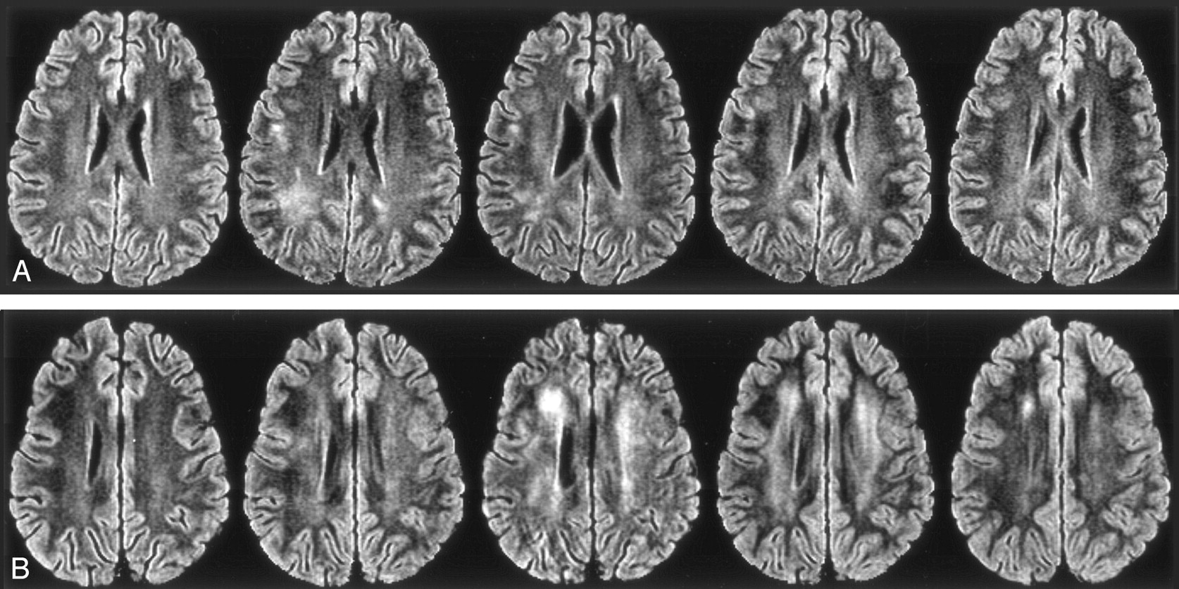
- Fig 5.
FLAIR images from a typical examination. Left to right, Sections after one course, four courses, and seven courses of high-dose IV MTX therapy; at the end of therapy; and at 2 years after the completion of therapy.
A, Transient LE. Although the first image is normal, the second and third show LE, which resolves by the fourth and remains normal on the final image.
B, Persistent LE. First image is normal, but all others, including the 2-year follow-up image, show LE.


Tables
- TABLE 1:
Prevalence of LE in patients with ALL treated with intrathecal MTX and CRT prophylaxis
Study Modality Prevalence of LE* Point in Therapy Chu et al, 200314 MR imaging 0/2 (0) Baseline Chu et al, 200314 MR imaging 0/3 (0) 8 wk after diagnosis Chu et al, 200314 MR imaging 1/3 (33) 20 wk after diagnosis Paakko et al, 199610 MR imaging 0/12 (0) End Consolidation Ochs et al, 19835 CT 5/55 (9) Continuation Paakko et al, 200012 MR imaging 0/16 (0) During McIntosh et al, 19773 CT 10/30 (33) 0.5–6 y after induction Peylan-Ramu et al, 19784 CT 5/32 (16) 3.5 y after diagnosis Chu et al, 200314 MR imaging 1/3 (33) 1 year after therapy Chu et al, 200314 MR imaging 1/2 (50) 2 y after therapy Paakko et al, 19928 MR imaging 3/16 (19) 3 y after therapy Chu et al, 200314 MR imaging 2/2 (100) 3 y after therapy Brouwers and Poplack 19906 CT 5/23 (22) 4 y after therapy Ochs et al, 19917 CT 8/23 (35) 6 y after therapy Hertzberg et al, 199711 CT, MR imaging 23/41 (56) 6 y after therapy Kingma et al, 19939 MR imaging 24/35 (69) 8 y after diagnosis Hertzberg et al, 199711 CT, MR imaging 23/38 (61) 9 y after therapy Kingma et al, 200113 MR imaging 15/24 (63) 10 y after diagnosis * Data in parentheses are percentages.
Study Modality Prevalence of LE* Point in Therapy Wilson et al, 199115 MR imaging 0/21 (0) Baseline Chu et al, 200314 MR imaging 0/17 (0) Baseline Asato et al, 199216 MR imaging 6/16 (38) End Induction Chu et al, 200314 MR imaging 0/20 (0) 8 wk after diagnosis Wilson et al, 199115 MR imaging 0/23 (0) Beginning of consolidation Chu et al, 200314 MR imaging 4/19 (21) 20 wk after diagnosis Wilson et al, 199115 MR imaging 15/25 (60) Middle of consolidation Paakko et al, 199610 MR imaging 2/6 (33) End of consolidation Wilson et al, 199115 MR imaging 15/25 (60) Begin of maintenance Ochs et al, 19835 CT 10/53 (19) Continuation Paakko et al, 200012 MR imaging 3/17 (18) During therapy Wilson et al, 199115 MR imaging 12/23 (52) 1 year after diagnosis Mahoney et al, 199818 CT, MR imaging 57/75 (76) 1 year after diagnosis Chu et al, 200314 MR imaging 1/19 (5) 1 year after therapy Chu et al, 200314 MR imaging 1/17 (6) 2 y after therapy Paakko et al, 19928 MR imaging 1/11 (9) 3 y after therapy Chu et al, 200314 MR imaging 2/16 (13) 3 y after therapy Bakke et al, 199317 MR imaging 8/15 (53) 5 y after therapy Ochs et al, 19917 CT 7/25 (28) 6 y after therapy Hertzberg et al, 199711 CT, MR imaging 15/39 (39) 7 y after therapy Kingma et al, 200113 MR imaging 6/16 (38) 10 y after diagnosis * Data in parentheses are percentages.
Data Low Risk Standard or High Risk No. of subjects After 1 course of IV MTX 21 23 After 4 courses of IV MTX 20 21 After 7 courses of IV MTX 21 21 End of Therapy 20 17 Sex Male 10 11 Female 12 12 Age at diagnosis (y)* 5.0±2.7 9.2±4.8 * Data are the mean ± standard deviation.
- TABLE 4:
Estimated coefficients for the general linear model to predict probability of LE during therapy
Coefficient Estimate Standard Error P Value α (intercept) −1.77 0.625 .004 β1 dose (g/m2) 5.0 0.219 0.832 .792 2.5 0.000 β2 exam First 0.000 Second 2.181 0.632 <.001 Third 2.473 0.662 <.001 Fourth 1.423 0.548 .009 β3 exam∗dose (g/m2) First and 5.0 0.000 First and 2.5 0.000 Second and 5.0 −0.269 0.850 .752 Second and 2.5 0.000 Third and 5.0 0.950 1.020 .351 Third and 2.5 0.000 Fourth and 5.0 0.338 0.793 .670 Fourth and 2.5 0.000
In this issue


Jump to section
Related Articles
Cited By...
- The Impact of Persistent Leukoencephalopathy on Brain White Matter Microstructure in Long-Term Survivors of Acute Lymphoblastic Leukemia Treated with Chemotherapy Only
- Methotrexate-Induced Neurotoxicity and Leukoencephalopathy in Childhood Acute Lymphoblastic Leukemia
- Voxel-Based Analysis of T2 Hyperintensities in White Matter during Treatment of Childhood Leukemia
- High-dose compared with intermediate-dose methotrexate in children with a first relapse of acute lymphoblastic leukemia