
Article Figures & Data
Figures
- Fig 1.
Example of a DSA false-positive finding for basilar occlusion in a patient with low-flow state due to a severe stenosis of the left vertebral artery.
A and B, Frontal (A) and lateral (B) middle arterial phase DSA images show selective injection of dominant left vertebral artery (shown in anatomic orientation). Note the severe stenosis of the left vertebral artery (long arrow) and a small amount of reflux down the nondominant right vertebral artery (short curved arrow in A). The basilar artery distal to the origin of the left anterior inferior cerebellar artery is not opacified and therefore appears occluded (open arrow), even on late arterial and venous images (not shown).
C, Lateral projection, left ICA injection, middle arterial phase DSA image shows minimal retrograde filling of the distal basilar artery (arrow) through the posterior communicating artery to the level of the superior cerebellar arteries, suggesting segmental occlusion of the midbasilar artery.
D, Corresponding volume-rendered 3D CTA image in anatomic orientation. CTA image was obtained 3 days before DSA and shows a severe left vertebral artery stenosis (black arrow) associated with heavy calcific atheromatous plaque. However, CTA depicts the basilar artery as patent. In addition, the CTA image demonstrates two tandem stenoses of the distal basilar artery (white arrows), which may have contributed to impaired retrograde flow into the basilar artery via the posterior communicating artery upon anterior circulation injection at DSA. There was no change in patient symptoms during the intervening period between the CTA and DSA studies to suggest interval arterial thrombosis.
- Fig 2.
Example of a DSA false-positive finding for basilar occlusion in a patient with a low- or balanced-flow state due to a severe stenosis of the basilar artery.
A and B, Frontal (A) and lateral (B) middle arterial phase DSA images show selective injection of a dominant left vertebral artery (DSA images shown in anatomic orientation). Note absent cephalad flow in the basilar artery distal to the midbasilar segment (arrow), suggesting basilar artery occlusion.
C, Lateral projection, right ICA injection, middle arterial phase DSA image shows minimal retrograde filling of the distal basilar artery (arrow) through the posterior communicating artery. The midbasilar segment is not visualized, suggesting segmental occlusion of the midbasilar artery.
D, CTA was performed 12 days before DSA. This volume-rendered 3D CTA image, in anatomic orientation, shows a severe, eccentric, focal midbasilar stenosis (arrow); however, the basilar artery is clearly patent. This was verified on the gray-scale 2D source image.
E, Axial 2D curved oblique CTA reformation image, frontal projection, shows focal midbasilar artery stenosis (arrow) with 79% stenosis severity.
F, TOF MRA image with frontal oblique MIP shows a focal flow gap in the midbasilar artery (arrow).
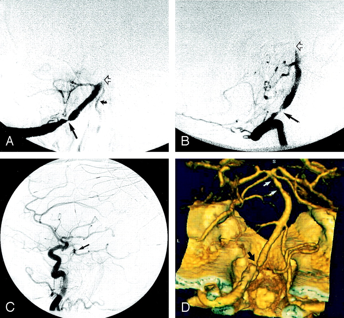
- Fig 3.
Example of a DSA false-positive finding for basilar occlusion in a patient with a relatively isolated posterior circulation due to bilateral vertebral artery occlusion, a hypoplastic right posterior communicating artery, and a significant left proximal P1 stenosis.
A, Frontal late arterial phase DSA image shows selective injection of a dominant right vertebral artery. The basilar artery is not opacified. The right vertebral artery appears to terminate in the right posterior inferior cerebellar artery (arrow). Note that the patent right posterior inferior cerebellar artery supplies some blood flow to the right anterior inferior cerebellar artery and a small tonsillar loop branch to the contralateral left posterior inferior cerebellar artery.
B, Frontal middle phase DSA image shows selective injection of the left vertebral artery, which terminates in an extracranial muscular branch near the skull base (arrow). No flow is seen in the intracranial segment of the left vertebral artery indicating occlusion of this vessel segment.
C, Lateral projection, right ICA injection, middle arterial phase DSA image shows minimal retrograde filling of the distal basilar artery (arrow) through the posterior communicating artery, but there is absence of flow in the remainder of the basilar artery, suggesting segmental occlusion of this vessel.
D, CTA was performed 1 day after DSA. This volume-rendered 3D CTA image in posteroanterior projection (anatomic orientation) shows bilateral distal vertebral artery occlusions (solid black arrows) and significant focal origin stenoses of both the left proximal P1 and the left superior cerebellar artery (white arrow). CTA image also demonstrates minimal flow in what appears to be a small segment of a hypoplastic distal right intracranial vertebral artery, distal to the origin of the right posterior inferior cerebellar artery. The CTA image shows that the basilar artery is entirely patent and stenosis-free (open black arrow). This was verified on the gray-scale 2D source image.
E, Volume-rendered 3D CTA image, craniocaudal projection with anatomic orientation, demonstrates a prominent left posterior communicating artery (arrow) and absent right posterior communicating artery. The left P1 is small in caliber.
F, Targeted volume-rendered 3D CTA image in the anteroposterior projection demonstrates significant proximal focal left P1 and superior cerebellar artery stenoses (arrow).
G, TOF MRA image with frontal MIP shows absent flow signal (arrow) in the expected region of the basilar artery, suggesting occlusion.


Tables
Artery No. (%) of Lesions (n = 115) Internal carotid 27 (23) Vertebral 26 (23) Posterior cerebral 24 (21) Basilar 17 (15) Middle cerebral 17 (15) Anterior cerebral 4 (3) Modality No. (%) of Reader Errors (n=672) DSA 25 (4) CTA 7 (1) MRA 17 (3) Patient No. Occlusion Location at DSA Stenosis Location at CTA Probable DSA Error 3 Vertebral Vertebral False-positive 4 Vertebral Vertebral False-positive 20 Basilar Basilar False-positive 22 Basilar Basilar False-positive 24 Basilar Basilar False-positive 28 Basilar Basilar False-positive Performance Measure CTA MRA Sensitivity 71 (100) 81 (87) Specificity 100 (100) 98 (98) Positive predictive value 100 (100) 61 (59) Negative predictive value 99 (100) 99 (99.5) Note.—Data are percentages using DSA as the reference standard; numbers in parentheses are percentages corrected for DSA false-positive occlusion by consensus reading. See Tables 6 and 8 for raw data.
Performance Measure CTA MRA Sensitivity 98 (98) 70 (70) Specificity 98 (99) 99 (97) Positive predictive value 78 (93) 63 (65) Negative predictive value 100 (100) 98 (98) Note.—Data are percentages using DSA as the reference standard; numbers in parentheses are percentages corrected for DSA false-positive stenosis by consensus reading. See Tables 7 and 9 for raw data.
CTA Occlusion DSA Occlusion Total Yes No Yes 20 0 20 No 0 634 634 Total 20 634 654 Note.—Data are number of vessel segments and relate to the CTA performance measures in parentheses in Table 4.
CTA Stenosis DSA Stenosis Total Yes No Yes 42 3 45 No 1 595 596 Total 43 598 641 Note.—Data are number of vessel segments and relate to the CTA performance measures in parentheses in Table 5.
MRA Occlusion DSA Occlusion Total Yes No Yes 20 14 34 No 3 634 637 Total 23 648 671 Note.—Data are number of vessel segments and relate to the MRA performance measures in parentheses in Table 4.
MRA Stenosis DSA Stenosis Total Yes No Yes 30 16 46 No 13 595 608 Total 43 611 654 Note.—Data are number of vessel segments and relate to the MRA performance measures in parentheses in Table 5.
In this issue


Jump to section
Related Articles
Cited By...
- Comprehensive imaging analysis of intracranial atherosclerosis
- Chinese Stroke Association guidelines for clinical management of ischaemic cerebrovascular diseases: executive summary and 2023 update
- USe of Diagnostic sUbtraction angiography in The isCHemic stroke setting (US DUTCH study)
- Characteristics for Machine Learning Detection of Large Vessel Occlusion on Computed Tomography Angiography
- Prognostic Role of Hypertriglyceridemia in Patients With Stroke of Atherothrombotic Origin
- Intracranial Atherosclerotic Disease: Current Concepts in Medical and Surgical Management
- Correlation between intracranial vertebral artery stenosis diameter measured by digital subtraction angiography and cross-sectional area measured by optical coherence tomography
- Carotid Vessel Wall Imaging on CTA
- 3D Black-Blood Luminal Angiography Derived from High-Resolution MR Vessel Wall Imaging in Detecting MCA Stenosis: A Preliminary Study
- Value of Contrast-Enhanced MRA versus Time-of-Flight MRA in Acute Ischemic Stroke MRI
- Site and Rate of Occlusive Disease in Cervicocerebral Arteries: A CT Angiography Study of 2209 Patients with Acute Ischemic Stroke
- Quantifying Intracranial Internal Carotid Artery Stenosis on MR Angiography
- Acute Ischemic Stroke Therapy Overview
- Centripetal Propagation of Vasoconstriction at the Time of Headache Resolution in Patients with Reversible Cerebral Vasoconstriction Syndrome
- Hemorrhagic Reversible Cerebral Vasoconstriction Syndrome: Features and Mechanisms
- Comparing Vessel Imaging: Noncontrast Computed Tomography/Computed Tomographic Angiography Should Be the New Minimum Standard in Acute Disabling Stroke
- Multimodal Diagnostic Imaging for Hyperacute Stroke
- Prediction of Infarction and Reperfusion in Stroke by Flow- and Volume-Weighted Collateral Signal in MR Angiography
- Gadolinium Enhancement of Atherosclerotic Plaque in the Middle Cerebral Artery: Relation to Symptoms and Degree of Stenosis
- Imaging Intracranial Vessel Wall Pathology With Magnetic Resonance Imaging: Current Prospects and Future Directions
- Imaging and Treatment of Patients with Acute Stroke: An Evidence-Based Review
- Posterior circulation ischaemic stroke
- Current State of Acute Stroke Imaging
- Imaging Recommendations for Acute Stroke and Transient Ischemic Attack Patients: A Joint Statement by the American Society of Neuroradiology, the American College of Radiology, and the Society of NeuroInterventional Surgery
- Prevalence and long-term clinical significance of intracranial atherosclerosis after ischaemic stroke or transient ischaemic attack: a cohort study
- Acute Stroke Imaging Research Roadmap II
- Computed Tomography Angiography in Hyperacute Ischemic Stroke: Prognostic Implications and Role in Decision-Making
- Imaging-based selection for intra-arterial stroke therapies
- Guidelines for the Early Management of Patients With Acute Ischemic Stroke: A Guideline for Healthcare Professionals From the American Heart Association/American Stroke Association
- Intracranial Steno-Occlusive Arterial Disease and its Associations in Egyptian Ischemic Stroke Patients
- Feasibility of Intravenous Flat Panel Detector CT Angiography for Intracranial Arterial Stenosis
- Standard of practice: endovascular treatment of intracranial atherosclerosis
- Severity of Leukoaraiosis in Large Vessel Atherosclerotic Disease
- MR Angiography and Imaging for the Evaluation of Middle Cerebral Artery Atherosclerotic Disease
- Multimodal Imaging of Reversible Cerebral Vasoconstriction Syndrome: A Series of 6 Cases
- Intracranial Vessel Wall Imaging at 7.0-T MRI
- Detection of Intracranial Arterial Stenosis Using Transcranial Color-Coded Duplex Sonography, Computed Tomographic Angiography, and Digital Subtraction Angiography
- Abnormalities of cervical arteries in children with arterial ischemic stroke
- Reporting standards for angioplasty and stent-assisted angioplasty for intracranial atherosclerosis
- Assessment of Intracranial Arterial Stenosis with Multidetector Row CT Angiography: A Postprocessing Techniques Comparison
- Recommendations for Imaging of Acute Ischemic Stroke: A Scientific Statement From the American Heart Association
- Reporting Standards for Angioplasty and Stent-Assisted Angioplasty for Intracranial Atherosclerosis
- How Accurate Is CT Angiography in Evaluating Intracranial Atherosclerotic Disease?
- Noninvasive Detection of Diffuse Intracranial Disease
- Imaging of vertebral artery stenosis: a systematic review
- ACCF/AHA 2007 Clinical Competence Statement on Vascular Imaging With Computed Tomography and Magnetic Resonance: A Report of the American College of Cardiology Foundation/American Heart Association/American College of Physicians Task Force on Clinical Competence and Training Developed in Collaboration With the Society of Atherosclerosis Imaging and Prevention, the Society for Cardiovascular Angiography and Interventions, the Society of Cardiovascular Computed Tomography, the Society for Cardiovascular Magnetic Resonance, and the Society for Vascular Medicine and Biology
- The Stroke Outcomes and Neuroimaging of Intracranial Atherosclerosis (SONIA) Trial
- Noninvasive imaging is improving but digital subtraction angiography remains the gold standard