
Article Figures & Data
Figures
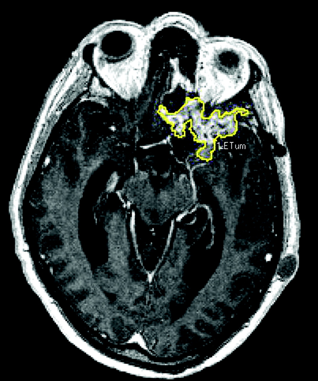
- Fig 1.
Image showing software used (developed for Alkermes, Inc., by Evergreen Technologies, Inc., 00000, ME) by neuroradiologist to outline areas of enhancement manually on each transverse section to determine tumor volume.
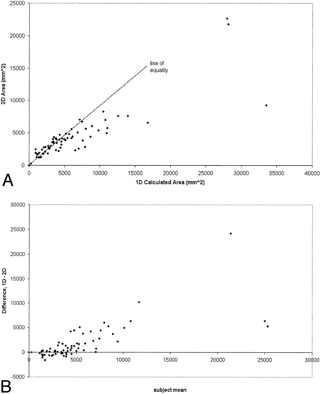
- Fig 2.
Tumor size measured using 1D and 2D techniques. A, 1D versus 2D technique plotted showing line of equality (r = 0.85; P < .05), B, Difference versus mean of tumor size measured by the two methods.
- Fig 3.
Tumor size measured using 1D and 3D techniques. A, 1D versus 3D technique plotted showing line of equality (r = 0.44; P < .05). B, Difference versus mean of tumor size measured by the two methods.
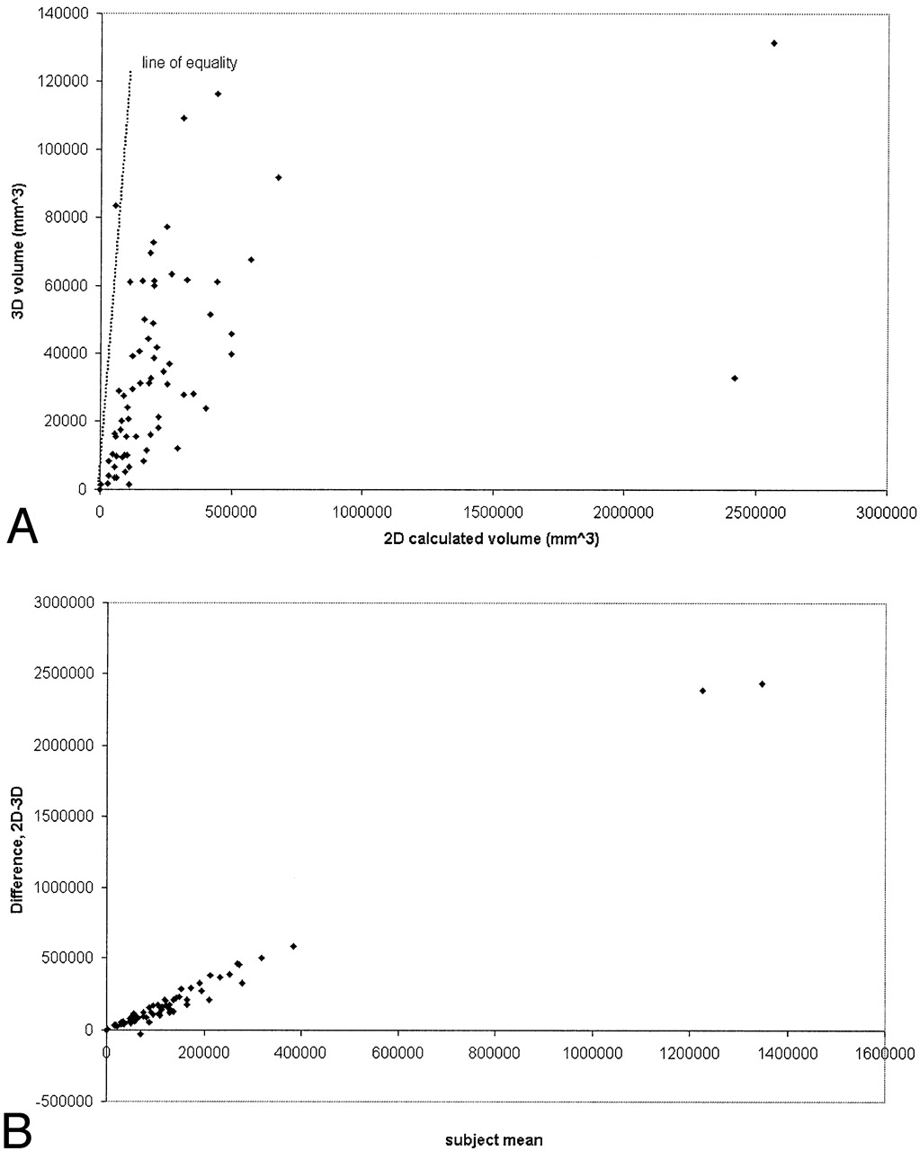
- Fig 4.
Tumor size measured by 2D and 3D techniques. A, 2D versus 3D technique plotted showing line of equality (r = 0.47; P < .05). B, Difference versus mean of tumor size measured by the two methods.


Tables
1D (mm) 2D (mm2) 3D (mm3) 1D→2D (mm2) 1D→3D (mm3) 2D→3D (mm3) Minimum 2.8 22.5 53.5 6.2 11.6 80.3 Maximum 206.6* 22645.4* 131387.4 33501.7* 4613963.1* 2564157.0* Mean 78.6 4240.2 34416.5 5922.4 454253.8 253035.7 * Such large maximum values originate from images with several regions of contrast enhancement outlined separately and consequent summation of the appropriate diameters resulting in a large 1D measurement and subsequent conversions.
Covariate Univariate Analysis Multivariate Analysis Coefficient (bi) HR [exp(bi)] 95% CI P-value Coefficient (bi) HR [exp(bi)] 95% CI P-value Age 0.036 1.039 (1.014–1.064) .002 0.036 1.037 (1.013–1.062) .002 Sex .015 .045 Female (0.00) (1.00) (0.00) (1.00) Male 0.725 2.070 (1.153–3.715) 0.625 1.868 (1.015–3.436) Previous .096 .545 Chemotherapy No (0.00) (1.00) (0.00) (1.00) Yes 0.433 1.542 (0.926–2.566) 0.164 1.178 (0.684–2.000) 1D Size 0.067 1.069 (0.997–1.147) .062 0.052 1.053 (0.182–1.129) .146 HR = hazard ratio, CI = confidence interval
Covariate Univariate Analysis Multivariate Analysis Coefficient (bi) HR [exp(bi)] 95% CI P-value Coefficient (bi) HR [exp(bi)] 95% CI P-value Age 0.038 1.039 (1.014–1.064) .002 0.036 1.037 (1.013–1.062) .003 Sex .015 .039 Female (0.00) (1.00) (0.00) (1.00) Male 0.725 2.070 (1.153–3.715) 0.648 1.912 (1.033–3.539) Previous .096 .601 Chemotherapy No (0.00) (1.00) (0.00) (1.00) Yes 0.433 1.542 (0.926–2.566) 0.144 1.155 (0.673–1.082) 2D Size 0.006 1.006 (1.000–1.012) .046 0.004 1.004 (0.999–1.010) .140 HR = hazard ratio, CI = confidence interval
Covariate Univariate Analysis Multivariate Analysis Coefficient (bi) HR [exp(bi)] 95% CI P-value Coefficient (bi) HR [exp(bi)] 95% CI P-value Age 0.038 1.039 (1.014–1.064) .002 0.034 1.034 (1.010–1.058) .005 Sex .015 .003 Female (0.00) (1.00) (0.00) (1.00) Male 0.725 2.070 (1.153–3.715) 0.936 2.551 (1.361–4.781) Previous .096 .115 Chemotherapy No (0.00) (1.00) (0.00) (1.00) Yes 0.433 1.542 (0.926–2.566) 0.430 1.537 (0.900–2.626) 3D Size 0.019 1.019 (1.009–1.029) .000 0.025 1.026 (0.015–1.036) .000 HR = hazard ratio, CI = confidence interval
In this issue


Jump to section
Related Articles
Cited By...
- Development and Evaluation of Automated Artificial Intelligence-Based Brain Tumor Response Assessment in Patients with Glioblastoma
- Development and Practical Implementation of a Deep Learning-Based Pipeline for Automated Pre- and Postoperative Glioma Segmentation
- Quantitative Delta T1 (dT1) as a Replacement for Adjudicated Central Reader Analysis of Contrast-Enhancing Tumor Burden: A Subanalysis of the American College of Radiology Imaging Network 6677/Radiation Therapy Oncology Group 0625 Multicenter Brain Tumor Trial
- Shape Features of the Lesion Habitat to Differentiate Brain Tumor Progression from Pseudoprogression on Routine Multiparametric MRI: A Multisite Study
- Current whole-body MRI applications in the neurofibromatoses: NF1, NF2, and schwannomatosis
- Semiautomated Volumetric Measurement on Postcontrast MR Imaging for Analysis of Recurrent and Residual Disease in Glioblastoma Multiforme
- Conventional MRI evaluation of gliomas
- Functional Diffusion Map As an Early Imaging Biomarker for High-Grade Glioma: Correlation With Conventional Radiologic Response and Overall Survival
- Brain Metastases: The HER2 Paradigm