Advertisement

Main menu

  • Home
  • Content
    • Current Issue
    • Accepted Manuscripts
    • Article Preview
    • Past Issue Archive
    • Video Articles
    • AJNR Case Collection
    • Case of the Week Archive
    • Case of the Month Archive
    • Classic Case Archive
  • Special Collections
    • AJNR Awards
    • Low-Field MRI
    • Alzheimer Disease
    • ASNR Foundation Special Collection
    • Photon-Counting CT
    • View All
  • Multimedia
    • AJNR Podcasts
    • AJNR SCANtastic
    • Trainee Corner
    • MRI Safety Corner
    • Imaging Protocols
  • For Authors
    • Submit a Manuscript
    • Submit a Video Article
    • Submit an eLetter to the Editor/Response
    • Manuscript Submission Guidelines
    • Statistical Tips
    • Fast Publishing of Accepted Manuscripts
    • Graphical Abstract Preparation
    • Imaging Protocol Submission
    • Author Policies
  • About Us
    • About AJNR
    • Editorial Board
    • Editorial Board Alumni
  • More
    • Become a Reviewer/Academy of Reviewers
    • Subscribers
    • Permissions
    • Alerts
    • Feedback
    • Advertisers
    • ASNR Home

User menu

  • Alerts
  • Log in

Search

  • Advanced search
American Journal of Neuroradiology

American Journal of Neuroradiology

ASHNR American Society of Functional Neuroradiology ASHNR American Society of Pediatric Neuroradiology ASSR
  • Alerts
  • Log in

Advanced Search

  • Home
  • Content
    • Current Issue
    • Accepted Manuscripts
    • Article Preview
    • Past Issue Archive
    • Video Articles
    • AJNR Case Collection
    • Case of the Week Archive
    • Case of the Month Archive
    • Classic Case Archive
  • Special Collections
    • AJNR Awards
    • Low-Field MRI
    • Alzheimer Disease
    • ASNR Foundation Special Collection
    • Photon-Counting CT
    • View All
  • Multimedia
    • AJNR Podcasts
    • AJNR SCANtastic
    • Trainee Corner
    • MRI Safety Corner
    • Imaging Protocols
  • For Authors
    • Submit a Manuscript
    • Submit a Video Article
    • Submit an eLetter to the Editor/Response
    • Manuscript Submission Guidelines
    • Statistical Tips
    • Fast Publishing of Accepted Manuscripts
    • Graphical Abstract Preparation
    • Imaging Protocol Submission
    • Author Policies
  • About Us
    • About AJNR
    • Editorial Board
    • Editorial Board Alumni
  • More
    • Become a Reviewer/Academy of Reviewers
    • Subscribers
    • Permissions
    • Alerts
    • Feedback
    • Advertisers
    • ASNR Home
  • Follow AJNR on Twitter
  • Visit AJNR on Facebook
  • Follow AJNR on Instagram
  • Join AJNR on LinkedIn
  • RSS Feeds

AJNR Awards, New Junior Editors, and more. Read the latest AJNR updates

OtherBRAIN

Cerebral Embolism from Left Atrial Myxoma Leading to Cerebral and Retinal Aneurysms: A Case Report

Martin Herbst, Mike Peter Wattjes, Horst Urbach, Claudia Inhetvin-Hutter, Dietmar Becker, Thomas Klockgether and Alexander Hartmann
American Journal of Neuroradiology March 2005, 26 (3) 666-669;
Martin Herbst
  • Find this author on Google Scholar
  • Find this author on PubMed
  • Search for this author on this site
Mike Peter Wattjes
  • Find this author on Google Scholar
  • Find this author on PubMed
  • Search for this author on this site
Horst Urbach
  • Find this author on Google Scholar
  • Find this author on PubMed
  • Search for this author on this site
Claudia Inhetvin-Hutter
  • Find this author on Google Scholar
  • Find this author on PubMed
  • Search for this author on this site
Dietmar Becker
  • Find this author on Google Scholar
  • Find this author on PubMed
  • Search for this author on this site
Thomas Klockgether
  • Find this author on Google Scholar
  • Find this author on PubMed
  • Search for this author on this site
Alexander Hartmann
  • Find this author on Google Scholar
  • Find this author on PubMed
  • Search for this author on this site
  • Article
  • Figures & Data
  • Info & Metrics
  • Responses
  • References
  • PDF
Loading

Abstract

Summary: Left atrial myxoma commonly leads to cerebral embolic ischemic stroke. The subsequent formation of cerebral aneurysms due to myxomatous emboli is a phenomenon that may lead to severe neurologic complications such as intracerebral hemorrhage. In addition to the formation of aneurysms in cerebral arteries, we report here the unique picture of a retinal involvement consisting in microaneurysm formation associated with myxomatous embolism.

Atrial myxomas represent approximately 50% of all cardiac tumors, occurring mainly in the 3rd–6th decade of life (1). They originate from subendocardial mesenchymal cells mainly from the left atrium. Patients with left atrial myxoma usually present with signs of cardiac failure due to obstructed ventricular filling causing dyspnea, pulmonary edema, and right heart failure. In some cases, it leads to syncope, sudden death, or signs of systemic embolism.

The embolization of tumor particles or thrombotic material covered with tumor cells occurs in 30–45% of myxoma patients. In at least half of the cases cerebral arteries are affected, leading to embolic ischemic stroke (2). In contrast, the formation of intracranial aneurysms associated with left atrial myxomas is a less common phenomenon. Other rare neurologic complications include parenchymal brain metastases and intracerebral hemorrhage due to ruptured aneurysms (3, 4). We report the case of a young male patient who presented with cerebral embolic ischemic stroke and showed the angiographic picture of multiple cerebral aneurysms. In addition, microaneurysms of retinal arterioles were demonstrated by fluorescein angiography.

Case Report

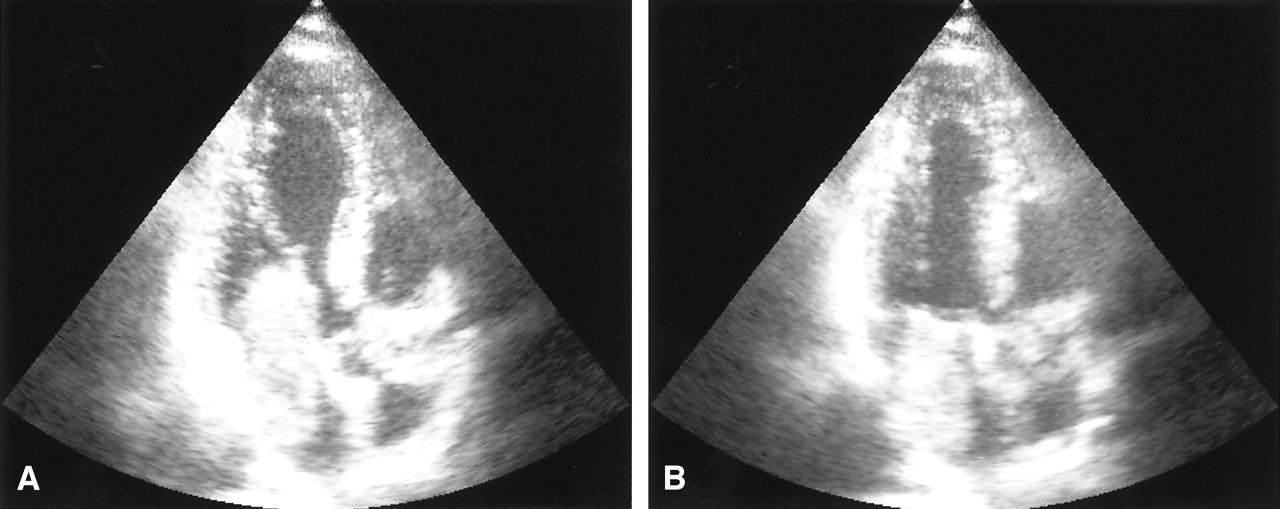
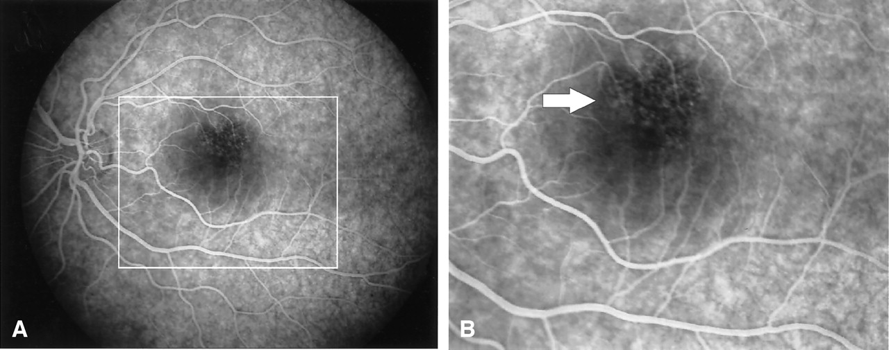
A 31-year-old man was admitted to our hospital with acute onset of dizziness, nausea, blurred vision of his left eye, and gait disturbance. On physical examination, he showed postural imbalance with tendency to fall to the right, dysarthria, skew deviation with a lower left bulb, and mild left-sided facial weakness. On cardiac examination, he had a regular rate and rhythm. No murmurs, rubs, or gallops were audible on auscultation. Duplex sonography gave normal results for his carotid, basilar, and vertebral arteries. Electrocardiography demonstrated signs of left atrial enlargement; a chest radiograph was normal. Holter electrocardiogram showed few ventricular and supraventricular extrasystoles but was otherwise regular. CSF results were normal, with no signs of hemorrhage or cytological abnormalities. Erythrocyte sedimentation rate was 30 mm after 1 hour. A preoperatively performed Coombs test was positive, with detection of cold agglutinins (anti-P1 antibody). All other laboratory results were normal. Cerebral MR imaging revealed multiple cerebellar and brain stem infarctions (Fig 1A, -B) as well as some smaller bihemispheric subcortical white matter lesions on T2-weighted images (not shown). There were no signs of intracranial bleeding. Gadolinium-enhanced T1-weighted sequences showed multiple small enhancing lesions probably corresponding to dilated blood vessels (Fig 1C, -D). Subsequent transthoracic and transesophageal echocardiography demonstrated a large mass with a suspected size of 2.5 × 2.5 × 7 cm in the left atrium, which prolapsed through the mitral valve into the left ventricle, suggesting the diagnosis of atrial myxoma (Fig 2). Cerebral angiography showed multiple intracranial microaneurysms up to 6 mm in diameter mainly localized in peripheral branches of middle, anterior, and posterior cerebral arteries; a few aneurysms were seen in branches of the vertebrobasilar arteries (Fig 3). One week after the initial neurologic event, fundoscopy was normal. A retinal fluorescein angiography revealed parafoveal teleangiectasias and microaneurysms in both eyes (Fig 4). Open heart surgery was performed, entirely removing the gelatinous tumor. Histologic examination confirmed the diagnosis of myxoma. After surgery, the patient was discharged from hospital without any specific treatment and recovered completely. Cerebral MR imaging 2 weeks after the operation showed the residues of cerebral infarctions and the cerebral aneurysm unchanged compared with the prior MR images. No new cerebral ischemic lesions were detected. Two years after the initial event, he was evaluated in the neurologic outpatient clinic and classified as being well, without any residual or newly developed symptoms.

Fig 1.
  • Download figure
  • Open in new tab
  • Download powerpoint
Fig 1.

MR images showing infarction and dilated blood vessels. A and B, Coronal diffusion-weighted MR imaging (TR, 5000 ms; TE, 120 ms; Td, 85 ms; section thickness, 5 mm) shows multiple cerebellar and brain stem infarcts. C and D, Axial T1-weighted spin-echo MR imaging following gadolinium-DTPA administration (TR, 560 ms; TE, 14 ms; section thickness, 5 mm; interslice gap, 1 mm) demonstrates dilated peripheral middle cerebral artery branches suggestive of fusiform dilatations (arrow).

Fig 2.
  • Download figure
  • Open in new tab
  • Download powerpoint
Fig 2.

Echocardiography demonstrating left atrial myxoma. Transesophageal echocardiogram in the two chamber view in diastole (A) and systole (B). A large intraatrial mass which prolapses through the mitral valve into the left ventricle in diastole (A) can be seen.

Fig 3.
  • Download figure
  • Open in new tab
  • Download powerpoint
Fig 3.

Digital subtraction angiography confirming aneurysm formation. The largest aneurysm is located in the precentral branch of the left middle cerebral artery (arrow).

Fig 4.
  • Download figure
  • Open in new tab
  • Download powerpoint
Fig 4.

Fluorescein angiography of the left eye with parafoveal teleangiectasis or microaneurysms. A and B, Early-phase fluorescein angiogram demonstrates teleangiectasis of the upper perifoveal capillary network (arrow). Similar changes were observed in the right eye. A detail from panel A, marked by a rectangle, is shown in panel B. Visual acuity was 25/25.

Discussion

Atrial myxomas have been estimated to cause up to 0.5% of ischemic strokes (5). In a recently published series, the median delay between onset of symptoms and diagnosis in myxoma patients with neurologic manifestation—mainly transient ischemic attacks—was 36 months (6). Cerebral imaging often demonstrates multiple infarcts suggestive of an embolic cause, but in some cases it may show only small subcortical ischemic lesions mimicking lacunar disease (7). Transthoracic echocardiography has a sensitivity of around 90% in detection of left atrial myxoma; the sensitivity of transesophageal examination is even higher (8).

To the best of our knowledge, fewer than 20 cases with intracranial aneurysms associated with myxoma have been reported in the literature (4, 9–11). Several possible mechanisms of aneurysm formation have been proposed. Tumor cells may infiltrate cerebral vessels via vasa vasorum, thereby destroying the architecture of arterial walls similar to the mechanism of mycotic aneurysms. Vasa vasorum, however, are scarcely found in intracranial arteries of experimental animals or humans (12). Alternatively, perivascular damage may be due to vascular occlusion by tumor material with subsequent scarring and pseudoaneurysm formation. This explanation is, however, challenged by the observation that most reported aneurysms occurred without a previous history of cerebrovascular embolization. Another proposed mechanism is the direct transendothelial invasion of tumor cells into the arterial wall causing destruction of its architecture and subsequent aneurysm dilatation. Some histopathologic studies indeed detected myxoma cells in the wall of aneurysms, with one study directly demonstrating interruption of the internal elastic lamina by invading tumor cells (13). In our patient, aneurysms formed without previous symptoms of cerebral embolism. This supports the hypothesis that aneurysm formation and vascular occlusion are not necessarily associated but occur as independent complications of left atrial myxoma; however, clinically silent cerebrovascular occlusion and subsequent ischemic damage cannot be totally excluded.

A number of cases of myxomatous embolism to ocular arteries with a predilection for the left eye and in some cases with retinal hemorrhage have been reported (14). It is conceivable that the retinal vascular dilatations observed in our patient indeed represent microaneurysms corresponding to the intracranial aneurysms (Fig 3). To the best of our knowledge, this is the first report demonstrating retinal microaneurysm formation associated with atrial myxoma.

Conclusion

Because reports covering long-term follow-up of patients with cerebral myxomatous aneurysms associated are rare, recommendations for postoperative management for this subgroup are problematic. Although published case series show that immediate central nervous manifestations are often severe and may be fatal (15), they also indicate a good long-term prognosis with respect to recurrence of neurologic events (2, 5, 6, 15, 16); however, several cases of rapid growth of preexisting aneurysms or postoperative formation of new aneurysms, as well as recurrent cerebral hemorrhages up to years after atrial myxoma resection, have been reported (3, 13, 17, 18). Spontaneous thrombosis resulting in resolution of an aneurysm has been reported once (19). Altogether, these reports indicate that postoperative neurologic complications may occur even years after myxoma resection but are probably rare. Careful follow-up of patients with known aneurysms is recommended, and large aneurysms may require invasive treatment. In view of the persistent risk of embolism, aneurysm formation, and other systemic complications, on one hand, and the good postoperative prognosis, on the other hand, early diagnosis of cardiac myxoma is desirable (1, 16).

References

  1. ↵
    Reynen K. Cardiac myxomas. N Engl J Med 1995;333:1610–1617
    CrossRefPubMed
  2. ↵
    Pinede L, Duhaut P, Loire R. Clinical presentation of left atrial cardiac myxoma. A series of 112 consecutive cases. Medicine (Baltimore) 2001;80:159–172
    CrossRefPubMed
  3. ↵
    Ng HK, Poon WS. Cardiac myxoma metastasizing to the brain: case report. J Neurosurg 1990;72:295–298
    PubMed
  4. ↵
    Chen HJ, Liou CW, Chen L. Metastatic atrial myxoma presenting as intracranial aneurysms with hemorrhage: case report. Surg Neurol 1993;40:61–64
    CrossRefPubMed
  5. ↵
    Knepper LE, Biller J, Adams HP Jr, Bruno A. Neurologic manifestations of atrial myxoma. A 12-year experience and review. Stroke 1988;19:1435–1440
    Abstract/FREE Full Text
  6. ↵
    Alvarez-Sabin J, Lozano M, Sastre-Garriga J, et al. Transient ischaemic attack: a common initial manifestation of cardiac myxomas. Eur Neurol 2001;45:165–170
    CrossRefPubMed
  7. ↵
    Kawamura T, Muratani H, Inamura T, et al. Serial MRI of cerebral infarcts before and after removal of an atrial myxoma. Neuroradiology 1999;41:573–575
    PubMed
  8. ↵
    Mugge A, Daniel WG, Haverich A, Lichtlen PR. Diagnosis of noninfective cardiac mass lesions by two-dimensional echocardiography: comparison of the transthoracic and transesophageal approaches. Circulation 1991;83:70–78
    Abstract/FREE Full Text
  9. ↵
    Jean WC, Walski-Easton SM, Nussbaum ES. Multiple intracranial aneurysms as delayed complications of an atrial myxoma: case report. Neurosurgery 2001;49:200–202
    CrossRefPubMed
  10. Nucifora PG, Dillon WP. MR diagnosis of myxomatous aneurysms: report of two cases. AJNR Am J Neuroradiol 2001;22:1349–1352
    Abstract/FREE Full Text
  11. ↵
    Hwang BJ, Connelly MM, Lev MH. Distinctive MR imaging appearance of hemorrhagic cerebral aneurysms associated with atrial myxoma. AJR Am J Roentgenol 2001;177:925–927
    PubMed
  12. ↵
    Aydin F. Do human intracranial arteries lack vasa vasorum? A comparative immunohistochemical study of intracranial and systemic arteries. Acta Neuropathol 1998;96:22–28
    CrossRefPubMed
  13. ↵
    Furuya K, Sasaki T, Yoshimoto Y, et al. Histologically verified cerebral aneurysm formation secondary to embolism from cardiac myxoma: case report. J Neurosurg 1995;83:170–173
    PubMed
  14. ↵
    Porrini G, Scassellati-Sforzolini B, Mariotti C, et al. Plurifocal cilioretinal occlusion as the presenting symptom of cardiac myxoma. Retina 2000;20:550–552
    PubMed
  15. ↵
    DeSousa AL, Muller J, Campbell RL, et al. Atrial myxoma: a review of the neurological complications, metastases, and recurrences. J Neurol Neurosurg Psychiatry 1978;41:1119–1124
    Abstract/FREE Full Text
  16. ↵
    Reichmann H, Romberg-Hahnloser R, Hofmann E, et al. Neurological long-term follow-up in left atrial myxoma: are late complications frequent or rare? J Neurol 1992;239:170–174
    CrossRefPubMed
  17. ↵
    Walker MT, Kilani RK, Toye LR, Bird CR. Central and peripheral fusiform aneurysms six years after left atrial myxoma resection. J Neurol Neurosurg Psychiatry 2003;74:277–282
    FREE Full Text
  18. ↵
    Suzuki T, Nagai R, Yamazaki T, et al. Rapid growth of intracranial aneurysms secondary to cardiac myxoma. Neurology 1994;44:570–571
    FREE Full Text
  19. ↵
    Branch CL Jr, Laster DW, Kelly DL Jr. Left atrial myxoma with cerebral emboli. Neurosurgery 1985;16:675–680
    PubMed
  • Received April 15, 2004.
  • Accepted after revision June 23, 2004.
  • Copyright © American Society of Neuroradiology
View Abstract
PreviousNext
Back to top

In this issue

American Journal of Neuroradiology: 26 (3)
American Journal of Neuroradiology
Vol. 26, Issue 3
1 Mar 2005
  • Table of Contents
  • Index by author
Advertisement
Print
Download PDF
Email Article

Thank you for your interest in spreading the word on American Journal of Neuroradiology.

NOTE: We only request your email address so that the person you are recommending the page to knows that you wanted them to see it, and that it is not junk mail. We do not capture any email address.

Enter multiple addresses on separate lines or separate them with commas.
Cerebral Embolism from Left Atrial Myxoma Leading to Cerebral and Retinal Aneurysms: A Case Report
(Your Name) has sent you a message from American Journal of Neuroradiology
(Your Name) thought you would like to see the American Journal of Neuroradiology web site.
CAPTCHA
This question is for testing whether or not you are a human visitor and to prevent automated spam submissions.
Cite this article
Martin Herbst, Mike Peter Wattjes, Horst Urbach, Claudia Inhetvin-Hutter, Dietmar Becker, Thomas Klockgether, Alexander Hartmann
Cerebral Embolism from Left Atrial Myxoma Leading to Cerebral and Retinal Aneurysms: A Case Report
American Journal of Neuroradiology Mar 2005, 26 (3) 666-669;

Citation Manager Formats

  • BibTeX
  • Bookends
  • EasyBib
  • EndNote (tagged)
  • EndNote 8 (xml)
  • Medlars
  • Mendeley
  • Papers
  • RefWorks Tagged
  • Ref Manager
  • RIS
  • Zotero
0 Responses
Respond to this article
Share
Bookmark this article
Cerebral Embolism from Left Atrial Myxoma Leading to Cerebral and Retinal Aneurysms: A Case Report
Martin Herbst, Mike Peter Wattjes, Horst Urbach, Claudia Inhetvin-Hutter, Dietmar Becker, Thomas Klockgether, Alexander Hartmann
American Journal of Neuroradiology Mar 2005, 26 (3) 666-669;
del.icio.us logo Twitter logo Facebook logo Mendeley logo
  • Tweet Widget
  • Facebook Like
  • Google Plus One

Jump to section

  • Article
    • Abstract
    • Case Report
    • Discussion
    • Conclusion
    • References
  • Figures & Data
  • Info & Metrics
  • Responses
  • References
  • PDF

Related Articles

  • No related articles found.
  • PubMed
  • Google Scholar

Cited By...

  • Rare presentation of intracranial vascular blowout after tumor resection and radiation therapy
  • Rare presentation of intracranial vascular blowout after tumor resection and radiation therapy
  • Recurrent non-aneurysmal, metastatic intraparenchymal haemorrhages following resection of atrial myxoma - case report and literature review
  • Crossref
  • Google Scholar

This article has not yet been cited by articles in journals that are participating in Crossref Cited-by Linking.

More in this TOC Section

  • Optimal MRI Sequence for Identifying Occlusion Location in Acute Stroke: Which Value of Time-Resolved Contrast-Enhanced MRA?
  • Evaluating the Effects of White Matter Multiple Sclerosis Lesions on the Volume Estimation of 6 Brain Tissue Segmentation Methods
  • Quiet PROPELLER MRI Techniques Match the Quality of Conventional PROPELLER Brain Imaging Techniques
Show more BRAIN

Similar Articles

Advertisement

Indexed Content

  • Current Issue
  • Accepted Manuscripts
  • Article Preview
  • Past Issues
  • Editorials
  • Editor's Choice
  • Fellows' Journal Club
  • Letters to the Editor
  • Video Articles

Cases

  • Case Collection
  • Archive - Case of the Week
  • Archive - Case of the Month
  • Archive - Classic Case

More from AJNR

  • Trainee Corner
  • Imaging Protocols
  • MRI Safety Corner
  • Book Reviews

Multimedia

  • AJNR Podcasts
  • AJNR Scantastics

Resources

  • Turnaround Time
  • Submit a Manuscript
  • Submit a Video Article
  • Submit an eLetter to the Editor/Response
  • Manuscript Submission Guidelines
  • Statistical Tips
  • Fast Publishing of Accepted Manuscripts
  • Graphical Abstract Preparation
  • Imaging Protocol Submission
  • Evidence-Based Medicine Level Guide
  • Publishing Checklists
  • Author Policies
  • Become a Reviewer/Academy of Reviewers
  • News and Updates

About Us

  • About AJNR
  • Editorial Board
  • Editorial Board Alumni
  • Alerts
  • Permissions
  • Not an AJNR Subscriber? Join Now
  • Advertise with Us
  • Librarian Resources
  • Feedback
  • Terms and Conditions
  • AJNR Editorial Board Alumni

American Society of Neuroradiology

  • Not an ASNR Member? Join Now

© 2025 by the American Society of Neuroradiology All rights, including for text and data mining, AI training, and similar technologies, are reserved.
Print ISSN: 0195-6108 Online ISSN: 1936-959X

Powered by HighWire