Advertisement

Main menu

  • Home
  • Content
    • Current Issue
    • Accepted Manuscripts
    • Article Preview
    • Past Issue Archive
    • Video Articles
    • AJNR Case Collection
    • Case of the Week Archive
    • Case of the Month Archive
    • Classic Case Archive
  • Special Collections
    • AJNR Awards
    • Low-Field MRI
    • Alzheimer Disease
    • ASNR Foundation Special Collection
    • Photon-Counting CT
    • View All
  • Multimedia
    • AJNR Podcasts
    • AJNR SCANtastic
    • Trainee Corner
    • MRI Safety Corner
    • Imaging Protocols
  • For Authors
    • Submit a Manuscript
    • Submit a Video Article
    • Submit an eLetter to the Editor/Response
    • Manuscript Submission Guidelines
    • Statistical Tips
    • Fast Publishing of Accepted Manuscripts
    • Graphical Abstract Preparation
    • Imaging Protocol Submission
    • Author Policies
  • About Us
    • About AJNR
    • Editorial Board
    • Editorial Board Alumni
  • More
    • Become a Reviewer/Academy of Reviewers
    • Subscribers
    • Permissions
    • Alerts
    • Feedback
    • Advertisers
    • ASNR Home

User menu

  • Alerts
  • Log in

Search

  • Advanced search
American Journal of Neuroradiology

American Journal of Neuroradiology

ASHNR American Society of Functional Neuroradiology ASHNR American Society of Pediatric Neuroradiology ASSR
  • Alerts
  • Log in

Advanced Search

  • Home
  • Content
    • Current Issue
    • Accepted Manuscripts
    • Article Preview
    • Past Issue Archive
    • Video Articles
    • AJNR Case Collection
    • Case of the Week Archive
    • Case of the Month Archive
    • Classic Case Archive
  • Special Collections
    • AJNR Awards
    • Low-Field MRI
    • Alzheimer Disease
    • ASNR Foundation Special Collection
    • Photon-Counting CT
    • View All
  • Multimedia
    • AJNR Podcasts
    • AJNR SCANtastic
    • Trainee Corner
    • MRI Safety Corner
    • Imaging Protocols
  • For Authors
    • Submit a Manuscript
    • Submit a Video Article
    • Submit an eLetter to the Editor/Response
    • Manuscript Submission Guidelines
    • Statistical Tips
    • Fast Publishing of Accepted Manuscripts
    • Graphical Abstract Preparation
    • Imaging Protocol Submission
    • Author Policies
  • About Us
    • About AJNR
    • Editorial Board
    • Editorial Board Alumni
  • More
    • Become a Reviewer/Academy of Reviewers
    • Subscribers
    • Permissions
    • Alerts
    • Feedback
    • Advertisers
    • ASNR Home
  • Follow AJNR on Twitter
  • Visit AJNR on Facebook
  • Follow AJNR on Instagram
  • Join AJNR on LinkedIn
  • RSS Feeds

AJNR Awards, New Junior Editors, and more. Read the latest AJNR updates

OtherBRAIN

MR Imaging of Hyperacute Subarachnoid and Intraventricular Hemorrhage at 3T: A Preliminary Report of Gradient Echo T2*-Weighted Sequences

Chul-Ho Sohn, Seung-Kug Baik, Hee-Jung Lee, Sung-Moon Lee, Il-Man Kim, Man-Bin Yim, Jae-Suk Hwang, M. Louis Lauzon and Robert J. Sevick
American Journal of Neuroradiology March 2005, 26 (3) 662-665;
Chul-Ho Sohn
  • Find this author on Google Scholar
  • Find this author on PubMed
  • Search for this author on this site
Seung-Kug Baik
  • Find this author on Google Scholar
  • Find this author on PubMed
  • Search for this author on this site
Hee-Jung Lee
  • Find this author on Google Scholar
  • Find this author on PubMed
  • Search for this author on this site
Sung-Moon Lee
  • Find this author on Google Scholar
  • Find this author on PubMed
  • Search for this author on this site
Il-Man Kim
  • Find this author on Google Scholar
  • Find this author on PubMed
  • Search for this author on this site
Man-Bin Yim
  • Find this author on Google Scholar
  • Find this author on PubMed
  • Search for this author on this site
Jae-Suk Hwang
  • Find this author on Google Scholar
  • Find this author on PubMed
  • Search for this author on this site
M. Louis Lauzon
  • Find this author on Google Scholar
  • Find this author on PubMed
  • Search for this author on this site
Robert J. Sevick
  • Find this author on Google Scholar
  • Find this author on PubMed
  • Search for this author on this site
  • Article
  • Figures & Data
  • Info & Metrics
  • Responses
  • References
  • PDF
Loading

Article Figures & Data

Figures

  • Fig 1.
    • Download figure
    • Open in new tab
    • Download powerpoint
    Fig 1.

    Case 1, a 65-year-old woman with hyperacute SAH and IVH due to a ruptured left anterior communicating artery aneurysm.

    A, CT scan (7.3 hours postictus) shows an SAH and IVH in the both Sylvian fissures, the anterior interhemispheric fissure, and the occipital horns of both lateral ventricles.

    B–D, MR images obtained 10.3 hours postictus. Axial FLAIR image (B) shows high signal intensity in the anterior interhemispheric fissure and both Sylvian fissures (black arrows). A layered area mildly increased signal intensity is evident in occipital horn of the left lateral ventricle (white arrow). Axial GRE T2*-weighted image (C) shows decreased signal intensity in the interhemispheric and both Sylvian fissures, 3rd ventricle, and occipital horns of both lateral ventricles (arrows). Axial fast spin-echo T2-weighted image (D) shows linear low signal intensity in the anterior interhemispheric fissure (arrow) and blood-CSF layered pattern of the IVH (arrowheads) in the occipital horns of both lateral ventricles.
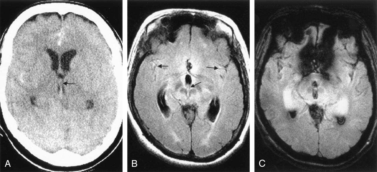
  • Fig 2.
    • Download figure
    • Open in new tab
    • Download powerpoint
    Fig 2.

    Case 2, a 52-year-old woman with hyperacute SAH and IVH due to a ruptured left anterior communicating artery aneurysm.

    A, CT scan (1.3 hours postictus) shows extensive SAHs and IVHs in the 3rd ventricle (arrow). IVH is not evident in the lateral ventricle.

    B and C, MR images obtained 5.1 hours postictus. Axial FLAIR image (B) shows increased signal intensity in both Sylvian fissures and 3rd ventricle (arrows). IVH in lateral ventricle is not visualized. Axial GRE T2*-weighted image (C) clearly shows the SAH and IVH in the Sylvian fissures bilaterally as well as 3rd ventricle and occipital horn of both lateral ventricles.

PreviousNext
Back to top

In this issue

American Journal of Neuroradiology: 26 (3)
American Journal of Neuroradiology
Vol. 26, Issue 3
1 Mar 2005
  • Table of Contents
  • Index by author
Advertisement
Print
Download PDF
Email Article

Thank you for your interest in spreading the word on American Journal of Neuroradiology.

NOTE: We only request your email address so that the person you are recommending the page to knows that you wanted them to see it, and that it is not junk mail. We do not capture any email address.

Enter multiple addresses on separate lines or separate them with commas.
MR Imaging of Hyperacute Subarachnoid and Intraventricular Hemorrhage at 3T: A Preliminary Report of Gradient Echo T2*-Weighted Sequences
(Your Name) has sent you a message from American Journal of Neuroradiology
(Your Name) thought you would like to see the American Journal of Neuroradiology web site.
CAPTCHA
This question is for testing whether or not you are a human visitor and to prevent automated spam submissions.
Cite this article
Chul-Ho Sohn, Seung-Kug Baik, Hee-Jung Lee, Sung-Moon Lee, Il-Man Kim, Man-Bin Yim, Jae-Suk Hwang, M. Louis Lauzon, Robert J. Sevick
MR Imaging of Hyperacute Subarachnoid and Intraventricular Hemorrhage at 3T: A Preliminary Report of Gradient Echo T2*-Weighted Sequences
American Journal of Neuroradiology Mar 2005, 26 (3) 662-665;

Citation Manager Formats

  • BibTeX
  • Bookends
  • EasyBib
  • EndNote (tagged)
  • EndNote 8 (xml)
  • Medlars
  • Mendeley
  • Papers
  • RefWorks Tagged
  • Ref Manager
  • RIS
  • Zotero
0 Responses
Respond to this article
Share
Bookmark this article
MR Imaging of Hyperacute Subarachnoid and Intraventricular Hemorrhage at 3T: A Preliminary Report of Gradient Echo T2*-Weighted Sequences
Chul-Ho Sohn, Seung-Kug Baik, Hee-Jung Lee, Sung-Moon Lee, Il-Man Kim, Man-Bin Yim, Jae-Suk Hwang, M. Louis Lauzon, Robert J. Sevick
American Journal of Neuroradiology Mar 2005, 26 (3) 662-665;
del.icio.us logo Twitter logo Facebook logo Mendeley logo
  • Tweet Widget
  • Facebook Like
  • Google Plus One

Jump to section

  • Article
    • Abstract
    • Case Reports
    • Discussion
    • Conclusion
    • References
  • Figures & Data
  • Info & Metrics
  • Responses
  • References
  • PDF

Related Articles

  • No related articles found.
  • PubMed
  • Google Scholar

Cited By...

  • Imaging suggestive, but symptoms atypical
  • Detection of aneurysmal subarachnoid hemorrhage 3 months after initial bleeding: evaluation of T2* and FLAIR MR sequences at 3 T in comparison with initial non-enhanced CT as a gold standard
  • 3D Fluid-Attenuated Inversion Recovery Imaging: Reduced CSF Artifacts and Enhanced Sensitivity and Specificity for Subarachnoid Hemorrhage
  • Role of Iron in Brain Injury After Intraventricular Hemorrhage
  • Evaluation of Traumatic Subarachnoid Hemorrhage Using Susceptibility-Weighted Imaging
  • Crossref
  • Google Scholar

This article has not yet been cited by articles in journals that are participating in Crossref Cited-by Linking.

More in this TOC Section

  • Evaluating the Effects of White Matter Multiple Sclerosis Lesions on the Volume Estimation of 6 Brain Tissue Segmentation Methods
  • Quiet PROPELLER MRI Techniques Match the Quality of Conventional PROPELLER Brain Imaging Techniques
  • Predictors of Reperfusion in Patients with Acute Ischemic Stroke
Show more BRAIN

Similar Articles

Advertisement

Indexed Content

  • Current Issue
  • Accepted Manuscripts
  • Article Preview
  • Past Issues
  • Editorials
  • Editor's Choice
  • Fellows' Journal Club
  • Letters to the Editor
  • Video Articles

Cases

  • Case Collection
  • Archive - Case of the Week
  • Archive - Case of the Month
  • Archive - Classic Case

More from AJNR

  • Trainee Corner
  • Imaging Protocols
  • MRI Safety Corner

Multimedia

  • AJNR Podcasts
  • AJNR Scantastics

Resources

  • Turnaround Time
  • Submit a Manuscript
  • Submit a Video Article
  • Submit an eLetter to the Editor/Response
  • Manuscript Submission Guidelines
  • Statistical Tips
  • Fast Publishing of Accepted Manuscripts
  • Graphical Abstract Preparation
  • Imaging Protocol Submission
  • Evidence-Based Medicine Level Guide
  • Publishing Checklists
  • Author Policies
  • Become a Reviewer/Academy of Reviewers
  • News and Updates

About Us

  • About AJNR
  • Editorial Board
  • Editorial Board Alumni
  • Alerts
  • Permissions
  • Not an AJNR Subscriber? Join Now
  • Advertise with Us
  • Librarian Resources
  • Feedback
  • Terms and Conditions
  • AJNR Editorial Board Alumni

American Society of Neuroradiology

  • Not an ASNR Member? Join Now

© 2025 by the American Society of Neuroradiology All rights, including for text and data mining, AI training, and similar technologies, are reserved.
Print ISSN: 0195-6108 Online ISSN: 1936-959X

Powered by HighWire