Advertisement

Main menu

  • Home
  • Content
    • Current Issue
    • Accepted Manuscripts
    • Article Preview
    • Past Issue Archive
    • Video Articles
    • AJNR Case Collection
    • Case of the Week Archive
    • Case of the Month Archive
    • Classic Case Archive
  • Special Collections
    • AJNR Awards
    • Low-Field MRI
    • Alzheimer Disease
    • ASNR Foundation Special Collection
    • Photon-Counting CT
    • View All
  • Multimedia
    • AJNR Podcasts
    • AJNR SCANtastic
    • Trainee Corner
    • MRI Safety Corner
    • Imaging Protocols
  • For Authors
    • Submit a Manuscript
    • Submit a Video Article
    • Submit an eLetter to the Editor/Response
    • Manuscript Submission Guidelines
    • Statistical Tips
    • Fast Publishing of Accepted Manuscripts
    • Graphical Abstract Preparation
    • Imaging Protocol Submission
    • Author Policies
  • About Us
    • About AJNR
    • Editorial Board
    • Editorial Board Alumni
  • More
    • Become a Reviewer/Academy of Reviewers
    • Subscribers
    • Permissions
    • Alerts
    • Feedback
    • Advertisers
    • ASNR Home

User menu

  • Alerts
  • Log in

Search

  • Advanced search
American Journal of Neuroradiology

American Journal of Neuroradiology

ASHNR American Society of Functional Neuroradiology ASHNR American Society of Pediatric Neuroradiology ASSR
  • Alerts
  • Log in

Advanced Search

  • Home
  • Content
    • Current Issue
    • Accepted Manuscripts
    • Article Preview
    • Past Issue Archive
    • Video Articles
    • AJNR Case Collection
    • Case of the Week Archive
    • Case of the Month Archive
    • Classic Case Archive
  • Special Collections
    • AJNR Awards
    • Low-Field MRI
    • Alzheimer Disease
    • ASNR Foundation Special Collection
    • Photon-Counting CT
    • View All
  • Multimedia
    • AJNR Podcasts
    • AJNR SCANtastic
    • Trainee Corner
    • MRI Safety Corner
    • Imaging Protocols
  • For Authors
    • Submit a Manuscript
    • Submit a Video Article
    • Submit an eLetter to the Editor/Response
    • Manuscript Submission Guidelines
    • Statistical Tips
    • Fast Publishing of Accepted Manuscripts
    • Graphical Abstract Preparation
    • Imaging Protocol Submission
    • Author Policies
  • About Us
    • About AJNR
    • Editorial Board
    • Editorial Board Alumni
  • More
    • Become a Reviewer/Academy of Reviewers
    • Subscribers
    • Permissions
    • Alerts
    • Feedback
    • Advertisers
    • ASNR Home
  • Follow AJNR on Twitter
  • Visit AJNR on Facebook
  • Follow AJNR on Instagram
  • Join AJNR on LinkedIn
  • RSS Feeds

AJNR Awards, New Junior Editors, and more. Read the latest AJNR updates

Research ArticleBRAIN

Reversible MR Imaging Abnormalities following Cerebral Venous Thrombosis

Carina Röttger, Susan Trittmacher, Tibo Gerriets, Franz Blaes, Manfred Kaps and Erwin Stolz
American Journal of Neuroradiology March 2005, 26 (3) 607-613;
Carina Röttger
  • Find this author on Google Scholar
  • Find this author on PubMed
  • Search for this author on this site
Susan Trittmacher
  • Find this author on Google Scholar
  • Find this author on PubMed
  • Search for this author on this site
Tibo Gerriets
  • Find this author on Google Scholar
  • Find this author on PubMed
  • Search for this author on this site
Franz Blaes
  • Find this author on Google Scholar
  • Find this author on PubMed
  • Search for this author on this site
Manfred Kaps
  • Find this author on Google Scholar
  • Find this author on PubMed
  • Search for this author on this site
Erwin Stolz
  • Find this author on Google Scholar
  • Find this author on PubMed
  • Search for this author on this site
  • Article
  • Figures & Data
  • Info & Metrics
  • Responses
  • References
  • PDF
Loading

Article Figures & Data

Figures

  • Tables
  • Fig 1.
    • Download figure
    • Open in new tab
    • Download powerpoint
    Fig 1.

    Time course of the T2 lesion volume reduction in the individual patients.

  • Fig 2.
    • Download figure
    • Open in new tab
    • Download powerpoint
    Fig 2.

    Venous cerebellar infarct due to transverse sinus thrombosis in a 56-year-old woman with partial obstruction of the superior sagittal sinus and partial thrombosis of the transverse sinuses.

    A and B, T2-weighted MR image (A) and 3D time-of-flight MR angiogram (B) obtained on admission show the venous cerebellar infarct and transverse sinus thrombosis, respectively.

    C and D, Follow-up MR images obtained after 6 months. Despite persistent partial obstruction of the transverse sinus (D), near complete resolution is noted of the volume of the T2 lesion (C).
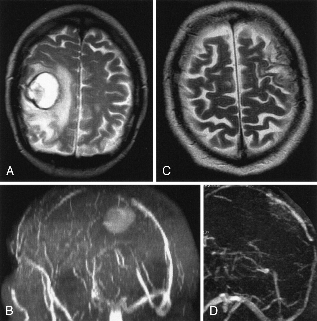
  • Fig 3.
    • Download figure
    • Open in new tab
    • Download powerpoint
    Fig 3.

    Venous temporal infarct in a 37-year-old woman.

    A and B, T2-weighted MR image (A) and 3D time-of-flight MR angiogram (B) obtained on admission show a venous infarct in the temporal lobe due to thrombosis of the transverse sinus. Thrombosis of the superior sagittal sinus caused no infarct.

    C–E, Follow-up MR images obtained after 12 months show that despite persistent occlusion of the transverse sinus (D and E) and only partial recanalization of the superior sagittal sinus, the volume of the venous infarct decreased substantially (C) at follow-up.

  • Fig 4.
    • Download figure
    • Open in new tab
    • Download powerpoint
    Fig 4.

    Venous infarct in the temporo-occipital region in a 43-year-old woman.

    A and B, T2-weighted MR image (A) and 3D time-of-flight MR angiogram (B) obtained on admission. The MR angiogram shows a thrombosis of the transverse sinus.

    C and D, Follow-up MR images obtained after 3 months show total resolution of the parenchymal lesion (C) despite persistent occlusion of the transverse sinus (D).
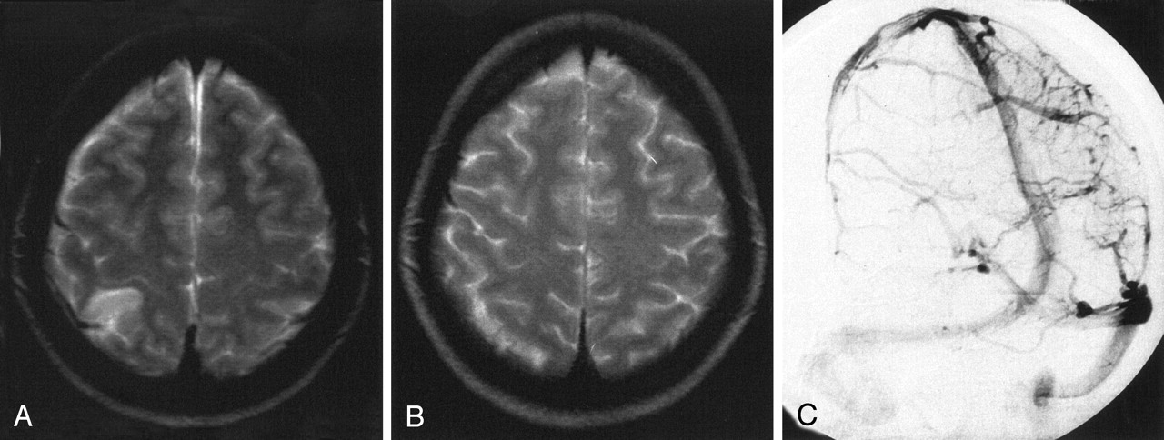
  • Fig 5.
    • Download figure
    • Open in new tab
    • Download powerpoint
    Fig 5.

    Venous infarct due to cortical vein thrombosis in a 27-year-old woman.

    A, T2-weighted MR image obtained on admission shows the venous infarct due to cortical vein thrombosis.

    B, Conventional angiography on admission shows a right parietal cortical vein thrombosis.

    C, Follow-up T2-weighted MR image obtained after 3 months shows total resolution of the initial lesion. In this case, no analysis regarding recanalization was performed.

  • Fig 6.
    • Download figure
    • Open in new tab
    • Download powerpoint
    Fig 6.

    Hemorrhagic venous infarct due to thrombosis of the superior sagittal sinus in a 66-year-old man.

    A and B, T2-weighted MR image (A) and MR angiogram (B) show a parenchymal lesion due to a superior sagittal sinus thrombosis. The patient also had a second infarct (not shown).

    C and D, Follow-up MR images show recanalization of the superior sagittal sinus (D) and decrease of total lesion volume by 74% (C).

Tables

  • Figures
  • Patient demographic data, affected sinuses and veins, and reduction of lesion volume at follow-up

    Patient No./Age (y)/SexLocation of ThrombosisRecanalization on First Follow-upFollow-up Time (mo)Parenchymal DamageHemorrhageReduction of Lesion Volume (%) Compared to Initial Study
    T1-WeightedT2-Weighted
    O1O2O3O1O2O3
    1/27/FCortical veins–9ParietalNo100100100100100100
    2/44/FSSS, TSNo6FrontoparietalYes82100–29100–
    3/43/FSTNo9Occipital, cerebellarYes10010010093100100
    4/29/FSSSNo9FrontalNo––100––100
    5/18/FSSS, STNo9OccipitalNo–100100–100100
    6/56/FSSS, STNo6CerebellarYes6896–8994–
    7/26/FCortical veins–8ParietalNo–100100–100100
    8/35/FSSS, STNo10FrontoparietalNo43901002359100
    9/33/FSSS, ST, SSYes<1Frontal, occipitalNo100––100––
    10/39/FICV, SRYes4ThalamusNo–100––100–
    11/30/FSTYes11Occipital, cerebellarNo100100100100100100
    12/37/FSSS, STNo16TemporalYes45–8030–62
    13/66/MSSS, ST, SSNo18Parietal, occipitalYes63–6073–74
    14/78/FICVYes23Basal gangliaNo5010010064100100
    15/29/FSTYes10CerebellarNo54–7254–72
    • Note.—SSS indicates superior sagittal sinus; TS, transverse sinus; SS, sigmoid sinus; ICV, inner cerebral veins; SR, straight sinus; O1–3, observation periods after initial MR imaging (O1, days 1–30; O2, days 90–240; O3, >240 days).

PreviousNext
Back to top

In this issue

American Journal of Neuroradiology: 26 (3)
American Journal of Neuroradiology
Vol. 26, Issue 3
1 Mar 2005
  • Table of Contents
  • Index by author
Advertisement
Print
Download PDF
Email Article

Thank you for your interest in spreading the word on American Journal of Neuroradiology.

NOTE: We only request your email address so that the person you are recommending the page to knows that you wanted them to see it, and that it is not junk mail. We do not capture any email address.

Enter multiple addresses on separate lines or separate them with commas.
Reversible MR Imaging Abnormalities following Cerebral Venous Thrombosis
(Your Name) has sent you a message from American Journal of Neuroradiology
(Your Name) thought you would like to see the American Journal of Neuroradiology web site.
CAPTCHA
This question is for testing whether or not you are a human visitor and to prevent automated spam submissions.
Cite this article
Carina Röttger, Susan Trittmacher, Tibo Gerriets, Franz Blaes, Manfred Kaps, Erwin Stolz
Reversible MR Imaging Abnormalities following Cerebral Venous Thrombosis
American Journal of Neuroradiology Mar 2005, 26 (3) 607-613;

Citation Manager Formats

  • BibTeX
  • Bookends
  • EasyBib
  • EndNote (tagged)
  • EndNote 8 (xml)
  • Medlars
  • Mendeley
  • Papers
  • RefWorks Tagged
  • Ref Manager
  • RIS
  • Zotero
0 Responses
Respond to this article
Share
Bookmark this article
Reversible MR Imaging Abnormalities following Cerebral Venous Thrombosis
Carina Röttger, Susan Trittmacher, Tibo Gerriets, Franz Blaes, Manfred Kaps, Erwin Stolz
American Journal of Neuroradiology Mar 2005, 26 (3) 607-613;
del.icio.us logo Twitter logo Facebook logo Mendeley logo
  • Tweet Widget
  • Facebook Like
  • Google Plus One

Jump to section

  • Article
    • Abstract
    • Methods
    • Results
    • Discussion
    • Conclusion
    • References
  • Figures & Data
  • Info & Metrics
  • Responses
  • References
  • PDF

Related Articles

  • No related articles found.
  • PubMed
  • Google Scholar

Cited By...

  • Diagnosis and Management of Cerebral Venous Thrombosis: A Statement for Healthcare Professionals From the American Heart Association/American Stroke Association
  • Intrasinus Catheter-Directed Heparin Infusion in the Treatment of Dural Venous Sinus Thrombosis
  • MR Imaging Features of Isolated Cortical Vein Thrombosis: Diagnosis and Follow-Up
  • Crossref
  • Google Scholar

This article has not yet been cited by articles in journals that are participating in Crossref Cited-by Linking.

More in this TOC Section

  • Multimodal CT Provides Improved Performance for Lacunar Infarct Detection
  • Optimal MRI Sequence for Identifying Occlusion Location in Acute Stroke: Which Value of Time-Resolved Contrast-Enhanced MRA?
  • Evaluating the Effects of White Matter Multiple Sclerosis Lesions on the Volume Estimation of 6 Brain Tissue Segmentation Methods
Show more BRAIN

Similar Articles

Advertisement

Indexed Content

  • Current Issue
  • Accepted Manuscripts
  • Article Preview
  • Past Issues
  • Editorials
  • Editor's Choice
  • Fellows' Journal Club
  • Letters to the Editor
  • Video Articles

Cases

  • Case Collection
  • Archive - Case of the Week
  • Archive - Case of the Month
  • Archive - Classic Case

More from AJNR

  • Trainee Corner
  • Imaging Protocols
  • MRI Safety Corner
  • Book Reviews

Multimedia

  • AJNR Podcasts
  • AJNR Scantastics

Resources

  • Turnaround Time
  • Submit a Manuscript
  • Submit a Video Article
  • Submit an eLetter to the Editor/Response
  • Manuscript Submission Guidelines
  • Statistical Tips
  • Fast Publishing of Accepted Manuscripts
  • Graphical Abstract Preparation
  • Imaging Protocol Submission
  • Evidence-Based Medicine Level Guide
  • Publishing Checklists
  • Author Policies
  • Become a Reviewer/Academy of Reviewers
  • News and Updates

About Us

  • About AJNR
  • Editorial Board
  • Editorial Board Alumni
  • Alerts
  • Permissions
  • Not an AJNR Subscriber? Join Now
  • Advertise with Us
  • Librarian Resources
  • Feedback
  • Terms and Conditions
  • AJNR Editorial Board Alumni

American Society of Neuroradiology

  • Not an ASNR Member? Join Now

© 2025 by the American Society of Neuroradiology All rights, including for text and data mining, AI training, and similar technologies, are reserved.
Print ISSN: 0195-6108 Online ISSN: 1936-959X

Powered by HighWire