Advertisement

Main menu

  • Home
  • Content
    • Current Issue
    • Accepted Manuscripts
    • Article Preview
    • Past Issue Archive
    • Video Articles
    • AJNR Case Collection
    • Case of the Week Archive
    • Case of the Month Archive
    • Classic Case Archive
  • Special Collections
    • AJNR Awards
    • Low-Field MRI
    • Alzheimer Disease
    • ASNR Foundation Special Collection
    • Photon-Counting CT
    • View All
  • Multimedia
    • AJNR Podcasts
    • AJNR SCANtastic
    • Trainee Corner
    • MRI Safety Corner
    • Imaging Protocols
  • For Authors
    • Submit a Manuscript
    • Submit a Video Article
    • Submit an eLetter to the Editor/Response
    • Manuscript Submission Guidelines
    • Statistical Tips
    • Fast Publishing of Accepted Manuscripts
    • Graphical Abstract Preparation
    • Imaging Protocol Submission
    • Author Policies
  • About Us
    • About AJNR
    • Editorial Board
    • Editorial Board Alumni
  • More
    • Become a Reviewer/Academy of Reviewers
    • Subscribers
    • Permissions
    • Alerts
    • Feedback
    • Advertisers
    • ASNR Home

User menu

  • Alerts
  • Log in

Search

  • Advanced search
American Journal of Neuroradiology

American Journal of Neuroradiology

ASHNR American Society of Functional Neuroradiology ASHNR American Society of Pediatric Neuroradiology ASSR
  • Alerts
  • Log in

Advanced Search

  • Home
  • Content
    • Current Issue
    • Accepted Manuscripts
    • Article Preview
    • Past Issue Archive
    • Video Articles
    • AJNR Case Collection
    • Case of the Week Archive
    • Case of the Month Archive
    • Classic Case Archive
  • Special Collections
    • AJNR Awards
    • Low-Field MRI
    • Alzheimer Disease
    • ASNR Foundation Special Collection
    • Photon-Counting CT
    • View All
  • Multimedia
    • AJNR Podcasts
    • AJNR SCANtastic
    • Trainee Corner
    • MRI Safety Corner
    • Imaging Protocols
  • For Authors
    • Submit a Manuscript
    • Submit a Video Article
    • Submit an eLetter to the Editor/Response
    • Manuscript Submission Guidelines
    • Statistical Tips
    • Fast Publishing of Accepted Manuscripts
    • Graphical Abstract Preparation
    • Imaging Protocol Submission
    • Author Policies
  • About Us
    • About AJNR
    • Editorial Board
    • Editorial Board Alumni
  • More
    • Become a Reviewer/Academy of Reviewers
    • Subscribers
    • Permissions
    • Alerts
    • Feedback
    • Advertisers
    • ASNR Home
  • Follow AJNR on Twitter
  • Visit AJNR on Facebook
  • Follow AJNR on Instagram
  • Join AJNR on LinkedIn
  • RSS Feeds

AJNR Awards, New Junior Editors, and more. Read the latest AJNR updates

Research ArticleBRAIN

MR Imaging of Normal Perivascular Space Expansion at Midbrain

Naokatsu Saeki, Motoki Sato, Motoo Kubota, Yoshio Uchino, Hisayuki Murai, Yuichiro Nagai, Hiroshi Ishikura, Seitaro Nomura, Iichiro Matsuura and Akira Yamaura
American Journal of Neuroradiology March 2005, 26 (3) 566-571;
Naokatsu Saeki
  • Find this author on Google Scholar
  • Find this author on PubMed
  • Search for this author on this site
Motoki Sato
  • Find this author on Google Scholar
  • Find this author on PubMed
  • Search for this author on this site
Motoo Kubota
  • Find this author on Google Scholar
  • Find this author on PubMed
  • Search for this author on this site
Yoshio Uchino
  • Find this author on Google Scholar
  • Find this author on PubMed
  • Search for this author on this site
Hisayuki Murai
  • Find this author on Google Scholar
  • Find this author on PubMed
  • Search for this author on this site
Yuichiro Nagai
  • Find this author on Google Scholar
  • Find this author on PubMed
  • Search for this author on this site
Hiroshi Ishikura
  • Find this author on Google Scholar
  • Find this author on PubMed
  • Search for this author on this site
Seitaro Nomura
  • Find this author on Google Scholar
  • Find this author on PubMed
  • Search for this author on this site
Iichiro Matsuura
  • Find this author on Google Scholar
  • Find this author on PubMed
  • Search for this author on this site
Akira Yamaura
  • Find this author on Google Scholar
  • Find this author on PubMed
  • Search for this author on this site
  • Article
  • Figures & Data
  • Info & Metrics
  • Responses
  • References
  • PDF
Loading

Article Figures & Data

Figures

  • Tables
  • Fig 1.
    • Download figure
    • Open in new tab
    • Download powerpoint
    Fig 1.

    MR images obtained in a 32-year-old woman who presented with headache and progressive right hemiparesis; MR images revealed multilobular CSF-filled expanding spaces at the MDJ and ventricular dilatation. She underwent endoscopic fenestration of the cyst wall and became free of the neurologic deficits. (Reproduced with permission from reference 12.)

    T1-weighted axial (A), coronal (B), and sagittal (C) images reveal multilobular, CSF-filled, expanding spaces at the MDJ.
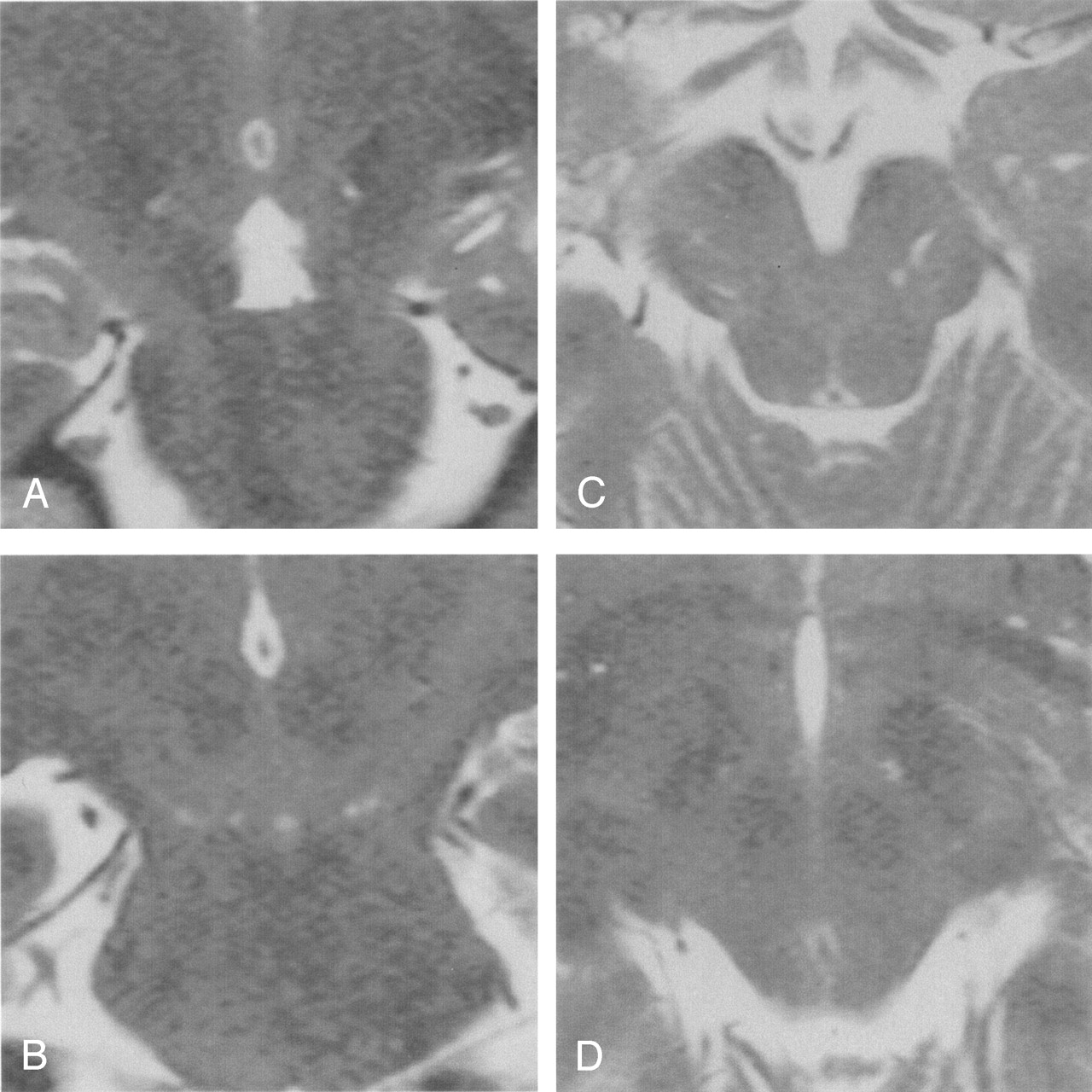
  • Fig 2.
    • Download figure
    • Open in new tab
    • Download powerpoint
    Fig 2.

    MR images obtained in a 48-year-old-man who presented with double vision due to the left nerve palsy.

    A, Coronal section at the anterior midbrain. Expanded PV spaces are visible as round or ovoid hypointense lesions at the PMJ and the MDJ.

    B, Coronal section at the cerebral peduncle. Expanded PV spaces are visible at the level of the PMJ, corresponding to the tentorial margin. The largest segment of the PV space is graded 4.

    C, Axial section at the lower midbrain. Multiple ovoid and round PV spaces are visible between the cerebral peduncle and substantia nigra and in the cerebral peduncle.

    D, Axial section, two sections rostral to Figure 2C. Small PV spaces are visible behind the cerebral peduncle.

    E, Sagittal section, two sections lateral to the midline. Expanded PV spaces are visible between the pons and midbrain.

    F, Axial section, four sections lateral to the midline. Large and small PV spaces are visible at the PMJ and MDJ.

  • Fig 3.
    • Download figure
    • Open in new tab
    • Download powerpoint
    Fig 3.

    A 19-year-old female patient who complained of retro-orbital pain.

    A, Coronal section at the cerebral peduncle. Round and ovoid PV spaces are visible at the level of the MDJ. The largest size is graded 1.

    B, Coronal section two sections posterior to Figure 2A. Small PV spaces are visible at the PMJ and at the level of tentorial margin. The largest one is grade 0.

    C, Axial section at the lower midbrain. Ovoid and linear PV spaces are visible between the cerebral peduncle and substantia nigra and in the cerebral peduncle.

    D, Axial section, two sections rostral to Figure 3C. Small PV spaces are visible behind the cerebral peduncle.
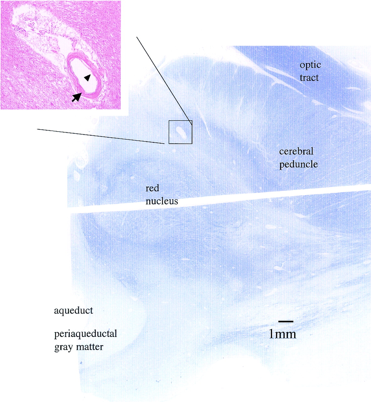
  • Fig 4.
    • Download figure
    • Open in new tab
    • Download powerpoint
    Fig 4.

    A, Axial section of the right upper midbrain. Kluver-Barrera stain (magnification ×4). Section line between ventral and dorsal halves is noted in the center. Aqueduct, periaqueductal gray matter, red nucleus, cerebral peduncle, and optic tract are demonstrated. Behind the cerebral peduncle, there is an ovoid space.

    B, Magnified axial section demonstrating the PV space at the upper midbrain. H&E stain (magnification ×50). The ovoid space includes a vessel (arrow) and is lined by a pial layer (arrowhead). No necrotic or ischemic changes were visible in the surrounding brain tissue in this magnified view. Histologic findings histologically a PV space (1–5).

Tables

  • Figures
  • Perivascular spaces at the PMJ and MDJ among age subgroups

    Age (y) SubgroupCase (No.)PVS at PMJPVS at MDJ
    presentnonepresentnone
    –201211193
    21–40423572418
    41–6034313268
    61–272341413
    total 115100 (87%)15 (13%)73 (63%)42 (37%)
    • Note.—PVS indicates perivascular spaces; PMJ, ponto–mesencephalic junction; MDJ, mesenecphalo–diencephalic junction.

PreviousNext
Back to top

In this issue

American Journal of Neuroradiology: 26 (3)
American Journal of Neuroradiology
Vol. 26, Issue 3
1 Mar 2005
  • Table of Contents
  • Index by author
Advertisement
Print
Download PDF
Email Article

Thank you for your interest in spreading the word on American Journal of Neuroradiology.

NOTE: We only request your email address so that the person you are recommending the page to knows that you wanted them to see it, and that it is not junk mail. We do not capture any email address.

Enter multiple addresses on separate lines or separate them with commas.
MR Imaging of Normal Perivascular Space Expansion at Midbrain
(Your Name) has sent you a message from American Journal of Neuroradiology
(Your Name) thought you would like to see the American Journal of Neuroradiology web site.
CAPTCHA
This question is for testing whether or not you are a human visitor and to prevent automated spam submissions.
Cite this article
Naokatsu Saeki, Motoki Sato, Motoo Kubota, Yoshio Uchino, Hisayuki Murai, Yuichiro Nagai, Hiroshi Ishikura, Seitaro Nomura, Iichiro Matsuura, Akira Yamaura
MR Imaging of Normal Perivascular Space Expansion at Midbrain
American Journal of Neuroradiology Mar 2005, 26 (3) 566-571;

Citation Manager Formats

  • BibTeX
  • Bookends
  • EasyBib
  • EndNote (tagged)
  • EndNote 8 (xml)
  • Medlars
  • Mendeley
  • Papers
  • RefWorks Tagged
  • Ref Manager
  • RIS
  • Zotero
0 Responses
Respond to this article
Share
Bookmark this article
MR Imaging of Normal Perivascular Space Expansion at Midbrain
Naokatsu Saeki, Motoki Sato, Motoo Kubota, Yoshio Uchino, Hisayuki Murai, Yuichiro Nagai, Hiroshi Ishikura, Seitaro Nomura, Iichiro Matsuura, Akira Yamaura
American Journal of Neuroradiology Mar 2005, 26 (3) 566-571;
del.icio.us logo Twitter logo Facebook logo Mendeley logo
  • Tweet Widget
  • Facebook Like
  • Google Plus One

Jump to section

  • Article
    • Abstract
    • Methods
    • Results
    • Discussion
    • Conclusion
    • References
  • Figures & Data
  • Info & Metrics
  • Responses
  • References
  • PDF

Related Articles

  • No related articles found.
  • PubMed
  • Google Scholar

Cited By...

  • No citing articles found.
  • Crossref
  • Google Scholar

This article has not yet been cited by articles in journals that are participating in Crossref Cited-by Linking.

More in this TOC Section

  • Progression of Microstructural Damage in Spinocerebellar Ataxia Type 2: A Longitudinal DTI Study
  • SWI or T2*: Which MRI Sequence to Use in the Detection of Cerebral Microbleeds? The Karolinska Imaging Dementia Study
  • Statin Therapy Does Not Affect the Radiographic and Clinical Profile of Patients with TIA and Minor Stroke
Show more BRAIN

Similar Articles

Advertisement

Indexed Content

  • Current Issue
  • Accepted Manuscripts
  • Article Preview
  • Past Issues
  • Editorials
  • Editor's Choice
  • Fellows' Journal Club
  • Letters to the Editor
  • Video Articles

Cases

  • Case Collection
  • Archive - Case of the Week
  • Archive - Case of the Month
  • Archive - Classic Case

More from AJNR

  • Trainee Corner
  • Imaging Protocols
  • MRI Safety Corner

Multimedia

  • AJNR Podcasts
  • AJNR Scantastics

Resources

  • Turnaround Time
  • Submit a Manuscript
  • Submit a Video Article
  • Submit an eLetter to the Editor/Response
  • Manuscript Submission Guidelines
  • Statistical Tips
  • Fast Publishing of Accepted Manuscripts
  • Graphical Abstract Preparation
  • Imaging Protocol Submission
  • Evidence-Based Medicine Level Guide
  • Publishing Checklists
  • Author Policies
  • Become a Reviewer/Academy of Reviewers
  • News and Updates

About Us

  • About AJNR
  • Editorial Board
  • Editorial Board Alumni
  • Alerts
  • Permissions
  • Not an AJNR Subscriber? Join Now
  • Advertise with Us
  • Librarian Resources
  • Feedback
  • Terms and Conditions
  • AJNR Editorial Board Alumni

American Society of Neuroradiology

  • Not an ASNR Member? Join Now

© 2025 by the American Society of Neuroradiology All rights, including for text and data mining, AI training, and similar technologies, are reserved.
Print ISSN: 0195-6108 Online ISSN: 1936-959X

Powered by HighWire