Advertisement

Main menu

  • Home
  • Content
    • Current Issue
    • Accepted Manuscripts
    • Article Preview
    • Past Issue Archive
    • Video Articles
    • AJNR Case Collection
    • Case of the Week Archive
    • Case of the Month Archive
    • Classic Case Archive
  • Special Collections
    • AJNR Awards
    • Low-Field MRI
    • Alzheimer Disease
    • ASNR Foundation Special Collection
    • Photon-Counting CT
    • View All
  • Multimedia
    • AJNR Podcasts
    • AJNR SCANtastic
    • Trainee Corner
    • MRI Safety Corner
    • Imaging Protocols
  • For Authors
    • Submit a Manuscript
    • Submit a Video Article
    • Submit an eLetter to the Editor/Response
    • Manuscript Submission Guidelines
    • Statistical Tips
    • Fast Publishing of Accepted Manuscripts
    • Graphical Abstract Preparation
    • Imaging Protocol Submission
    • Author Policies
  • About Us
    • About AJNR
    • Editorial Board
    • Editorial Board Alumni
  • More
    • Become a Reviewer/Academy of Reviewers
    • Subscribers
    • Permissions
    • Alerts
    • Feedback
    • Advertisers
    • ASNR Home

User menu

  • Alerts
  • Log in

Search

  • Advanced search
American Journal of Neuroradiology

American Journal of Neuroradiology

ASHNR American Society of Functional Neuroradiology ASHNR American Society of Pediatric Neuroradiology ASSR
  • Alerts
  • Log in

Advanced Search

  • Home
  • Content
    • Current Issue
    • Accepted Manuscripts
    • Article Preview
    • Past Issue Archive
    • Video Articles
    • AJNR Case Collection
    • Case of the Week Archive
    • Case of the Month Archive
    • Classic Case Archive
  • Special Collections
    • AJNR Awards
    • Low-Field MRI
    • Alzheimer Disease
    • ASNR Foundation Special Collection
    • Photon-Counting CT
    • View All
  • Multimedia
    • AJNR Podcasts
    • AJNR SCANtastic
    • Trainee Corner
    • MRI Safety Corner
    • Imaging Protocols
  • For Authors
    • Submit a Manuscript
    • Submit a Video Article
    • Submit an eLetter to the Editor/Response
    • Manuscript Submission Guidelines
    • Statistical Tips
    • Fast Publishing of Accepted Manuscripts
    • Graphical Abstract Preparation
    • Imaging Protocol Submission
    • Author Policies
  • About Us
    • About AJNR
    • Editorial Board
    • Editorial Board Alumni
  • More
    • Become a Reviewer/Academy of Reviewers
    • Subscribers
    • Permissions
    • Alerts
    • Feedback
    • Advertisers
    • ASNR Home
  • Follow AJNR on Twitter
  • Visit AJNR on Facebook
  • Follow AJNR on Instagram
  • Join AJNR on LinkedIn
  • RSS Feeds

AJNR Awards, New Junior Editors, and more. Read the latest AJNR updates

OtherINTERVENTIONAL

Emergency Carotid Artery Stenting in Persistent Hemodynamic Deficit Associated with Severe Carotid Stenosis

Benjamin S. Geisler, Joachim Röther, Thomas Kucinski, Hermann Zeumer and Bernd Eckert
American Journal of Neuroradiology March 2005, 26 (3) 549-552;
Benjamin S. Geisler
  • Find this author on Google Scholar
  • Find this author on PubMed
  • Search for this author on this site
Joachim Röther
  • Find this author on Google Scholar
  • Find this author on PubMed
  • Search for this author on this site
Thomas Kucinski
  • Find this author on Google Scholar
  • Find this author on PubMed
  • Search for this author on this site
Hermann Zeumer
  • Find this author on Google Scholar
  • Find this author on PubMed
  • Search for this author on this site
Bernd Eckert
  • Find this author on Google Scholar
  • Find this author on PubMed
  • Search for this author on this site
  • Article
  • Figures & Data
  • Info & Metrics
  • Responses
  • References
  • PDF
Loading

Abstract

Summary: MR imaging in a 73-year-old man presenting with hemispheric stroke revealed a subcortical diffusion-weighted imaging lesion associated with a high-grade stenosis of the proximal internal carotid artery, which suggested hemodynamic infarction. Collateral supply was absent. After failure of conservative therapy, successful unprotected carotid artery stent placement (CAS) was performed followed by a complete neurologic recovery. Emergency CAS should be considered in acute stroke patients with severe hemodynamic impairment if conservative treatment options are not successful.

Elective carotid artery stent placement (CAS) may be an alternative to surgery in secondary stroke prevention. Ongoing trials (1) such as SPACE (stent-protected angioplasty of carotid artery versus endarterectomy) are evaluating the clinical benefit of CAS in comparison with carotid endarterectomy (CEA). Emergency CAS in acute stroke entails increased risks due to hyperperfusion injury, bleeding into infarcted areas, and secondary embolism.

In the rare case of severe carotid artery stenosis in combination with poor collateral supply, ischemic symptoms may be caused by persistent hemodynamic impairment rather than by thromboembolism. Acute MR imaging including diffusion-weighted (DW) imaging and perfusion-weighted imaging (PWI) sequences, as well as time-of-flight (TOF) and contrast-enhanced MR angiography (CE-MRA), delivers information about stroke pathogenesis and helps detect these patients. We present a case of acute hemodynamic stroke where emergency CAS was the appropriate treatment option.

Case Description

A 73-year-old man presented with acute left hemiplegia, dysarthria, gaze palsy to the right, and anosognosia. The National Institutes of Health Stroke Scale (NIHSS) on admission was 15.

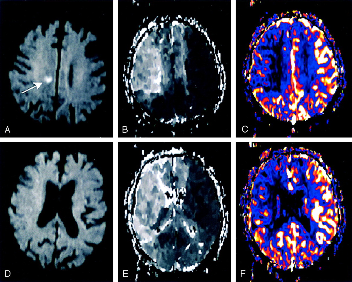
MR diagnostics 2 hours after the onset of stroke were performed on a 1.5-T system (Siemens Magnetom Sonata, Erlangen, Germany) including fluid-attenuated inversion recovery (FLAIR), T2*-weighted imaging, DW imaging, PWI (time-to-peak [TTP] and cerebral blood flow [CBF]), TOF, and CE-MRA. Total acquisition time for MR imaging was 25 minutes. The DW imaging revealed a small lesion in the subcortical white matter of the right hemisphere without any cortical lesions (Fig 1A). Intracranial TOF only faintly visualized the right distal internal carotid artery (ICA) and the right middle cerebral artery (MCA), which suggested a proximal ICA occlusion. Supplementary CE-MRA confirmed a high-grade stenosis (>90%) of the proximal right ICA (Fig 2A) and a moderate stenosis (50%) of the left ICA. Intracranially, the A1 segment of the left anterior cerebral artery (ACA) and the right posterior communicating artery were absent. These findings suggested a dominant perfusion of both ACA territories by the stenotic right ICA. A subsequent PWI (TTP and CBF) demonstrated a substantial delay and decrease of perfusion in almost the entire right MCA territory, as well as in both ACA territories (Fig 1B, -C, -E, -F). In summary, MR imaging findings revealed hemodynamic stroke origin without peripheral embolic occlusions. Therefore, intravenous thrombolysis was not performed.

Fig 1.
  • Download figure
  • Open in new tab
  • Download powerpoint
Fig 1.

MR imaging 2 hours after the onset of stroke.

A and D, Acute MR imaging with two representative sections of DW imaging revealing a small lesion in the subcortical white matter (A, arrow), suggesting hemodynamic infarction.

B and E, Acute MR imaging with the same sections of TTP, demonstrating a perfusion delay in almost the entire right MCA territory and in both ACA territories.

C and F, Acute MR imaging with the same sections of CBF showing a decrease of cerebral blood flow in almost the entire right MCA territory and in both ACA territories, matching the TTP results.

Fig 2.
  • Download figure
  • Open in new tab
  • Download powerpoint
Fig 2.

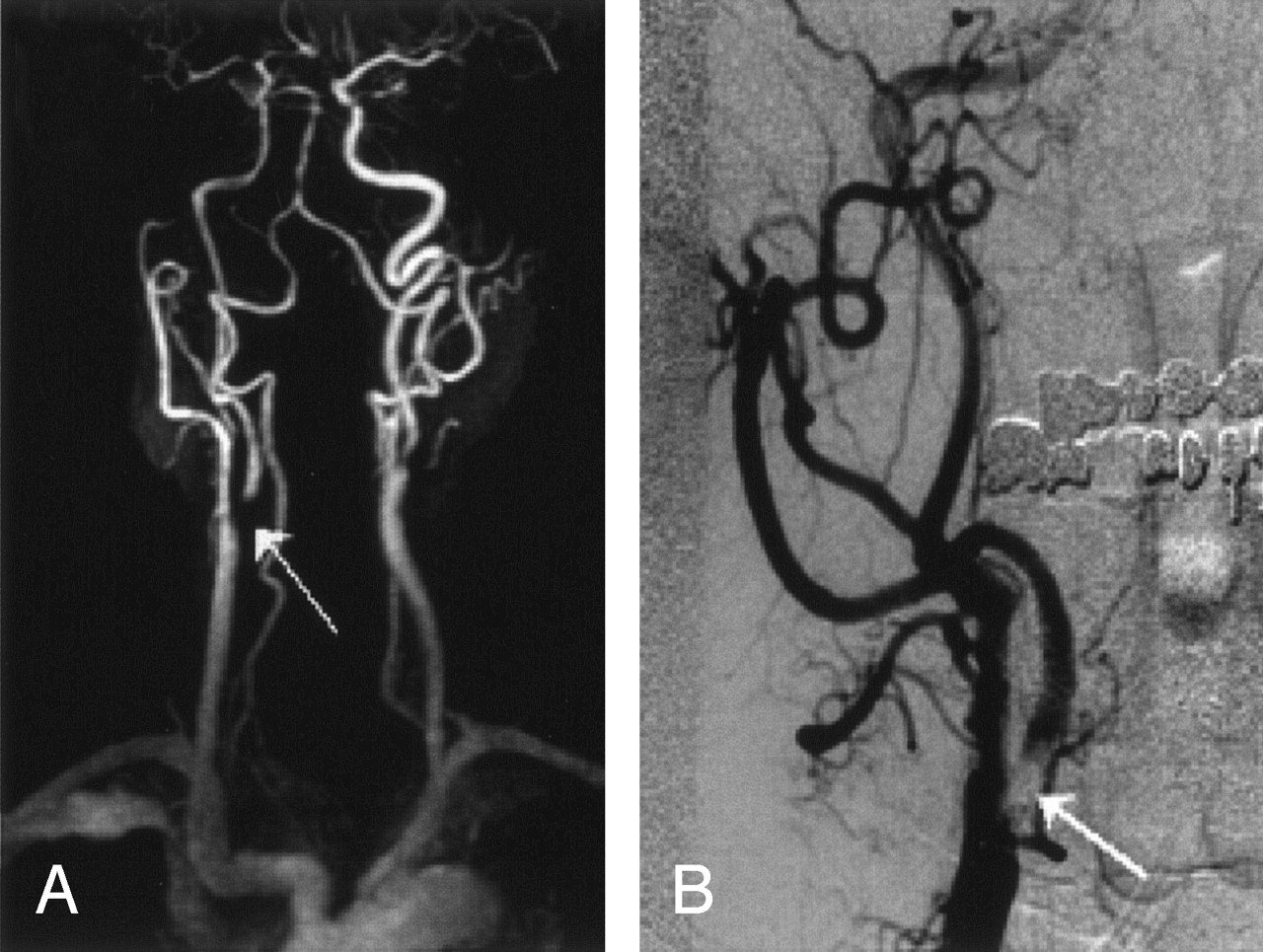
Before CAS placement.

A, Initial CE-MRA displaying a high-grade stenosis of the proximal right ICA (arrow), a moderate stenosis of the left ICA, and lack of an A1-segment of the left ACA.

B, Diagnostic angiography of the right ICA (anteroposterior) after selective injection in the right CCA, revealing the >95% stenosis of the proximal right ICA (arrow).

In a 1-hour attempt to increase systemic blood pressure by hypervolemic and hypertensive therapy, clinical symptoms did not improve. The persistent neurologic deficit and risk of ICA occlusion with a severe infarction of the right hemisphere and potentially even the left ACA territory led to the decision for CAS. Informed consent was obtained from the patient.

Diagnostic angiography revealed a >95% stenosis of the proximal right ICA (Fig 2B). The A1 segment of the left ACA and right posterior communicating artery were lacking. Because of progressing neurologic symptoms, endotracheal intubation was required in the course of the diagnostic angiography. To prevent thromboembolism, intravenous Abciximab was administered (bolus of 0.25 mg/kg, followed by 0.125 μg/kg/min continuously over a period of 12 hours). Neither acetyl salicylic acid (ASA) nor Clopidogrel was given before CAS.

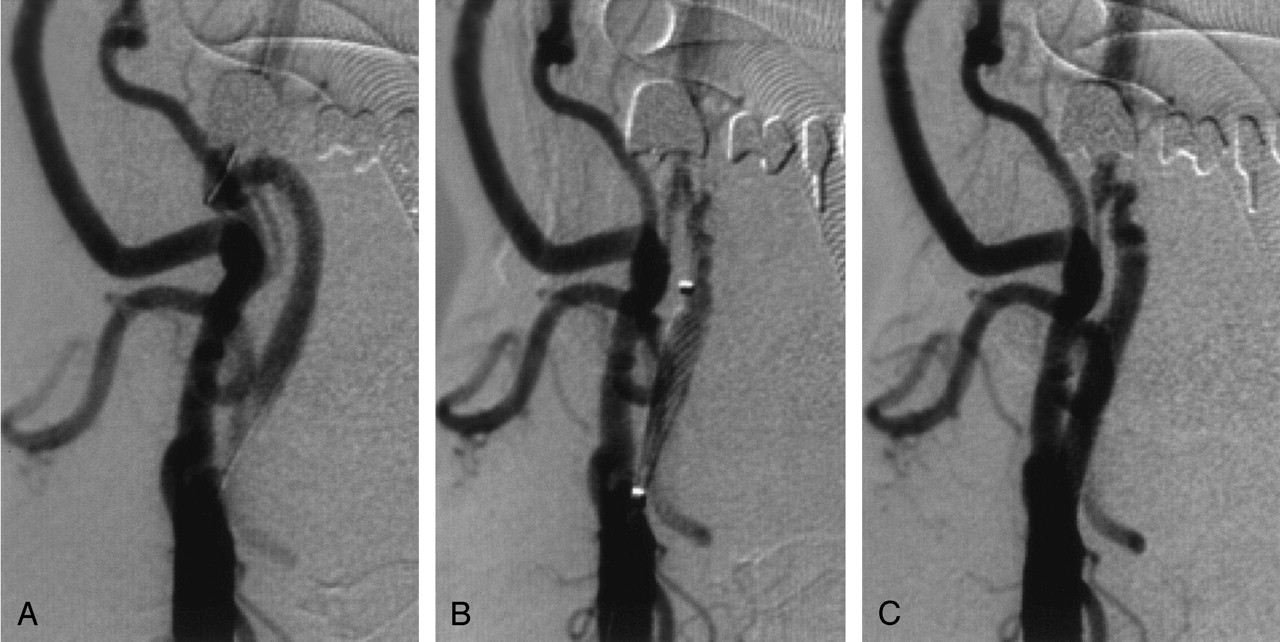
CAS was performed according to the protocol used in our neuroradiology department ( 2). Protection devices were not used. After placing a guidewire (Balance Heavyweight, Guidant, Santa Clara) through the stenosis (Fig 3A), a primary stent placement without predilatation was performed (Fig 3B) by using a 7 × 21 mm stent (Carotid Wallstent Monorail, Boston Scientific, Galway, Ireland). Postdilatation was carried out with a 5.5 × 20 mm balloon catheter (Maverick XL, Boston Scientific, Maple Grove, MN) inflated to a pressure of 12 bar. CAS was completed 5 hours after the onset of stroke. The control angiogram demonstrated a complete recanalization of the proximal right ICA (Fig 3C) with normalized filling of the right MCA and both ACA territories.

Fig 3.
  • Download figure
  • Open in new tab
  • Download powerpoint
Fig 3.

Three angiographic depictions during CAS (after selective CCA injection; anteroposterior views).

A, Guidewire placed through the stenosis.

B, Stent deployment along the stenosis.

C, Complete recanalization of the proximal right ICA after treatment.

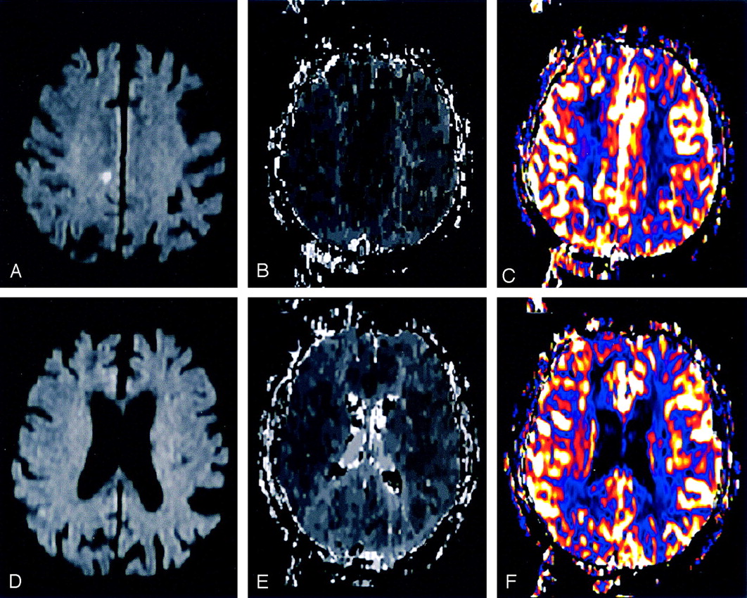
The patient was transferred to the neurologic intensive care unit for extubation and further treatment. The following day (18 hours after extubation) a complete clinical neurologic recovery was observed. Follow-up MR imaging 24 hours after CAS revealed no new ischemic lesions (Fig 4A–D) or intracranial bleeding. The TOF angiography showed a restored flow void of the right ICA, the right MCA, and both ACAs. Correspondingly, the PWI (TTP and CBF) showed a timely reperfusion of the right MCA and both ACA territories (Fig 4B, -C, -E, -F). The NIHSS at discharge (12 days after admission) was 0.

Fig 4.
  • Download figure
  • Open in new tab
  • Download powerpoint
Fig 4.

MR imaging at follow-up.

A and D, MR imaging 24 hours after treatment with corresponding sections of DW imaging without any new cortical or subcortical ischemic lesions.

B and E, MR imaging 24 hours after treatment with the same sections of TTP demonstrating a timely reperfusion of the right MCA and both ACA territories.

C and F, MR imaging 24 hours after treatment with the same sections of CBF showing a normalized cerebral blood flow, matching the TTP results.

Discussion

Persistent hemodynamic deficit is a rare cause of acute hemispheric stroke (3,,4). Acute stroke MR imaging—including DW imaging, PWI, TOF, and CE-MRA—may differentiate rare cases of pure hemodynamic stroke from embolic stroke. A typical constellation regarding the hemodynamic stroke subtype is a severe proximal stenosis associated with abnormalities in the circle of Willis (5). With a patient selection based on CT data according to the National Institute of Neurological Disorders and Stroke protocol (6) the presented patient (stable deficit, exclusion of hemorrhage) would have been a candidate for intravenous thrombolysis. Although we cannot prove our hypothesis, it seems very unlikely that this patient might have had a benefit from intravenous thrombolysis. On the basis of the MR imaging diagnosis of pure hemodynamic stroke was confirmed and because of the persistent neurologic deficit despite of aggressive attempts to increase the blood pressure, we decided to perform emergency CAS.

In this acute phase, fresh thrombotic material may cover the surface of the stenosis, increasing the risk of CAS induced embolic complication. Therefore, a complete and immediate blockade of platelet aggregation is required. This can be achieved with an intravenous administered glycoprotein IIb/IIIa-receptor antagonist, such as Abciximab (7,8) in the presented case.

The use of protection devices in CAS is another matter of debate (9,10). We preferred primary unprotected stent placement with periprocedural application of Abciximab to a protection system requiring a predilatation in this tight stenosis. Emergency CEA may be an alternative treatment (11,12), but it implies a considerable time delay. The endovascular approach, in contrast to CEA, ensures the visualization of the intracranial vessels during the procedure.

Conclusion

Modern MR imaging acute stroke diagnostics may select rare cases of pure hemodynamic stroke in differentiation from embolic stroke. In case of hemodynamic stroke, after failure of conservative treatment, CAS in the acute phase may be the treatment of choice.

References

  1. ↵
    Ringleb P, Kunze A, Allenberg JR, et al. Evaluation of stent-protected angioplasty for therapy of symptomatic stenoses of the carotid artery: SPACE and other randomized trials. Nervenarzt. 2003;74:482–428
    PubMed
  2. ↵
    Koch C, Kucinski T, Eckert B, et al. Endovascular therapy of high-degree stenoses of the neck vessels-stent-supported percutaneous angioplasty of the carotid artery without cerebral protection. Rofo Fortschr Geb Rontgenstr Neuen Bildgeb Verfahr 2002;174:1506–1510
    PubMed
  3. ↵
    Mull M, Schwarz M, Thron A. Cerebral hemispheric low-flow infarcts in arterial occlusive disease: lesion patterns and angiomorphological conditions. Stroke 1997;28:118–123
    Abstract/FREE Full Text
  4. ↵
    Derdeyn CP, Grubb RL Jr, Powers WJ. Cerebral hemodynamic impairment: methods of measurement and association with stroke risk. Neurology 1999;53:251–259
    Abstract/FREE Full Text
  5. ↵
    Kluytmans M, van der Grond J, van Everdingen KJ, et al. Cerebral hemodynamics in relation to patterns of collateral flow. Stroke 1999;30:1432–1439
    Abstract/FREE Full Text
  6. ↵
    National Institute of Neurological Disorders and Stroke rt-PA Stroke Study Group. Tissue plasminogen activator for acute ischemic stroke. The National Institute of Neurological Disorders and Stroke rt-PA Stroke Study Group. N Engl J Med 1995;333:1581–1587
    CrossRefPubMed
  7. ↵
    Cecena FE, Hoelzinger DH, Miller JA. The platelet IIb/IIIa inhibitor Abciximab as adjunctive therapy in carotid stenting of potential thrombotic lesions. J Intervent Cardiol 1999;12:355–362
  8. ↵
    Qureshi AI, Suri MF, Khan J, et al. Abciximab as an adjunct to high-risk carotid or vertebrobasilar angioplasty: preliminary experience. Neurosurgery 2000;46:1316–1324; discussion 1324–1325
    CrossRefPubMed
  9. ↵
    Eckert B, Zeumer H. Editorial comment: carotid artery stenting with or without protection devices? Strong opinions, poor evidence! Stroke 2003;34:1941–1943
    FREE Full Text
  10. ↵
    Forsting M. Editorial comment: with or without protection? The second important question in carotid artery stenting. Stroke 2004;35:e20–21
    FREE Full Text
  11. ↵
    Eckstein HH, Schumacher H, Klemm K, et al. Emergency carotid endarterectomy. Cerebrovasc Dis 1999;9:270–281
    CrossRefPubMed
  12. ↵
    Pikus HJ, Heros RC. Stroke: indications for emergent surgical intervention. Clin Neurosurg 1999;45:113–127
    PubMed
  • Received May 18, 2004.
  • Accepted after revision June 26, 2004.
  • Copyright © American Society of Neuroradiology
PreviousNext
Back to top

In this issue

American Journal of Neuroradiology: 26 (3)
American Journal of Neuroradiology
Vol. 26, Issue 3
1 Mar 2005
  • Table of Contents
  • Index by author
Advertisement
Print
Download PDF
Email Article

Thank you for your interest in spreading the word on American Journal of Neuroradiology.

NOTE: We only request your email address so that the person you are recommending the page to knows that you wanted them to see it, and that it is not junk mail. We do not capture any email address.

Enter multiple addresses on separate lines or separate them with commas.
Emergency Carotid Artery Stenting in Persistent Hemodynamic Deficit Associated with Severe Carotid Stenosis
(Your Name) has sent you a message from American Journal of Neuroradiology
(Your Name) thought you would like to see the American Journal of Neuroradiology web site.
CAPTCHA
This question is for testing whether or not you are a human visitor and to prevent automated spam submissions.
Cite this article
Benjamin S. Geisler, Joachim Röther, Thomas Kucinski, Hermann Zeumer, Bernd Eckert
Emergency Carotid Artery Stenting in Persistent Hemodynamic Deficit Associated with Severe Carotid Stenosis
American Journal of Neuroradiology Mar 2005, 26 (3) 549-552;

Citation Manager Formats

  • BibTeX
  • Bookends
  • EasyBib
  • EndNote (tagged)
  • EndNote 8 (xml)
  • Medlars
  • Mendeley
  • Papers
  • RefWorks Tagged
  • Ref Manager
  • RIS
  • Zotero
0 Responses
Respond to this article
Share
Bookmark this article
Emergency Carotid Artery Stenting in Persistent Hemodynamic Deficit Associated with Severe Carotid Stenosis
Benjamin S. Geisler, Joachim Röther, Thomas Kucinski, Hermann Zeumer, Bernd Eckert
American Journal of Neuroradiology Mar 2005, 26 (3) 549-552;
del.icio.us logo Twitter logo Facebook logo Mendeley logo
  • Tweet Widget
  • Facebook Like
  • Google Plus One

Jump to section

  • Article
    • Abstract
    • Case Description
    • Discussion
    • Conclusion
    • References
  • Figures & Data
  • Info & Metrics
  • Responses
  • References
  • PDF

Related Articles

  • No related articles found.
  • PubMed
  • Google Scholar

Cited By...

  • Safety and effectiveness of emergency carotid artery stenting for a high-grade carotid stenosis with intraluminal thrombus under proximal flow control in hyperacute and acute stroke
  • Crossref
  • Google Scholar

This article has not yet been cited by articles in journals that are participating in Crossref Cited-by Linking.

More in this TOC Section

  • A Meta-analysis of Combined Aspiration Catheter and Stent Retriever versus Stent Retriever Alone for Large-Vessel Occlusion Ischemic Stroke
  • Follow-up of Intracranial Aneurysms Treated by Flow Diverters: Evaluation of Parent Artery Patency Using 3D-T1 Gradient Recalled-Echo Imaging with 2-Point Dixon in Combination with 3D-TOF-MRA with Compressed Sensing
  • CT-Guided C2 Dorsal Root Ganglion Radiofrequency Ablation for the Treatment of Cervicogenic Headache: Case Series and Clinical Outcomes
Show more INTERVENTIONAL

Similar Articles

Advertisement

Indexed Content

  • Current Issue
  • Accepted Manuscripts
  • Article Preview
  • Past Issues
  • Editorials
  • Editor's Choice
  • Fellows' Journal Club
  • Letters to the Editor
  • Video Articles

Cases

  • Case Collection
  • Archive - Case of the Week
  • Archive - Case of the Month
  • Archive - Classic Case

More from AJNR

  • Trainee Corner
  • Imaging Protocols
  • MRI Safety Corner

Multimedia

  • AJNR Podcasts
  • AJNR Scantastics

Resources

  • Turnaround Time
  • Submit a Manuscript
  • Submit a Video Article
  • Submit an eLetter to the Editor/Response
  • Manuscript Submission Guidelines
  • Statistical Tips
  • Fast Publishing of Accepted Manuscripts
  • Graphical Abstract Preparation
  • Imaging Protocol Submission
  • Evidence-Based Medicine Level Guide
  • Publishing Checklists
  • Author Policies
  • Become a Reviewer/Academy of Reviewers
  • News and Updates

About Us

  • About AJNR
  • Editorial Board
  • Editorial Board Alumni
  • Alerts
  • Permissions
  • Not an AJNR Subscriber? Join Now
  • Advertise with Us
  • Librarian Resources
  • Feedback
  • Terms and Conditions
  • AJNR Editorial Board Alumni

American Society of Neuroradiology

  • Not an ASNR Member? Join Now

© 2025 by the American Society of Neuroradiology All rights, including for text and data mining, AI training, and similar technologies, are reserved.
Print ISSN: 0195-6108 Online ISSN: 1936-959X

Powered by HighWire