Advertisement

Main menu

  • Home
  • Content
    • Current Issue
    • Accepted Manuscripts
    • Article Preview
    • Past Issue Archive
    • Video Articles
    • AJNR Case Collection
    • Case of the Week Archive
    • Case of the Month Archive
    • Classic Case Archive
  • Special Collections
    • AJNR Awards
    • Low-Field MRI
    • Alzheimer Disease
    • ASNR Foundation Special Collection
    • Photon-Counting CT
    • View All
  • Multimedia
    • AJNR Podcasts
    • AJNR SCANtastic
    • Trainee Corner
    • MRI Safety Corner
    • Imaging Protocols
  • For Authors
    • Submit a Manuscript
    • Submit a Video Article
    • Submit an eLetter to the Editor/Response
    • Manuscript Submission Guidelines
    • Statistical Tips
    • Fast Publishing of Accepted Manuscripts
    • Graphical Abstract Preparation
    • Imaging Protocol Submission
    • Author Policies
  • About Us
    • About AJNR
    • Editorial Board
    • Editorial Board Alumni
  • More
    • Become a Reviewer/Academy of Reviewers
    • Subscribers
    • Permissions
    • Alerts
    • Feedback
    • Advertisers
    • ASNR Home

User menu

  • Alerts
  • Log in

Search

  • Advanced search
American Journal of Neuroradiology

American Journal of Neuroradiology

ASHNR American Society of Functional Neuroradiology ASHNR American Society of Pediatric Neuroradiology ASSR
  • Alerts
  • Log in

Advanced Search

  • Home
  • Content
    • Current Issue
    • Accepted Manuscripts
    • Article Preview
    • Past Issue Archive
    • Video Articles
    • AJNR Case Collection
    • Case of the Week Archive
    • Case of the Month Archive
    • Classic Case Archive
  • Special Collections
    • AJNR Awards
    • Low-Field MRI
    • Alzheimer Disease
    • ASNR Foundation Special Collection
    • Photon-Counting CT
    • View All
  • Multimedia
    • AJNR Podcasts
    • AJNR SCANtastic
    • Trainee Corner
    • MRI Safety Corner
    • Imaging Protocols
  • For Authors
    • Submit a Manuscript
    • Submit a Video Article
    • Submit an eLetter to the Editor/Response
    • Manuscript Submission Guidelines
    • Statistical Tips
    • Fast Publishing of Accepted Manuscripts
    • Graphical Abstract Preparation
    • Imaging Protocol Submission
    • Author Policies
  • About Us
    • About AJNR
    • Editorial Board
    • Editorial Board Alumni
  • More
    • Become a Reviewer/Academy of Reviewers
    • Subscribers
    • Permissions
    • Alerts
    • Feedback
    • Advertisers
    • ASNR Home
  • Follow AJNR on Twitter
  • Visit AJNR on Facebook
  • Follow AJNR on Instagram
  • Join AJNR on LinkedIn
  • RSS Feeds

AJNR Awards, New Junior Editors, and more. Read the latest AJNR updates

OtherNeurointervention

Possible Electrical Stimulation on Rostral Ventrolateral Medulla That Causes Significant Hypertensive and Tachycardic Changes during Endovascular Treatment of a PICA Aneurysm Abutting the Left Medullary Sulcus

Harun Ozer, Omer Uzma, Sait Albayram, Hakan Selcuk, Civan Isilak and Naci Kocer
American Journal of Neuroradiology March 2005, 26 (3) 522-524;
Harun Ozer
  • Find this author on Google Scholar
  • Find this author on PubMed
  • Search for this author on this site
Omer Uzma
  • Find this author on Google Scholar
  • Find this author on PubMed
  • Search for this author on this site
Sait Albayram
  • Find this author on Google Scholar
  • Find this author on PubMed
  • Search for this author on this site
Hakan Selcuk
  • Find this author on Google Scholar
  • Find this author on PubMed
  • Search for this author on this site
Civan Isilak
  • Find this author on Google Scholar
  • Find this author on PubMed
  • Search for this author on this site
Naci Kocer
  • Find this author on Google Scholar
  • Find this author on PubMed
  • Search for this author on this site
  • Article
  • Figures & Data
  • Info & Metrics
  • Responses
  • References
  • PDF
Loading

Abstract

Summary: We report the case of a 43-year-old man who underwent endovascular treatment for posterior inferior cerebellar aneurysm. Significant hemodynamic changes were observed as electric stimulation was applied during coil detachment for a PICA aneurysm. We postulate that changes in heart rate and blood pressure during coil detachment were due to the electric stimulation of the tonic vasomotor center located in the rostral ventrolateral medulla, which was very close to the PICA aneurysm.

The rostral ventrolateral medulla (RVLM) has been known to be a major regulating center of sympathetic and cardiovascular activities. An association between blood pressure and the RVLM has been reported on the basis of animal experiments and clinical observations.

We present a case of interesting hemodynamic changes that occurred during endovascular treatment of a posterior inferior cerebellar artery aneurysm with Guglielmi detachable coils (GDCs). We discuss the underlying physiologic mechanism for these hemodynamic changes.

Case Reports

A 43-year-old man presented with acute onset of headache and meningismus. He had no significant medical history, including blood pressure and medication. A CT scan conducted at our institution revealed subarachnoid hemorrhages. Same-day diagnostic angiography was performed and revealed that the patient had left posterior communicating and left posterior inferior cerebellar artery aneurysms. Because of CT blood pattern and the morphologic appearance of the posterior communicating aneurysm, it was evaluated as suspicious for bleeding. It was then treated by the endovascular balloon remodeling technique without difficulty. During the coil detachment process and follow-up period, no abnormality regarding heart rate or blood pressure was seen. Two weeks later, endovascular treatment for the PICA aneurysm was performed while the patient was under general anesthesia with routine full heparinization. Arterial invasive monitoring was obtained from a femoral sheath introducer. First, a guiding catheter was introduced into the left vertebral artery, and then the PICA aneurysm sac was catheterized using an Excel 10 microcatheter (Target/Boston Scientific, Fremont, CA) and a Transend Guidewire 10 (Target/Boston Scientific) combination. For occlusion of aneurysm, one GDC 3D 6 × 10 mm (Boston Scientific), one Matrix soft 2D 3 × 4 mm, and three Matrix Ultrasoft 2 × 6, 2 × 4, 2 × 2 mm coils were placed into aneurysmal sac and detached by using standard GDC power supply system with 2-mA current applied about 25 seconds for each coil (21 seconds for 3D GDC; 25 seconds for Matrix soft; and 23, 29, and 32 seconds for the three Matrix Ultrasoft coils). During the first coil detachment, an abrupt increase of the heart rate was observed. Angiography was performed immediately to determine whether any bleeding occurred, but it revealed no extravasations from the aneurysm. During the other detachment of coils, heart rate and blood pressure were recorded. Both showed significant changes as long as the electric stimulation continued. Blood pressure and heart rate obtained during electric stimulation for coil detachment showed a mean increase from 150–180 mm Hg and 65–80 bpm, respectively (Fig 1A). These surprising hemodynamic changes repeated during every electric stimulation for coil detachment. After coil detachment, both blood pressure and heart rate returned to the previous values.

Fig 1.
  • Download figure
  • Open in new tab
  • Download powerpoint
Fig 1.

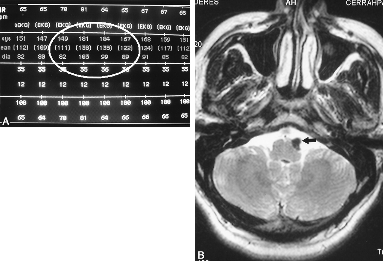
Images obtained in a 43-year-old man with left PICA aneurysm.

A, Heart rate and blood pressure recordings during the second coil detachment. Note that systolic blood pressure and heart rate show increases from 151–184 mm Hg and 65–81 bpm, respectively.

B, Axial T2-weighted MR images shows that left PICA aneurysm adjacent to and slightly compressed to the RVLM.

A control angiogram of the left vertebral artery was obtained after detachment of five detachable coils and showed complete occlusion of the aneurysm sac, whereupon the procedure was completed. Because significant hemodynamic changes were observed during the endovascular procedures, MR image was performed 2 days later. It showed no abnormality of the brain stem regarding infarct or any other pathologic signal intensity changes, except for slight vascular compression of occluded PICA aneurysm on the lateral medullary sulcus (Fig 1B). After the procedure, no subarachnoid hemorrhages were observed on the MR imaging examination. During the follow-up period, no blood pressure or pulse rate changes were observed.

Discussion

The RVLM has been known to be a major regulating center of sympathetic and cardiovascular activities. An association between blood pressure and the RVLM has been reported on the basis of animal experiments and clinical observations. In our case, during coil (GDC) detachment, prominent blood pressure and heart rate changes were observed probably because of the close location of the aneurysm to the lateral medullary sulcus.

Ross et al (1) studied the responses to electrical and chemical stimulation of the ventrolateral medulla in the rat. Elevations of arterial pressure (+81.6 ± 2.5 mm Hg) and cardio acceleration (+73 ± 13.6 bpm) were elicited with low current (five times threshold of 9.5 ± 1.1 mA) electrical stimulation in a region of rostral ventrolateral medullary reticular formation (RVLM). Electrical stimulation of the RVLM increased plasma catecholamines (16.8-fold for adrenaline, 5.3-fold for noradrenaline, and 1.9-fold for dopamine) and vasopressin (1.7-fold before spinal transection and 4.7-fold after). Electric stimulation of the RVLM with a 10-second stimulus train (100Hz, 10–50 mA) invariably resulted in elevation of arterial pressure and usually heart rate. Ninety percent of the peak response was obtained within 2 seconds of the onset of the stimulus, and in about 70% of the animals the response decayed within 1 or 2 seconds following its termination. The stimulus-locked rise in arterial pressure was accompanied by pure tachycardia in about 75% of the animals. At the end of the stimulation, the heart rate either remained elevated, gradually returning to control values, or was replaced by bradycardia. The pressor response to stimulation of the RVLM was stable and could be reproduced over several hours with repeated stimuli.

The findings about the vasomotor center in humans arise from the initial report by Jannetta and Gendell (2) in 1978, clinical and experimental observations, including MR imaging studies that have indicated an association between essential hypertension and neurovascular compression of the ventrolateral medulla (3, 4). Morimoto et al (5) proposed the association between essential hypertension and neurovascular compression of the RVLM. Jannetta and Gendell (2) reported that neurovascular compression of the RVLM was found in 51 of 53 hypertensive patients and in none of the 50 normotensive patients. High blood pressure returned to normal in 32 and improved in four of 42 patients who were treated with left microvascular decompression of the RVLM. On the other hand, blood pressure remained unchanged in the seven hypertensive patients who were treated with right microvascular decompression of the RVLM (6). Since then, several observations have indicated an association between essential hypertension and neurovascular compression of the RVLM (3, 7). Jannetta and Gendell and Morimoto et al (2, 8) also reported the development of hypertension by pulsatile compression on the left RVLM. Angiographic and pathologic studies further indicated that the neurovascular compression of the left RVLM was involved in essential hypertension (4, 9).

Our case presented with a left PICA aneurysm located very close to the lateral medullary sulcus. During GDC detachment—these coils need electric current for detachment from the pushing wire—we applied electrical stimulation to a nearby area and observed the significant changes in blood pressure and heart rate as long as the stimulation continued, an observation comparable to the results of the Ross et al study. From this observation, we were able to make several conclusions.

Conclusion

In this case, we observed that during every coil detachment the blood pressure and heart rate showed a mean increase from 150–180 mm Hg and 65–80 bpm, respectively, with two mA current applied for 25 seconds with each coiling. First, our observation—a kind of in vivo experiment—indicates that the RVLM could be the vasomotor center in humans also.

After our observation, it is not speculation to propose that electrically stimulated hypertensive changes may occur during coil detachment, specifically in cases of aneurysms located close to the RVLM. This abrupt hemodynamic changes in an aneurysm patient would complicate the clinical picture, so interventional neuroradiologists should be aware of this fact and precautions should be taken. In addition, because the procedures are performed under general anesthesia, we cannot observe possible other types of neurologic changes caused by electric stimulation.

References

  1. ↵
    Ross CA, Ruggiero DA, Park DH, et al. Tonic vasomotor control by the rostral ventrolateral medulla: effect of electrical or chemical stimulation of the area containing C1 adrenaline neurons on arterial pressure, heart rate, and plasma catecholamines and vasopressin. J Neurosci 1984;4:474–494
    Abstract
  2. ↵
    Jannetta PJ, Gendell HM. Neurovascular compression associated with essential hypertension. Neurosurgery 1978;2:165
    PubMed
  3. ↵
    Yamamoto I, Yamada S, Sato O. Microvascular decompression for hypertension: clinical and experimental study. Neurol Med Chir (Tokyo) 1991;31:1–6
    PubMed
  4. ↵
    Naraghi R, Gaab MR, Walter GF, Kleinberg B. Arterial hypertension and neurovascular compression at the ventrolateral medulla. J Neurosurg 1992;77:103–112
    PubMed
  5. ↵
    Morimoto S, Sasaki S, Miki S, et al. Neurovascular compression of the rostral ventrolateral medulla related to essential hypertension. Hypertension 1997;30:77–82
    Abstract/FREE Full Text
  6. ↵
    Jannetta PJ, Segal R, Wolfson SK. Neurogenic hypertension: etiology and surgical treatment. I. Observations in 53 patients. Ann Surg 1985;201:391–398
    PubMed
  7. ↵
    Akimura T, Furutani Y, Jimi Y, et al. Essential hypertension and neurovascular compression at the ventrolateral medulla oblongata: MR evaluation. AJNR Am J Neuroradiol 1995;16:401–405
    Abstract/FREE Full Text
  8. ↵
    Morimoto S, Sasaki S, Miki S, et al. Pulsatile compression of the rostral ventrolateral medulla in hypertension. Hypertension 1997;29:514
    Abstract/FREE Full Text
  9. ↵
    Kleineberg B, Becker H, Gaab MR. Neurovascular compression and essential hypertension: an angiographic study. Neuroradiology 1991;33:2–8
    CrossRefPubMed
  • Received April 28, 2004.
  • Accepted after revision June 16, 2004.
  • Copyright © American Society of Neuroradiology
View Abstract
PreviousNext
Back to top

In this issue

American Journal of Neuroradiology: 26 (3)
American Journal of Neuroradiology
Vol. 26, Issue 3
1 Mar 2005
  • Table of Contents
  • Index by author
Advertisement
Print
Download PDF
Email Article

Thank you for your interest in spreading the word on American Journal of Neuroradiology.

NOTE: We only request your email address so that the person you are recommending the page to knows that you wanted them to see it, and that it is not junk mail. We do not capture any email address.

Enter multiple addresses on separate lines or separate them with commas.
Possible Electrical Stimulation on Rostral Ventrolateral Medulla That Causes Significant Hypertensive and Tachycardic Changes during Endovascular Treatment of a PICA Aneurysm Abutting the Left Medullary Sulcus
(Your Name) has sent you a message from American Journal of Neuroradiology
(Your Name) thought you would like to see the American Journal of Neuroradiology web site.
CAPTCHA
This question is for testing whether or not you are a human visitor and to prevent automated spam submissions.
Cite this article
Harun Ozer, Omer Uzma, Sait Albayram, Hakan Selcuk, Civan Isilak, Naci Kocer
Possible Electrical Stimulation on Rostral Ventrolateral Medulla That Causes Significant Hypertensive and Tachycardic Changes during Endovascular Treatment of a PICA Aneurysm Abutting the Left Medullary Sulcus
American Journal of Neuroradiology Mar 2005, 26 (3) 522-524;

Citation Manager Formats

  • BibTeX
  • Bookends
  • EasyBib
  • EndNote (tagged)
  • EndNote 8 (xml)
  • Medlars
  • Mendeley
  • Papers
  • RefWorks Tagged
  • Ref Manager
  • RIS
  • Zotero
0 Responses
Respond to this article
Share
Bookmark this article
Possible Electrical Stimulation on Rostral Ventrolateral Medulla That Causes Significant Hypertensive and Tachycardic Changes during Endovascular Treatment of a PICA Aneurysm Abutting the Left Medullary Sulcus
Harun Ozer, Omer Uzma, Sait Albayram, Hakan Selcuk, Civan Isilak, Naci Kocer
American Journal of Neuroradiology Mar 2005, 26 (3) 522-524;
del.icio.us logo Twitter logo Facebook logo Mendeley logo
  • Tweet Widget
  • Facebook Like
  • Google Plus One

Jump to section

  • Article
    • Abstract
    • Case Reports
    • Discussion
    • Conclusion
    • References
  • Figures & Data
  • Info & Metrics
  • Responses
  • References
  • PDF

Related Articles

  • No related articles found.
  • PubMed
  • Google Scholar

Cited By...

  • No citing articles found.
  • Crossref
  • Google Scholar

This article has not yet been cited by articles in journals that are participating in Crossref Cited-by Linking.

More in this TOC Section

  • Optimizing Voxel Size in 3D Rotational Angiography
  • Neuroform Atlas Stent for Intracranial Aneurysms
  • Neurologic Complication in Transradial Angiography
Show more NEUROINTERVENTION

Similar Articles

Advertisement

Indexed Content

  • Current Issue
  • Accepted Manuscripts
  • Article Preview
  • Past Issues
  • Editorials
  • Editor's Choice
  • Fellows' Journal Club
  • Letters to the Editor
  • Video Articles

Cases

  • Case Collection
  • Archive - Case of the Week
  • Archive - Case of the Month
  • Archive - Classic Case

More from AJNR

  • Trainee Corner
  • Imaging Protocols
  • MRI Safety Corner

Multimedia

  • AJNR Podcasts
  • AJNR Scantastics

Resources

  • Turnaround Time
  • Submit a Manuscript
  • Submit a Video Article
  • Submit an eLetter to the Editor/Response
  • Manuscript Submission Guidelines
  • Statistical Tips
  • Fast Publishing of Accepted Manuscripts
  • Graphical Abstract Preparation
  • Imaging Protocol Submission
  • Evidence-Based Medicine Level Guide
  • Publishing Checklists
  • Author Policies
  • Become a Reviewer/Academy of Reviewers
  • News and Updates

About Us

  • About AJNR
  • Editorial Board
  • Editorial Board Alumni
  • Alerts
  • Permissions
  • Not an AJNR Subscriber? Join Now
  • Advertise with Us
  • Librarian Resources
  • Feedback
  • Terms and Conditions
  • AJNR Editorial Board Alumni

American Society of Neuroradiology

  • Not an ASNR Member? Join Now

© 2025 by the American Society of Neuroradiology All rights, including for text and data mining, AI training, and similar technologies, are reserved.
Print ISSN: 0195-6108 Online ISSN: 1936-959X

Powered by HighWire