Advertisement

Main menu

  • Home
  • Content
    • Current Issue
    • Accepted Manuscripts
    • Article Preview
    • Past Issue Archive
    • Video Articles
    • AJNR Case Collection
    • Case of the Week Archive
    • Case of the Month Archive
    • Classic Case Archive
  • Special Collections
    • AJNR Awards
    • Low-Field MRI
    • Alzheimer Disease
    • ASNR Foundation Special Collection
    • Photon-Counting CT
    • View All
  • Multimedia
    • AJNR Podcasts
    • AJNR SCANtastic
    • Trainee Corner
    • MRI Safety Corner
    • Imaging Protocols
  • For Authors
    • Submit a Manuscript
    • Submit a Video Article
    • Submit an eLetter to the Editor/Response
    • Manuscript Submission Guidelines
    • Statistical Tips
    • Fast Publishing of Accepted Manuscripts
    • Graphical Abstract Preparation
    • Imaging Protocol Submission
    • Author Policies
  • About Us
    • About AJNR
    • Editorial Board
    • Editorial Board Alumni
  • More
    • Become a Reviewer/Academy of Reviewers
    • Subscribers
    • Permissions
    • Alerts
    • Feedback
    • Advertisers
    • ASNR Home

User menu

  • Alerts
  • Log in

Search

  • Advanced search
American Journal of Neuroradiology

American Journal of Neuroradiology

ASHNR American Society of Functional Neuroradiology ASHNR American Society of Pediatric Neuroradiology ASSR
  • Alerts
  • Log in

Advanced Search

  • Home
  • Content
    • Current Issue
    • Accepted Manuscripts
    • Article Preview
    • Past Issue Archive
    • Video Articles
    • AJNR Case Collection
    • Case of the Week Archive
    • Case of the Month Archive
    • Classic Case Archive
  • Special Collections
    • AJNR Awards
    • Low-Field MRI
    • Alzheimer Disease
    • ASNR Foundation Special Collection
    • Photon-Counting CT
    • View All
  • Multimedia
    • AJNR Podcasts
    • AJNR SCANtastic
    • Trainee Corner
    • MRI Safety Corner
    • Imaging Protocols
  • For Authors
    • Submit a Manuscript
    • Submit a Video Article
    • Submit an eLetter to the Editor/Response
    • Manuscript Submission Guidelines
    • Statistical Tips
    • Fast Publishing of Accepted Manuscripts
    • Graphical Abstract Preparation
    • Imaging Protocol Submission
    • Author Policies
  • About Us
    • About AJNR
    • Editorial Board
    • Editorial Board Alumni
  • More
    • Become a Reviewer/Academy of Reviewers
    • Subscribers
    • Permissions
    • Alerts
    • Feedback
    • Advertisers
    • ASNR Home
  • Follow AJNR on Twitter
  • Visit AJNR on Facebook
  • Follow AJNR on Instagram
  • Join AJNR on LinkedIn
  • RSS Feeds

AJNR Awards, New Junior Editors, and more. Read the latest AJNR updates

Research ArticlePediatric Neuroimaging

Selecting the Best Index for Following the Temporal Evolution of Apparent Diffusion Coefficient and Diffusion Anisotropy After Hypoxic-Ischemic White Matter Injury in Neonates

Carola van Pul, Jan Buijs, Maurice J. A. Janssen, George F. Roos, Marinus T. Vlaardingerbroek and Pieter F. F. Wijn
American Journal of Neuroradiology March 2005, 26 (3) 469-481;
Carola van Pul
  • Find this author on Google Scholar
  • Find this author on PubMed
  • Search for this author on this site
Jan Buijs
  • Find this author on Google Scholar
  • Find this author on PubMed
  • Search for this author on this site
Maurice J. A. Janssen
  • Find this author on Google Scholar
  • Find this author on PubMed
  • Search for this author on this site
George F. Roos
  • Find this author on Google Scholar
  • Find this author on PubMed
  • Search for this author on this site
Marinus T. Vlaardingerbroek
  • Find this author on Google Scholar
  • Find this author on PubMed
  • Search for this author on this site
Pieter F. F. Wijn
  • Find this author on Google Scholar
  • Find this author on PubMed
  • Search for this author on this site
  • Article
  • Figures & Data
  • Info & Metrics
  • Responses
  • References
  • PDF
Loading

Article Figures & Data

Figures

  • Tables
  • Fig 1.
    • Download figure
    • Open in new tab
    • Download powerpoint
    Fig 1.

    Definition of the regions of interest in the adult brain. 1 indicates corpus callosum; 2, white matter.

  • Fig 2.
    • Download figure
    • Open in new tab
    • Download powerpoint
    Fig 2.

    Eigenvalue space plots as defined by Bahn (33), plotted for anisotropy index FA. FA is high (bright) for both λ3/λ1 and λ2/λ1 close to zero, which corresponds to a cigar-shaped diffusion ellipse. But FA is still relatively large for λ2/λ1 close to 1, the pancake-shaped diffusion ellipse.
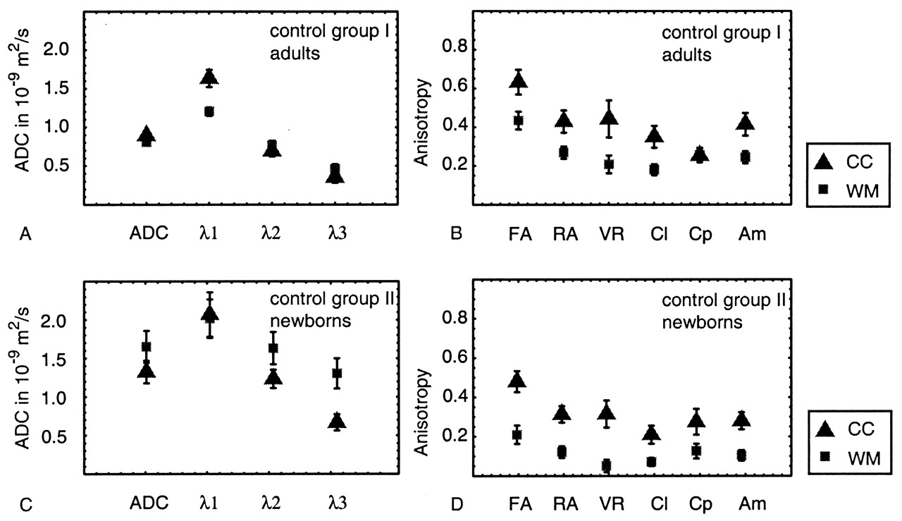
  • Fig 3.
    • Download figure
    • Open in new tab
    • Download powerpoint
    Fig 3.

    A–D, Average ADC, eigenvalues, and anisotropy indexes in frontal white matter (WM) and in the corpus callosum (CC), measured in 16 adult volunteers (A and B) and in 10 full-term neonates with normal MR images (C and D).
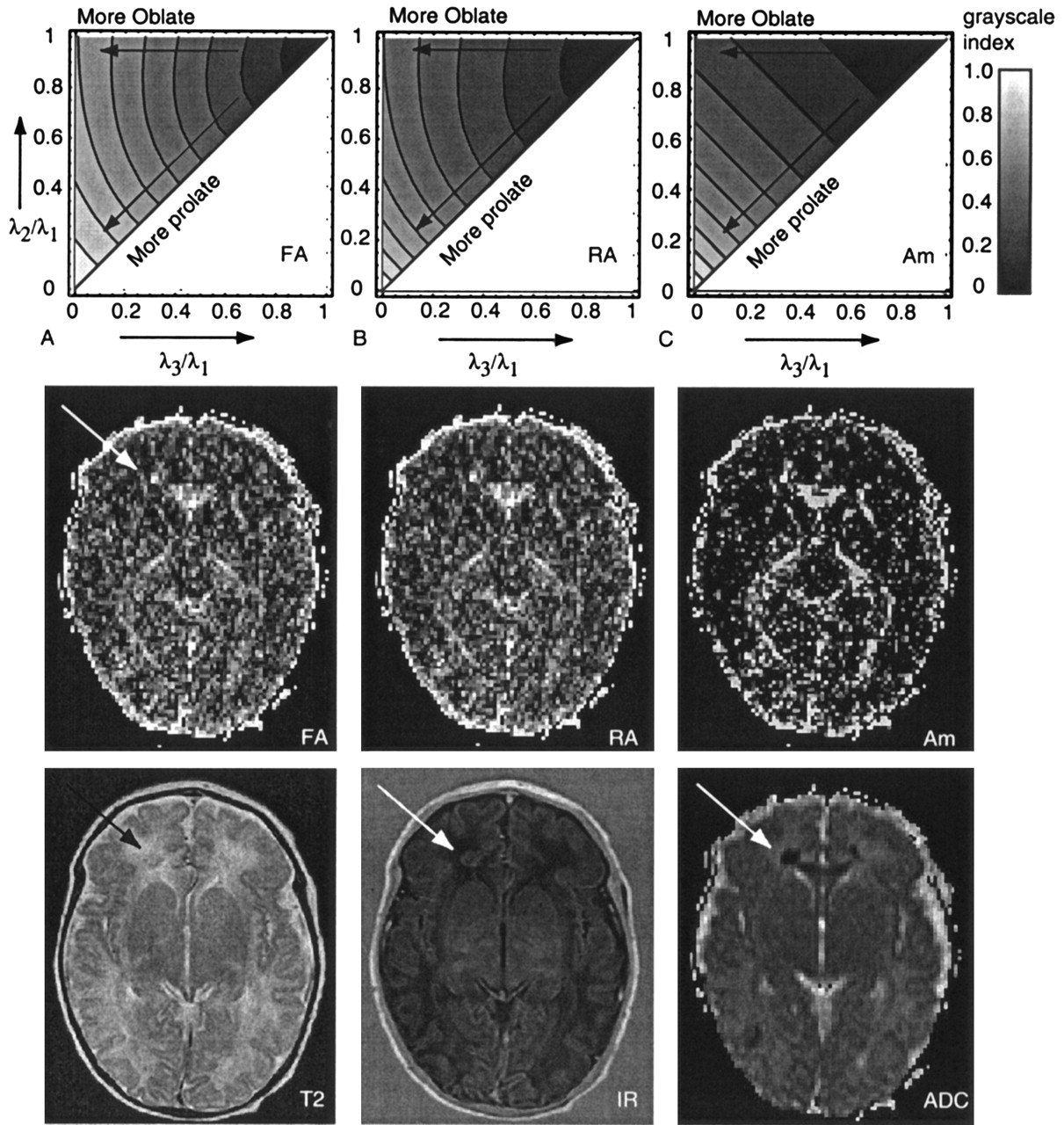
  • Fig 4.
    • Download figure
    • Open in new tab
    • Download powerpoint
    Fig 4.

    A–C, Eigenvalue space plots, as introduced by Bahn (33) (explained in Fig 2), for FA, RA, and Am. In the middle row, the corresponding anisotropy maps are shown for a neonate with lesions (arrow) in the frontal white matter and occipital white matter. Note the increased anisotropy corresponding to the low ADC area. In the bottom row, T2-weighted image, IR image, and ADC map in the same neonate are shown.

  • Fig 5.
    • Download figure
    • Open in new tab
    • Download powerpoint
    Fig 5.

    A–D, Results of simulations of the effect of random noise on ADC and eigenvalues (A and C) and anisotropy (B and D) for the adult case (A and B) and neonate case (C and D). Open symbols indicate the input values (Table 2); filled symbols, the values after noise was added. The error bars show the standard deviation of the simulated results.
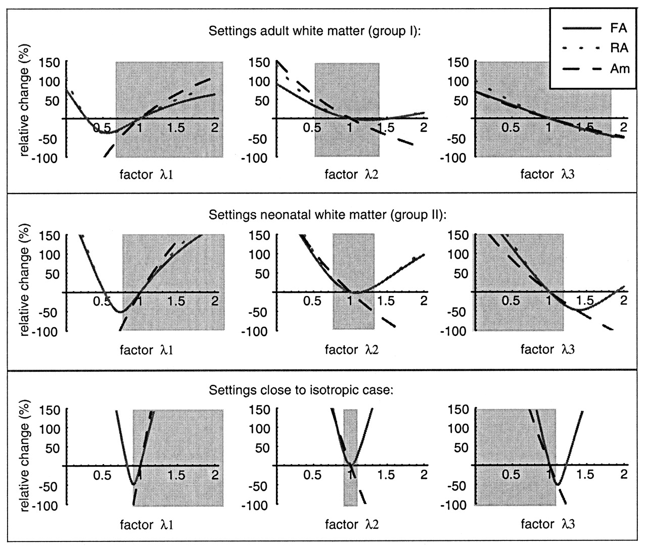
  • Fig 6.
    • Download figure
    • Open in new tab
    • Download powerpoint
    Fig 6.

    Relative change in FA, RA, and Am as a function of a change in one of the eigenvalues λ1, λ2, and λ3. The change is defined as a factor times the original value; the factor is displayed on the x-axis. The relative change is shown for three input settings, corresponding to adult white matter (top row), neonatal white matter (middle row), and a case close to isotropic (bottom row). The gray area is the area in which the function is valid, that is where λ1 > λ2 > λ3.

  • Fig 7.
    • Download figure
    • Open in new tab
    • Download powerpoint
    Fig 7.

    Average absolute values for the ADC (top image), the three eigenvalues λ1, λ2, and λ3 (middle row), and the three anisotropy indexes FA, RA, and Am (bottom row) as a function of time after the onset of symptoms for neonates with a white matter lesion.

  • Fig 8.
    • Download figure
    • Open in new tab
    • Download powerpoint
    Fig 8.

    A–C, Time evolution of the relative change (Change equation) in ADC values (A), the three eigenvalues (B), and the anisotropy indexes FA, RA, and Am (C) after hypoxic-ischemic brain injury in neonates. Trend lines are shown only if the trend was significant (P < .05).

Tables

  • Figures
    • View popup
    TABLE 1:

    Equations for the anisotropy indexes evaluated in this study and the authors who introduced them

    IndexIntroducing Authors (reference no.)Equation
    FABasser and Pierpaoli (12)Embedded Image
    RABasser and Pierpaoli (12)Embedded Image
    VRLe Bihan et al (14)Embedded Image
    ClPeled et al (31)Embedded Image
    CpPeled et al (31)Embedded Image
    AmConturo et al (32)Embedded Image
    • Note.—All indexes are scaled to make isotropic diffusion 0 and anisotropic diffusion 1. λ1 is the largest eigenvalue, λ3 the smallest.

    • View popup
    TABLE 2:

    Input eigenvalues for noise simulations

    Input SettingsEigenvaluesSNR
    λ1 (10−9 m2/s)λ2 (10−9 m2/s)λ3 (10−9 m2/s)
    Corpus callosum
        Adult1.60.70.3564
        Neonatal2.01.20.6535
    White matter
        Adult1.20.80.4564
        Neonatal2.01.61.335
    Isotropic ADC
        Adult0.880.880.8864
        Neonatal1.31.31.335
PreviousNext
Back to top

In this issue

American Journal of Neuroradiology: 26 (3)
American Journal of Neuroradiology
Vol. 26, Issue 3
1 Mar 2005
  • Table of Contents
  • Index by author
Advertisement
Print
Download PDF
Email Article

Thank you for your interest in spreading the word on American Journal of Neuroradiology.

NOTE: We only request your email address so that the person you are recommending the page to knows that you wanted them to see it, and that it is not junk mail. We do not capture any email address.

Enter multiple addresses on separate lines or separate them with commas.
Selecting the Best Index for Following the Temporal Evolution of Apparent Diffusion Coefficient and Diffusion Anisotropy After Hypoxic-Ischemic White Matter Injury in Neonates
(Your Name) has sent you a message from American Journal of Neuroradiology
(Your Name) thought you would like to see the American Journal of Neuroradiology web site.
CAPTCHA
This question is for testing whether or not you are a human visitor and to prevent automated spam submissions.
Cite this article
Carola van Pul, Jan Buijs, Maurice J. A. Janssen, George F. Roos, Marinus T. Vlaardingerbroek, Pieter F. F. Wijn
Selecting the Best Index for Following the Temporal Evolution of Apparent Diffusion Coefficient and Diffusion Anisotropy After Hypoxic-Ischemic White Matter Injury in Neonates
American Journal of Neuroradiology Mar 2005, 26 (3) 469-481;

Citation Manager Formats

  • BibTeX
  • Bookends
  • EasyBib
  • EndNote (tagged)
  • EndNote 8 (xml)
  • Medlars
  • Mendeley
  • Papers
  • RefWorks Tagged
  • Ref Manager
  • RIS
  • Zotero
0 Responses
Respond to this article
Share
Bookmark this article
Selecting the Best Index for Following the Temporal Evolution of Apparent Diffusion Coefficient and Diffusion Anisotropy After Hypoxic-Ischemic White Matter Injury in Neonates
Carola van Pul, Jan Buijs, Maurice J. A. Janssen, George F. Roos, Marinus T. Vlaardingerbroek, Pieter F. F. Wijn
American Journal of Neuroradiology Mar 2005, 26 (3) 469-481;
del.icio.us logo Twitter logo Facebook logo Mendeley logo
  • Tweet Widget
  • Facebook Like
  • Google Plus One

Jump to section

  • Article
    • Abstract
    • Methods
    • Results
    • Discussion
    • Conclusion
    • References
  • Figures & Data
  • Info & Metrics
  • Responses
  • References
  • PDF

Related Articles

  • No related articles found.
  • PubMed
  • Google Scholar

Cited By...

  • Diffusion tensor imaging in neonatal encephalopathy: a systematic review
  • Impact of therapeutic hypothermia on MRI diffusion changes in neonatal encephalopathy
  • Efficiency of Fractional Anisotropy and Apparent Diffusion Coefficient on Diffusion Tensor Imaging in Prognosis of Neonates with Hypoxic-Ischemic Encephalopathy: A Methodologic Prospective Pilot Study
  • Answering the Call: The Influence of Neuroimaging and Electrophysiological Evidence on Rehabilitation
  • Crossref
  • Google Scholar

This article has not yet been cited by articles in journals that are participating in Crossref Cited-by Linking.

More in this TOC Section

  • Clinical SVR of Fetal Brain MRI
  • FRACTURE MR in Congenital Vertebral Anomalies
  • Comparing MRI Perfusion in Pediatric Brain Tumors
Show more Pediatric Neuroimaging

Similar Articles

Advertisement

Indexed Content

  • Current Issue
  • Accepted Manuscripts
  • Article Preview
  • Past Issues
  • Editorials
  • Editor's Choice
  • Fellows' Journal Club
  • Letters to the Editor
  • Video Articles

Cases

  • Case Collection
  • Archive - Case of the Week
  • Archive - Case of the Month
  • Archive - Classic Case

More from AJNR

  • Trainee Corner
  • Imaging Protocols
  • MRI Safety Corner
  • Book Reviews

Multimedia

  • AJNR Podcasts
  • AJNR Scantastics

Resources

  • Turnaround Time
  • Submit a Manuscript
  • Submit a Video Article
  • Submit an eLetter to the Editor/Response
  • Manuscript Submission Guidelines
  • Statistical Tips
  • Fast Publishing of Accepted Manuscripts
  • Graphical Abstract Preparation
  • Imaging Protocol Submission
  • Evidence-Based Medicine Level Guide
  • Publishing Checklists
  • Author Policies
  • Become a Reviewer/Academy of Reviewers
  • News and Updates

About Us

  • About AJNR
  • Editorial Board
  • Editorial Board Alumni
  • Alerts
  • Permissions
  • Not an AJNR Subscriber? Join Now
  • Advertise with Us
  • Librarian Resources
  • Feedback
  • Terms and Conditions
  • AJNR Editorial Board Alumni

American Society of Neuroradiology

  • Not an ASNR Member? Join Now

© 2025 by the American Society of Neuroradiology All rights, including for text and data mining, AI training, and similar technologies, are reserved.
Print ISSN: 0195-6108 Online ISSN: 1936-959X

Powered by HighWire