Advertisement

Main menu

  • Home
  • Content
    • Current Issue
    • Accepted Manuscripts
    • Article Preview
    • Past Issue Archive
    • Video Articles
    • AJNR Case Collection
    • Case of the Week Archive
    • Case of the Month Archive
    • Classic Case Archive
  • Special Collections
    • AJNR Awards
    • Low-Field MRI
    • Alzheimer Disease
    • ASNR Foundation Special Collection
    • Photon-Counting CT
    • View All
  • Multimedia
    • AJNR Podcasts
    • AJNR SCANtastic
    • Trainee Corner
    • MRI Safety Corner
    • Imaging Protocols
  • For Authors
    • Submit a Manuscript
    • Submit a Video Article
    • Submit an eLetter to the Editor/Response
    • Manuscript Submission Guidelines
    • Statistical Tips
    • Fast Publishing of Accepted Manuscripts
    • Graphical Abstract Preparation
    • Imaging Protocol Submission
    • Author Policies
  • About Us
    • About AJNR
    • Editorial Board
    • Editorial Board Alumni
  • More
    • Become a Reviewer/Academy of Reviewers
    • Subscribers
    • Permissions
    • Alerts
    • Feedback
    • Advertisers
    • ASNR Home

User menu

  • Alerts
  • Log in

Search

  • Advanced search
American Journal of Neuroradiology

American Journal of Neuroradiology

ASHNR American Society of Functional Neuroradiology ASHNR American Society of Pediatric Neuroradiology ASSR
  • Alerts
  • Log in

Advanced Search

  • Home
  • Content
    • Current Issue
    • Accepted Manuscripts
    • Article Preview
    • Past Issue Archive
    • Video Articles
    • AJNR Case Collection
    • Case of the Week Archive
    • Case of the Month Archive
    • Classic Case Archive
  • Special Collections
    • AJNR Awards
    • Low-Field MRI
    • Alzheimer Disease
    • ASNR Foundation Special Collection
    • Photon-Counting CT
    • View All
  • Multimedia
    • AJNR Podcasts
    • AJNR SCANtastic
    • Trainee Corner
    • MRI Safety Corner
    • Imaging Protocols
  • For Authors
    • Submit a Manuscript
    • Submit a Video Article
    • Submit an eLetter to the Editor/Response
    • Manuscript Submission Guidelines
    • Statistical Tips
    • Fast Publishing of Accepted Manuscripts
    • Graphical Abstract Preparation
    • Imaging Protocol Submission
    • Author Policies
  • About Us
    • About AJNR
    • Editorial Board
    • Editorial Board Alumni
  • More
    • Become a Reviewer/Academy of Reviewers
    • Subscribers
    • Permissions
    • Alerts
    • Feedback
    • Advertisers
    • ASNR Home
  • Follow AJNR on Twitter
  • Visit AJNR on Facebook
  • Follow AJNR on Instagram
  • Join AJNR on LinkedIn
  • RSS Feeds

AJNR Awards, New Junior Editors, and more. Read the latest AJNR updates

OtherBRAIN

Volumetric Perfusion CT Using Prototype 256–Detector Row CT Scanner: Preliminary Study with Healthy Porcine Model

Shinichiro Mori, Takayuki Obata, Naoshi Nakajima, Nobutsune Ichihara and Masahiro Endo
American Journal of Neuroradiology November 2005, 26 (10) 2536-2541;
Shinichiro Mori
  • Find this author on Google Scholar
  • Find this author on PubMed
  • Search for this author on this site
Takayuki Obata
  • Find this author on Google Scholar
  • Find this author on PubMed
  • Search for this author on this site
Naoshi Nakajima
  • Find this author on Google Scholar
  • Find this author on PubMed
  • Search for this author on this site
Nobutsune Ichihara
  • Find this author on Google Scholar
  • Find this author on PubMed
  • Search for this author on this site
Masahiro Endo
  • Find this author on Google Scholar
  • Find this author on PubMed
  • Search for this author on this site
  • Article
  • Figures & Data
  • Supplemental
  • Info & Metrics
  • Responses
  • References
  • PDF
Loading

Article Figures & Data

Figures

  • Fig 1.
    • Download figure
    • Open in new tab
    • Download powerpoint
    Fig 1.

    3D volume rendering beginning 15 seconds after the start of injection (A) with and (B) without cranial bones. C, Shown 7 seconds after the start of injection. D, Slightly enhanced visualization of the superior sagittal sinus and inferior sagittal sinus (arrow) increasing gradually from 12 seconds after the start of injection. E, Optimum visualization of the superior sagittal sinus and inferior sagittal sinus 16 seconds after the start of injection. F, Decrease in enhancement of the superior sagittal sinus at 25 seconds after the start of injection. 1, dorsal cerebral veins; 2, carotid artery; 3, transverse facial artery; 4, sagittal venous sinus; 5, lingual artery; 6, caudal auricular artery; 7, infraorbital artery; 8, ophthalmic venous sinus; 9, supraorbital vein.
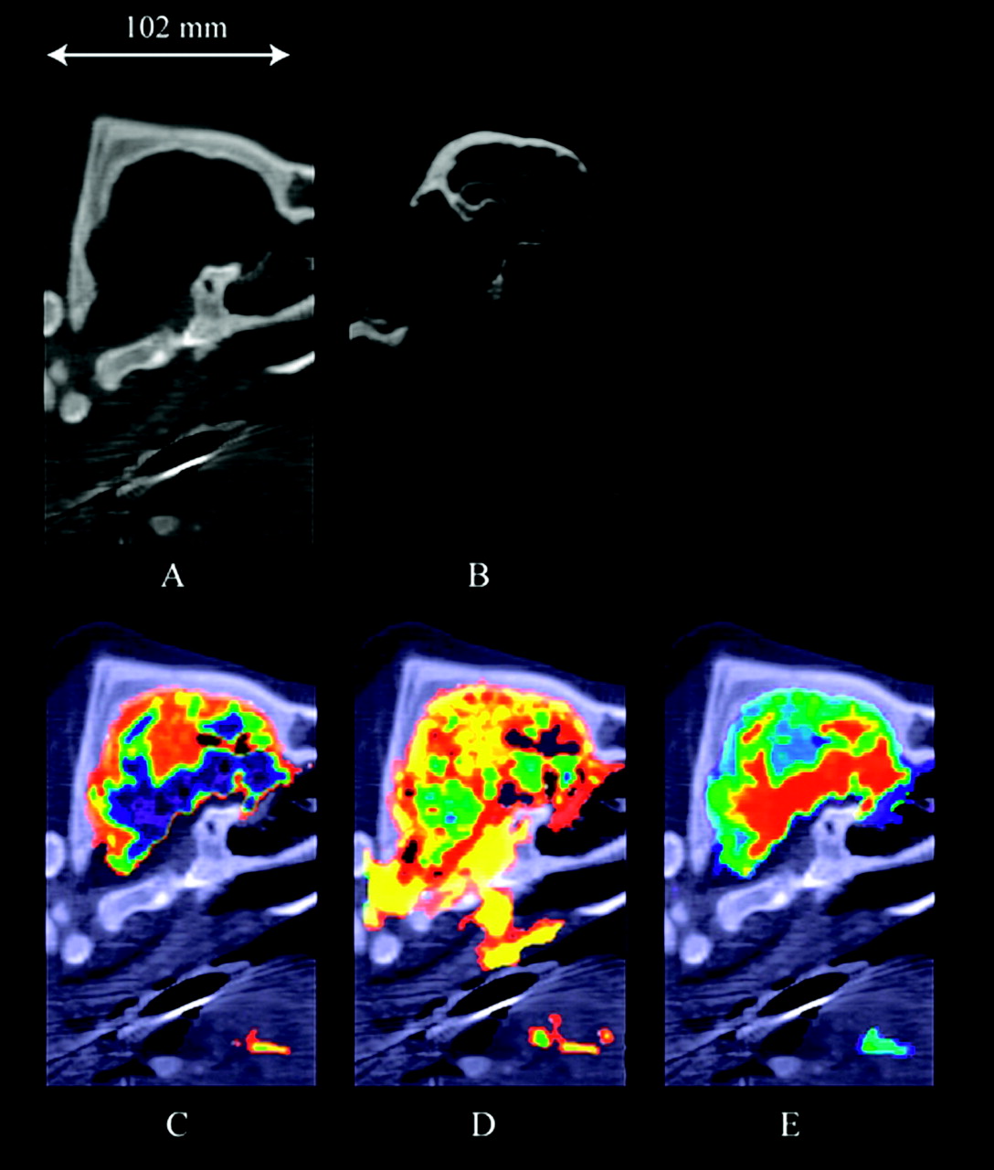
  • Fig 2.
    • Download figure
    • Open in new tab
    • Download powerpoint
    Fig 2.

    Results for a healthy domestic pig (pig 1). A, Nonenhanced sagittal image with a 6-mm section thickness. B, CTA in the sagittal plane at 14 seconds after the start of contrast injection. Various color ramps, selected according to user preference, were used to display the sagittal perfusion CT maps of (C) CBF, (D) CBV, and (E) MTT. These perfusion CT maps show enhancement of the lingual artery and vein in addition to the brain.
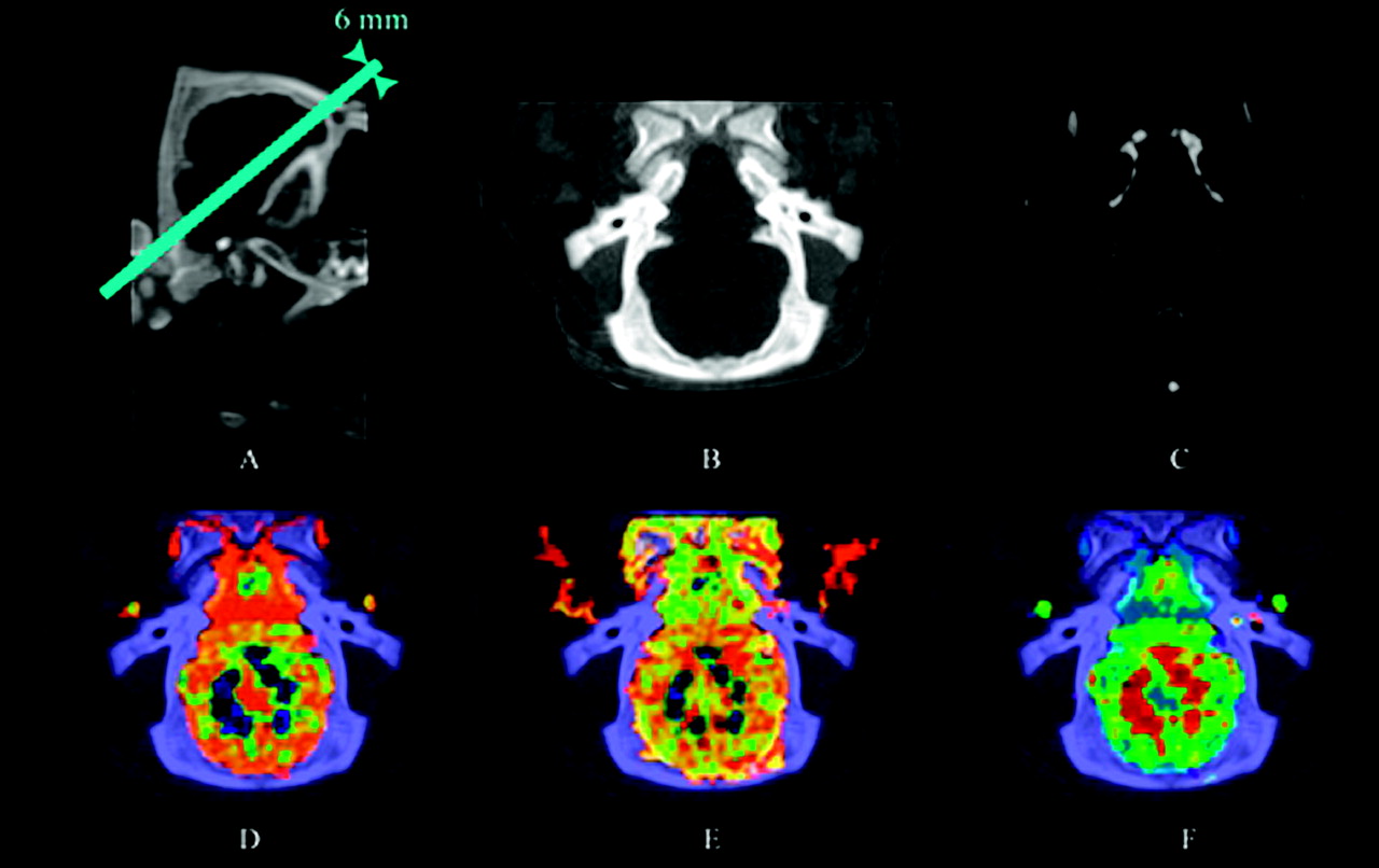
  • Fig 3.
    • Download figure
    • Open in new tab
    • Download powerpoint
    Fig 3.

    Results for a healthy domestic pig (pig 2). A, Nonenhanced sagittal image with a 6-mm section thickness. B, Oblique image at a 40° angle to the horizontal line shown in Fig 2A. C, CTA in an oblique plane at 14 seconds after the start of contrast injection. D, CBF. E, CBV. F, MTT.

PreviousNext
Back to top

In this issue

American Journal of Neuroradiology: 26 (10)
American Journal of Neuroradiology
Vol. 26, Issue 10
1 Nov 2005
  • Table of Contents
  • Index by author
Advertisement
Print
Download PDF
Email Article

Thank you for your interest in spreading the word on American Journal of Neuroradiology.

NOTE: We only request your email address so that the person you are recommending the page to knows that you wanted them to see it, and that it is not junk mail. We do not capture any email address.

Enter multiple addresses on separate lines or separate them with commas.
Volumetric Perfusion CT Using Prototype 256–Detector Row CT Scanner: Preliminary Study with Healthy Porcine Model
(Your Name) has sent you a message from American Journal of Neuroradiology
(Your Name) thought you would like to see the American Journal of Neuroradiology web site.
CAPTCHA
This question is for testing whether or not you are a human visitor and to prevent automated spam submissions.
Cite this article
Shinichiro Mori, Takayuki Obata, Naoshi Nakajima, Nobutsune Ichihara, Masahiro Endo
Volumetric Perfusion CT Using Prototype 256–Detector Row CT Scanner: Preliminary Study with Healthy Porcine Model
American Journal of Neuroradiology Nov 2005, 26 (10) 2536-2541;

Citation Manager Formats

  • BibTeX
  • Bookends
  • EasyBib
  • EndNote (tagged)
  • EndNote 8 (xml)
  • Medlars
  • Mendeley
  • Papers
  • RefWorks Tagged
  • Ref Manager
  • RIS
  • Zotero
0 Responses
Respond to this article
Share
Bookmark this article
Volumetric Perfusion CT Using Prototype 256–Detector Row CT Scanner: Preliminary Study with Healthy Porcine Model
Shinichiro Mori, Takayuki Obata, Naoshi Nakajima, Nobutsune Ichihara, Masahiro Endo
American Journal of Neuroradiology Nov 2005, 26 (10) 2536-2541;
del.icio.us logo Twitter logo Facebook logo Mendeley logo
  • Tweet Widget
  • Facebook Like
  • Google Plus One

Jump to section

  • Article
    • Abstract
    • Materials and Methods
    • Results
    • Discussion
    • Footnotes
    • References
  • Figures & Data
  • Supplemental
  • Info & Metrics
  • Responses
  • References
  • PDF

Related Articles

  • PubMed
  • Google Scholar

Cited By...

  • Focal Hypoperfusion in Acute Ischemic Stroke Perfusion CT: Clinical and Radiologic Predictors and Accuracy for Infarct Prediction
  • Evaluation of CT Perfusion in the Setting of Cerebral Ischemia: Patterns and Pitfalls
  • 320-slice CT neuroimaging: initial clinical experience and image quality evaluation
  • Conversion factor for CT dosimetry to assess patient dose using a 256-slice CT scanner
  • Crossref
  • Google Scholar

This article has not yet been cited by articles in journals that are participating in Crossref Cited-by Linking.

More in this TOC Section

  • Multimodal CT Provides Improved Performance for Lacunar Infarct Detection
  • Optimal MRI Sequence for Identifying Occlusion Location in Acute Stroke: Which Value of Time-Resolved Contrast-Enhanced MRA?
  • Evaluating the Effects of White Matter Multiple Sclerosis Lesions on the Volume Estimation of 6 Brain Tissue Segmentation Methods
Show more BRAIN

Similar Articles

Advertisement

Indexed Content

  • Current Issue
  • Accepted Manuscripts
  • Article Preview
  • Past Issues
  • Editorials
  • Editor's Choice
  • Fellows' Journal Club
  • Letters to the Editor
  • Video Articles

Cases

  • Case Collection
  • Archive - Case of the Week
  • Archive - Case of the Month
  • Archive - Classic Case

More from AJNR

  • Trainee Corner
  • Imaging Protocols
  • MRI Safety Corner
  • Book Reviews

Multimedia

  • AJNR Podcasts
  • AJNR Scantastics

Resources

  • Turnaround Time
  • Submit a Manuscript
  • Submit a Video Article
  • Submit an eLetter to the Editor/Response
  • Manuscript Submission Guidelines
  • Statistical Tips
  • Fast Publishing of Accepted Manuscripts
  • Graphical Abstract Preparation
  • Imaging Protocol Submission
  • Evidence-Based Medicine Level Guide
  • Publishing Checklists
  • Author Policies
  • Become a Reviewer/Academy of Reviewers
  • News and Updates

About Us

  • About AJNR
  • Editorial Board
  • Editorial Board Alumni
  • Alerts
  • Permissions
  • Not an AJNR Subscriber? Join Now
  • Advertise with Us
  • Librarian Resources
  • Feedback
  • Terms and Conditions
  • AJNR Editorial Board Alumni

American Society of Neuroradiology

  • Not an ASNR Member? Join Now

© 2025 by the American Society of Neuroradiology All rights, including for text and data mining, AI training, and similar technologies, are reserved.
Print ISSN: 0195-6108 Online ISSN: 1936-959X

Powered by HighWire