Advertisement

Main menu

  • Home
  • Content
    • Current Issue
    • Accepted Manuscripts
    • Article Preview
    • Past Issue Archive
    • Video Articles
    • AJNR Case Collection
    • Case of the Week Archive
    • Case of the Month Archive
    • Classic Case Archive
  • Special Collections
    • AJNR Awards
    • Low-Field MRI
    • Alzheimer Disease
    • ASNR Foundation Special Collection
    • Photon-Counting CT
    • View All
  • Multimedia
    • AJNR Podcasts
    • AJNR SCANtastic
    • Trainee Corner
    • MRI Safety Corner
    • Imaging Protocols
  • For Authors
    • Submit a Manuscript
    • Submit a Video Article
    • Submit an eLetter to the Editor/Response
    • Manuscript Submission Guidelines
    • Statistical Tips
    • Fast Publishing of Accepted Manuscripts
    • Graphical Abstract Preparation
    • Imaging Protocol Submission
    • Author Policies
  • About Us
    • About AJNR
    • Editorial Board
    • Editorial Board Alumni
  • More
    • Become a Reviewer/Academy of Reviewers
    • Subscribers
    • Permissions
    • Alerts
    • Feedback
    • Advertisers
    • ASNR Home

User menu

  • Alerts
  • Log in

Search

  • Advanced search
American Journal of Neuroradiology

American Journal of Neuroradiology

ASHNR American Society of Functional Neuroradiology ASHNR American Society of Pediatric Neuroradiology ASSR
  • Alerts
  • Log in

Advanced Search

  • Home
  • Content
    • Current Issue
    • Accepted Manuscripts
    • Article Preview
    • Past Issue Archive
    • Video Articles
    • AJNR Case Collection
    • Case of the Week Archive
    • Case of the Month Archive
    • Classic Case Archive
  • Special Collections
    • AJNR Awards
    • Low-Field MRI
    • Alzheimer Disease
    • ASNR Foundation Special Collection
    • Photon-Counting CT
    • View All
  • Multimedia
    • AJNR Podcasts
    • AJNR SCANtastic
    • Trainee Corner
    • MRI Safety Corner
    • Imaging Protocols
  • For Authors
    • Submit a Manuscript
    • Submit a Video Article
    • Submit an eLetter to the Editor/Response
    • Manuscript Submission Guidelines
    • Statistical Tips
    • Fast Publishing of Accepted Manuscripts
    • Graphical Abstract Preparation
    • Imaging Protocol Submission
    • Author Policies
  • About Us
    • About AJNR
    • Editorial Board
    • Editorial Board Alumni
  • More
    • Become a Reviewer/Academy of Reviewers
    • Subscribers
    • Permissions
    • Alerts
    • Feedback
    • Advertisers
    • ASNR Home
  • Follow AJNR on Twitter
  • Visit AJNR on Facebook
  • Follow AJNR on Instagram
  • Join AJNR on LinkedIn
  • RSS Feeds

AJNR Awards, New Junior Editors, and more. Read the latest AJNR updates

OtherBRAIN

Unique Anomalous Origin of the Left Anterior Cerebral Artery

Nicole S. Burbank and P. Pearse Morris
American Journal of Neuroradiology November 2005, 26 (10) 2533-2535;
Nicole S. Burbank
  • Find this author on Google Scholar
  • Find this author on PubMed
  • Search for this author on this site
P. Pearse Morris
  • Find this author on Google Scholar
  • Find this author on PubMed
  • Search for this author on this site
  • Article
  • Figures & Data
  • Info & Metrics
  • Responses
  • References
  • PDF
Loading

Abstract

Summary: We present the case of an anomalous origin of the left anterior cerebral artery (ACA) from the supraclinoid segment of the right internal carotid artery. Because of improved imaging quality, anomalies of the ACA–anterior communicating artery (AComA) complex are increasingly recognized on transaxial images. Although most of these anomalies are incidental findings of little clinical significance, some ACA-AComA complex anomalies are clinically significant. Recognition of these anomalies may be instrumental in developing a differential diagnosis or for improved surgical planning.

A 2-year-old infant was admitted to the hospital for evaluation and management of new-onset seizures. She had a known history of Chiari II malformation and spina bifida with related hydrocephalus, for which she was treated with a ventriculoperitoneal shunt. A head CT was obtained to evaluate for possible shunt dysfunction. The head CT demonstrated diffuse hypoattenuation of the left cerebral hemisphere. Therefore, MR imaging of the brain was performed to verify left cerebral ischemia.

Case Report

Diffusion-weighted MR images showed decreased perfusion to the entire left cerebral hemisphere consistent with infarction. In light of this distribution, cerebral MR angiography (MRA) was also performed to visualize the anterior and posterior cerebral vasculature. The MRA demonstrated an anomalous origin of the left anterior cerebral artery (ACA) from the supraclinoid segment of the right internal carotid artery (ICA; Figs 1 and 2). More specifically, the origin of the left A1 segment was adjacent to, but separate from, the origin of the right ophthalmic artery. The anomalous segment coursed anterior to the optic chiasm before running close to the A1 segment of the right ACA to form the anterior communicating artery (AComA; Fig 3). In view of the anomalous origin of the left A1 segment and the normal perfusion pattern of the territories supplied by the right ICA (including the left ACA territory), a vascular etiology for the left hemispheric infarct was much less likely.

Fig 1.
  • Download figure
  • Open in new tab
  • Download powerpoint
Fig 1.

Anteroposterior 3D time-of-flight MRA of the right ICA distribution demonstrates the origin of the left A1 segment (L ACA) near the origin of the ophthalmic artery. R ACA, right anterior cerebral artery; R MCA, right middle cerebral artery.

Fig 2.
  • Download figure
  • Open in new tab
  • Download powerpoint
Fig 2.

Anteroposterior 3D time-of-flight MRA of the left ICA distribution demonstrates the absence of the left ACA from the left ICA. L MCA, left middle cerebral artery.

Fig 3.
  • Download figure
  • Open in new tab
  • Download powerpoint
Fig 3.

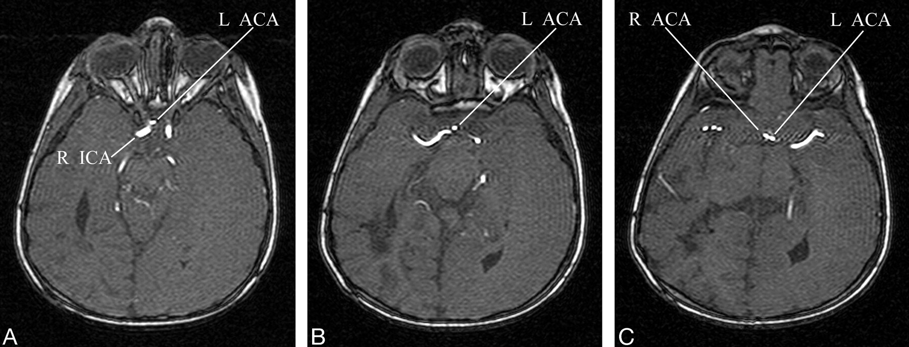
A–C, Selected axial images of the 3D time-of-flight MRA demonstrates the paraophthalmic origin of the left A1 segment (L ACA) from the right ICA. The anomalous vessel loops anterior to the optic chiasm before ascending to join the AComA.

On the basis of the imaging findings, we proposed that the cause of the left-sided cerebral infarct was prolonged seizure activity that was confined to that hemisphere by lack of the corpus callosum. Evaluation for other etiologies by echocardiogram, carotid duplex examination, ventriculoperitoneal shunt evaluation, and laboratory tests for infection and coagulopathy was unrevealing. Medical management of the seizure activity was optimized, and the patient began to receive baclofen to reduce right-sided spasticity. At the time of discharge, there was a high likelihood of left-sided cortical blindness, but the patient’s overall status continued to improve with rehabilitation.

Discussion

Padget described 8 stages in the development of the cerebral arteries, including the adult configuration (1). The first 7 stages occur during embryogenesis in the first trimester. When the embryo is approximately 40 mm in length (estimated ovulation age 52 days), cerebrovascular development corresponds with the seventh stage of development. At this stage, the circle of Willis is recognizable and the stems of all the cerebral arteries attain their adult configuration (1). In the final embryonic period of cerebrovascular development, the vessels branch in accordance with the developing parenchyma (1). This pattern of development has been termed “cerebrovascular ontogenic plasticity” (2). It is postulated that anomalous arteries and vascular malformations develop during this period (2).

In the adult configuration, the A1 segment usually arises from the ICA bifurcation. It then extends anteromedially toward the interhemispheric fissure, following a supraoptic course above the ipsilateral optic nerve or chiasm to join the AComA. Variation within the ACA-AComA complex is extremely common: some degree of asymmetry between the 2 A1 segments is identified in approximately 80% of patients (3). The most frequently reported anomalies include aplasia or hypoplasia of the A1 segment, fenestration or duplication of the AComA, and presence of 3 distal ACAs (4).

An anomalous origin of the ACA from the contralateral ICA is very rare. In addition to this case report, there has been only one other report of an anomalous origin of the ACA from the contralateral ICA (5). These cases differ in the course of the anomalous vessel as it ascends to its normal position.

Though still quite rare, an anomalous origin of the ACA from the paraclinoidal segment of the ipsilateral ICA is more common. There have been approximately 30 case reports of an infraoptic course of the A1 segment, which is associated with a low bifurcation of the ICA at or near the origin of the ophthalmic artery (4). One case of an anomalous A1 originating proximal to the ophthalmic artery has been reported (6). Anomalies with an infraoptic course are often associated with agenesis or hypoplasia of the contralateral A1. There are numerous variations in the ascent of the infraoptic A1 segment to the optic apparatus; most frequently, the infraoptic ACA ascends anterior to the chiasm between the optic nerves or pierces the ipsilateral optic nerve chiasm (3). A cavernous origin of the A1 segment has also been described (2).

Although these anomalies are frequently normal variants found incidentally during diagnostic studies, the incidence of coexistent abnormalities is higher in these patients. Specifically, the prevalence of cerebral aneurysms and arteriovenous malformations is increased in this patient population (2, 6). Cerebrovascular ontogenic plasticity theory suggests altered blood flow dynamics during embryogenesis may account for these coexistent anomalies. Other reported associated vascular anomalies include agenesis of the ICA, anomalous origin of the ipsilateral ophthalmic artery from the external circulation, fused pericallosal arteries, Moya-Moya disease, and symptoms related to compression of the optic apparatus.

Recognition of these anomalies is crucial when planning surgical or endovascular treatment of aneurysms. Characterization of these anomalies can be pivotal when developing a differential diagnosis, as shown in this case.

References

  1. ↵
    Padget DH. The development of the cranial arteries in the human embryo. Contrib Embryol 1948;32:207–262
  2. ↵
    Singer RJ, Abe T, Taylor WH, et al. Intracavernous anterior cerebral artery origin with associated arteriovenous malformations: a developmental analysis: case report. Neurosurgery 1997;40:829–831
    PubMed
  3. ↵
    Maurer J, Maurer E, Perneczky A. Surgically verified variations in the A1 segment of the anterior cerebral artery; report of two cases. J Neurosurg 1991;75:950–953
    PubMed
  4. ↵
    Spinnato S, Pasqualin A, Chioffi F, Da Pian R. Infraoptic course of the anterior cerebral artery associated with an anterior communicating artery aneurysm: anatomic case report and embryological considerations. Neurosurgery 1999;44:1315–1319
    CrossRefPubMed
  5. ↵
    Sincoff EH, Abdulrauf SI. Anomalous origin of the left A1 segment from the right internal carotid artery. J Neurosurg 2003;99:1101
    PubMed
  6. ↵
    Given CA, Morris PP. Recognition and importance of an infraoptic anterior cerebral artery: case report. AJNR Am J Neuroradiol 2002;23:452–454
    Abstract/FREE Full Text
  • Received February 15, 2005.
  • Accepted after revision March 25, 2005.
  • Copyright © American Society of Neuroradiology
View Abstract
PreviousNext
Back to top

In this issue

American Journal of Neuroradiology: 26 (10)
American Journal of Neuroradiology
Vol. 26, Issue 10
1 Nov 2005
  • Table of Contents
  • Index by author
Advertisement
Print
Download PDF
Email Article

Thank you for your interest in spreading the word on American Journal of Neuroradiology.

NOTE: We only request your email address so that the person you are recommending the page to knows that you wanted them to see it, and that it is not junk mail. We do not capture any email address.

Enter multiple addresses on separate lines or separate them with commas.
Unique Anomalous Origin of the Left Anterior Cerebral Artery
(Your Name) has sent you a message from American Journal of Neuroradiology
(Your Name) thought you would like to see the American Journal of Neuroradiology web site.
CAPTCHA
This question is for testing whether or not you are a human visitor and to prevent automated spam submissions.
Cite this article
Nicole S. Burbank, P. Pearse Morris
Unique Anomalous Origin of the Left Anterior Cerebral Artery
American Journal of Neuroradiology Nov 2005, 26 (10) 2533-2535;

Citation Manager Formats

  • BibTeX
  • Bookends
  • EasyBib
  • EndNote (tagged)
  • EndNote 8 (xml)
  • Medlars
  • Mendeley
  • Papers
  • RefWorks Tagged
  • Ref Manager
  • RIS
  • Zotero
0 Responses
Respond to this article
Share
Bookmark this article
Unique Anomalous Origin of the Left Anterior Cerebral Artery
Nicole S. Burbank, P. Pearse Morris
American Journal of Neuroradiology Nov 2005, 26 (10) 2533-2535;
del.icio.us logo Twitter logo Facebook logo Mendeley logo
  • Tweet Widget
  • Facebook Like
  • Google Plus One

Jump to section

  • Article
    • Abstract
    • Case Report
    • Discussion
    • References
  • Figures & Data
  • Info & Metrics
  • Responses
  • References
  • PDF

Related Articles

  • No related articles found.
  • PubMed
  • Google Scholar

Cited By...

  • No citing articles found.
  • Crossref
  • Google Scholar

This article has not yet been cited by articles in journals that are participating in Crossref Cited-by Linking.

More in this TOC Section

  • Multimodal CT Provides Improved Performance for Lacunar Infarct Detection
  • Optimal MRI Sequence for Identifying Occlusion Location in Acute Stroke: Which Value of Time-Resolved Contrast-Enhanced MRA?
  • Evaluating the Effects of White Matter Multiple Sclerosis Lesions on the Volume Estimation of 6 Brain Tissue Segmentation Methods
Show more BRAIN

Similar Articles

Advertisement

Indexed Content

  • Current Issue
  • Accepted Manuscripts
  • Article Preview
  • Past Issues
  • Editorials
  • Editor's Choice
  • Fellows' Journal Club
  • Letters to the Editor
  • Video Articles

Cases

  • Case Collection
  • Archive - Case of the Week
  • Archive - Case of the Month
  • Archive - Classic Case

More from AJNR

  • Trainee Corner
  • Imaging Protocols
  • MRI Safety Corner
  • Book Reviews

Multimedia

  • AJNR Podcasts
  • AJNR Scantastics

Resources

  • Turnaround Time
  • Submit a Manuscript
  • Submit a Video Article
  • Submit an eLetter to the Editor/Response
  • Manuscript Submission Guidelines
  • Statistical Tips
  • Fast Publishing of Accepted Manuscripts
  • Graphical Abstract Preparation
  • Imaging Protocol Submission
  • Evidence-Based Medicine Level Guide
  • Publishing Checklists
  • Author Policies
  • Become a Reviewer/Academy of Reviewers
  • News and Updates

About Us

  • About AJNR
  • Editorial Board
  • Editorial Board Alumni
  • Alerts
  • Permissions
  • Not an AJNR Subscriber? Join Now
  • Advertise with Us
  • Librarian Resources
  • Feedback
  • Terms and Conditions
  • AJNR Editorial Board Alumni

American Society of Neuroradiology

  • Not an ASNR Member? Join Now

© 2025 by the American Society of Neuroradiology All rights, including for text and data mining, AI training, and similar technologies, are reserved.
Print ISSN: 0195-6108 Online ISSN: 1936-959X

Powered by HighWire