Advertisement

Main menu

  • Home
  • Content
    • Current Issue
    • Accepted Manuscripts
    • Article Preview
    • Past Issue Archive
    • Video Articles
    • AJNR Case Collection
    • Case of the Week Archive
    • Case of the Month Archive
    • Classic Case Archive
  • Special Collections
    • Advancing NeuroMRI with High-Relaxivity Contrast Agents
    • AJNR Awards
    • Low-Field MRI
    • Alzheimer Disease
    • ASNR Foundation Special Collection
    • Photon-Counting CT
    • View All
  • Multimedia
    • AJNR Podcasts
    • AJNR SCANtastic
    • Trainee Corner
    • MRI Safety Corner
    • Imaging Protocols
  • For Authors
    • Submit a Manuscript
    • Submit a Video Article
    • Submit an eLetter to the Editor/Response
    • Manuscript Submission Guidelines
    • Statistical Tips
    • Fast Publishing of Accepted Manuscripts
    • Graphical Abstract Preparation
    • Imaging Protocol Submission
    • Author Policies
  • About Us
    • About AJNR
    • Editorial Board
    • Editorial Board Alumni
  • More
    • Become a Reviewer/Academy of Reviewers
    • Subscribers
    • Permissions
    • Alerts
    • Feedback
    • Advertisers
    • ASNR Home

User menu

  • Alerts
  • Log in

Search

  • Advanced search
American Journal of Neuroradiology

American Journal of Neuroradiology

ASHNR American Society of Functional Neuroradiology ASHNR American Society of Pediatric Neuroradiology ASSR
  • Alerts
  • Log in

Advanced Search

  • Home
  • Content
    • Current Issue
    • Accepted Manuscripts
    • Article Preview
    • Past Issue Archive
    • Video Articles
    • AJNR Case Collection
    • Case of the Week Archive
    • Case of the Month Archive
    • Classic Case Archive
  • Special Collections
    • Advancing NeuroMRI with High-Relaxivity Contrast Agents
    • AJNR Awards
    • Low-Field MRI
    • Alzheimer Disease
    • ASNR Foundation Special Collection
    • Photon-Counting CT
    • View All
  • Multimedia
    • AJNR Podcasts
    • AJNR SCANtastic
    • Trainee Corner
    • MRI Safety Corner
    • Imaging Protocols
  • For Authors
    • Submit a Manuscript
    • Submit a Video Article
    • Submit an eLetter to the Editor/Response
    • Manuscript Submission Guidelines
    • Statistical Tips
    • Fast Publishing of Accepted Manuscripts
    • Graphical Abstract Preparation
    • Imaging Protocol Submission
    • Author Policies
  • About Us
    • About AJNR
    • Editorial Board
    • Editorial Board Alumni
  • More
    • Become a Reviewer/Academy of Reviewers
    • Subscribers
    • Permissions
    • Alerts
    • Feedback
    • Advertisers
    • ASNR Home
  • Follow AJNR on Twitter
  • Visit AJNR on Facebook
  • Follow AJNR on Instagram
  • Join AJNR on LinkedIn
  • RSS Feeds

AJNR Awards, New Junior Editors, and more. Read the latest AJNR updates


Improved Turnaround Times | Median time to first decision: 12 days

OtherBrain

Serial Evaluation of Diffusion Tensor Brain Fiber Tracking in a Patient with Severe Diffuse Axonal Injury

Shinji Naganawa, Chiho Sato, Shunichi Ishihra, Hisashi Kumada, Takeo Ishigaki, Shunichi Miura, Masao Watanabe, Katsuya Maruyama and Osamu Takizawa
American Journal of Neuroradiology October 2004, 25 (9) 1553-1556;
Shinji Naganawa
  • Find this author on Google Scholar
  • Find this author on PubMed
  • Search for this author on this site
Chiho Sato
  • Find this author on Google Scholar
  • Find this author on PubMed
  • Search for this author on this site
Shunichi Ishihra
  • Find this author on Google Scholar
  • Find this author on PubMed
  • Search for this author on this site
Hisashi Kumada
  • Find this author on Google Scholar
  • Find this author on PubMed
  • Search for this author on this site
Takeo Ishigaki
  • Find this author on Google Scholar
  • Find this author on PubMed
  • Search for this author on this site
Shunichi Miura
  • Find this author on Google Scholar
  • Find this author on PubMed
  • Search for this author on this site
Masao Watanabe
  • Find this author on Google Scholar
  • Find this author on PubMed
  • Search for this author on this site
Katsuya Maruyama
  • Find this author on Google Scholar
  • Find this author on PubMed
  • Search for this author on this site
Osamu Takizawa
  • Find this author on Google Scholar
  • Find this author on PubMed
  • Search for this author on this site
  • Article
  • Figures & Data
  • Info & Metrics
  • Responses
  • References
  • PDF
Loading

Article Figures & Data

Figures

  • Fig 1.
    • Download figure
    • Open in new tab
    • Download powerpoint
    Fig 1.

    A 27-year-old woman 4 days after a traffic accident.

    A, Axial T1-weighted image shows the intraventricular hemorrhage in left lateral ventricule (bold arrow) and right subdural effusion. A small hemorrhage in the corpus callosum is seen (thin arrow).

    B, Sagittal trace DW image (b = 1000) shows areas of high signal intensity in corpus callosum (thin arrow), especially in the splenium (bold arrow).

    C, Axial echo planar T2*-weighted image (b = 0 image from DTI sequence) shows a small hemorrhage in the corpus callosum as a low signal intensity area (arrow).

    D, Axial FLAIR image shows high signal intensity area in the corpus callosum (arrows). No significant damage is seen in other white matter area.

    E and F, Diffusion tensor fiber tracking from the seed area around the corpus callosum. Fibers are overlayed on FA map. The view from right side of the patient shows some fibers extending up to upper frontal and parietal white matter (E, arrows). On the frontal view, upward fibers in the left side are not seen (F, arrow).

  • Fig 2.
    • Download figure
    • Open in new tab
    • Download powerpoint
    Fig 2.

    A 27-year-old woman 24 days after a traffic accident.

    A, Axial T1-weighted image show slight enlargement of the lateral ventricules and right subdural bloody effusion (thin arrow).

    B, Sagittal trace DW image (b = 1000) shows that the areas of strong high signal intensity in the corpus callosum have moved anteriorly compared with the first MR image (thin arrow).

    C, Axial echo planar T2*-weighted image (b = 0 image from DTI sequence) shows no significant hemorrhage in the corpus callosum as a low signal intensity area (arrow).

    D, Axial FLAIR image shows high signal intensity area in the anterior part of the corpus callosum (arrows). No significant damage is seen in other white matter areas.

    E and F, Diffusion tensor fiber tracking from the seed area around the corpus callosum is shown. Fibers are overlayed on FA map. The view from right side of the patient shows almost no fibers extending up to the upper frontal and parietal white matter (arrows, E). On the frontal view, upward fibers in the right side have almost disappeared (arrow, F).
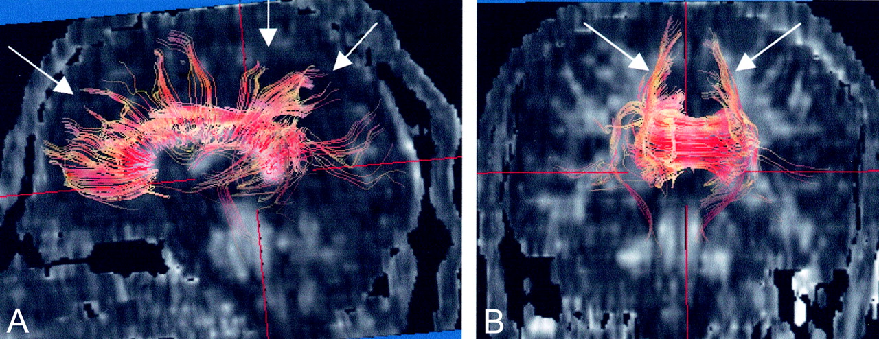
  • Fig 3.
    • Download figure
    • Open in new tab
    • Download powerpoint
    Fig 3.

    A 23-year-old male healthy volunteer for reference.

    Diffusion tensor fiber tracking from the seed area around the corpus callosum. Fibers are overlayed on an FA map. The view from right side of the volunteer shows many fibers extending upward to frontal and parietal white matter from the corpus callosum (arrows, A). On the frontal view, upward fibers in the both side were visualized (arrows, B).

PreviousNext
Back to top

In this issue

American Journal of Neuroradiology: 25 (9)
American Journal of Neuroradiology
Vol. 25, Issue 9
1 Oct 2004
  • Table of Contents
  • Index by author
Advertisement
Print
Download PDF
Email Article

Thank you for your interest in spreading the word on American Journal of Neuroradiology.

NOTE: We only request your email address so that the person you are recommending the page to knows that you wanted them to see it, and that it is not junk mail. We do not capture any email address.

Enter multiple addresses on separate lines or separate them with commas.
Serial Evaluation of Diffusion Tensor Brain Fiber Tracking in a Patient with Severe Diffuse Axonal Injury
(Your Name) has sent you a message from American Journal of Neuroradiology
(Your Name) thought you would like to see the American Journal of Neuroradiology web site.
CAPTCHA
This question is for testing whether or not you are a human visitor and to prevent automated spam submissions.
Cite this article
Shinji Naganawa, Chiho Sato, Shunichi Ishihra, Hisashi Kumada, Takeo Ishigaki, Shunichi Miura, Masao Watanabe, Katsuya Maruyama, Osamu Takizawa
Serial Evaluation of Diffusion Tensor Brain Fiber Tracking in a Patient with Severe Diffuse Axonal Injury
American Journal of Neuroradiology Oct 2004, 25 (9) 1553-1556;

Citation Manager Formats

  • BibTeX
  • Bookends
  • EasyBib
  • EndNote (tagged)
  • EndNote 8 (xml)
  • Medlars
  • Mendeley
  • Papers
  • RefWorks Tagged
  • Ref Manager
  • RIS
  • Zotero
0 Responses
Respond to this article
Share
Bookmark this article
Serial Evaluation of Diffusion Tensor Brain Fiber Tracking in a Patient with Severe Diffuse Axonal Injury
Shinji Naganawa, Chiho Sato, Shunichi Ishihra, Hisashi Kumada, Takeo Ishigaki, Shunichi Miura, Masao Watanabe, Katsuya Maruyama, Osamu Takizawa
American Journal of Neuroradiology Oct 2004, 25 (9) 1553-1556;
del.icio.us logo Twitter logo Facebook logo Mendeley logo
  • Tweet Widget
  • Facebook Like
  • Google Plus One

Jump to section

  • Article
    • Abstract
    • Case Report
    • Discussion
    • Conclusion
    • References
  • Figures & Data
  • Info & Metrics
  • Responses
  • References
  • PDF

Related Articles

  • No related articles found.
  • PubMed
  • Google Scholar

Cited By...

  • Diffusion Tensor Imaging Reliably Detects Experimental Traumatic Axonal Injury and Indicates Approximate Time of Injury
  • Crossref
  • Google Scholar

This article has not yet been cited by articles in journals that are participating in Crossref Cited-by Linking.

More in this TOC Section

  • Usefulness of Quantitative Susceptibility Mapping for the Diagnosis of Parkinson Disease
  • White Matter Alterations in the Brains of Patients with Active, Remitted, and Cured Cushing Syndrome: A DTI Study
  • Qualitative and Quantitative Analysis of MR Imaging Findings in Patients with Middle Cerebral Artery Stroke Implanted with Mesenchymal Stem Cells
Show more Brain

Similar Articles

Advertisement

Indexed Content

  • Current Issue
  • Accepted Manuscripts
  • Article Preview
  • Past Issues
  • Editorials
  • Editor's Choice
  • Fellows' Journal Club
  • Letters to the Editor
  • Video Articles

Cases

  • Case Collection
  • Archive - Case of the Week
  • Archive - Case of the Month
  • Archive - Classic Case

More from AJNR

  • Trainee Corner
  • Imaging Protocols
  • MRI Safety Corner
  • Book Reviews

Multimedia

  • AJNR Podcasts
  • AJNR Scantastics

Resources

  • Turnaround Time
  • Submit a Manuscript
  • Submit a Video Article
  • Submit an eLetter to the Editor/Response
  • Manuscript Submission Guidelines
  • Statistical Tips
  • Fast Publishing of Accepted Manuscripts
  • Graphical Abstract Preparation
  • Imaging Protocol Submission
  • Evidence-Based Medicine Level Guide
  • Publishing Checklists
  • Author Policies
  • Become a Reviewer/Academy of Reviewers
  • News and Updates

About Us

  • About AJNR
  • Editorial Board
  • Editorial Board Alumni
  • Alerts
  • Permissions
  • Not an AJNR Subscriber? Join Now
  • Advertise with Us
  • Librarian Resources
  • Feedback
  • Terms and Conditions
  • AJNR Editorial Board Alumni

American Society of Neuroradiology

  • Not an ASNR Member? Join Now

© 2025 by the American Society of Neuroradiology All rights, including for text and data mining, AI training, and similar technologies, are reserved.
Print ISSN: 0195-6108 Online ISSN: 1936-959X

Powered by HighWire