
Article Figures & Data
Figures
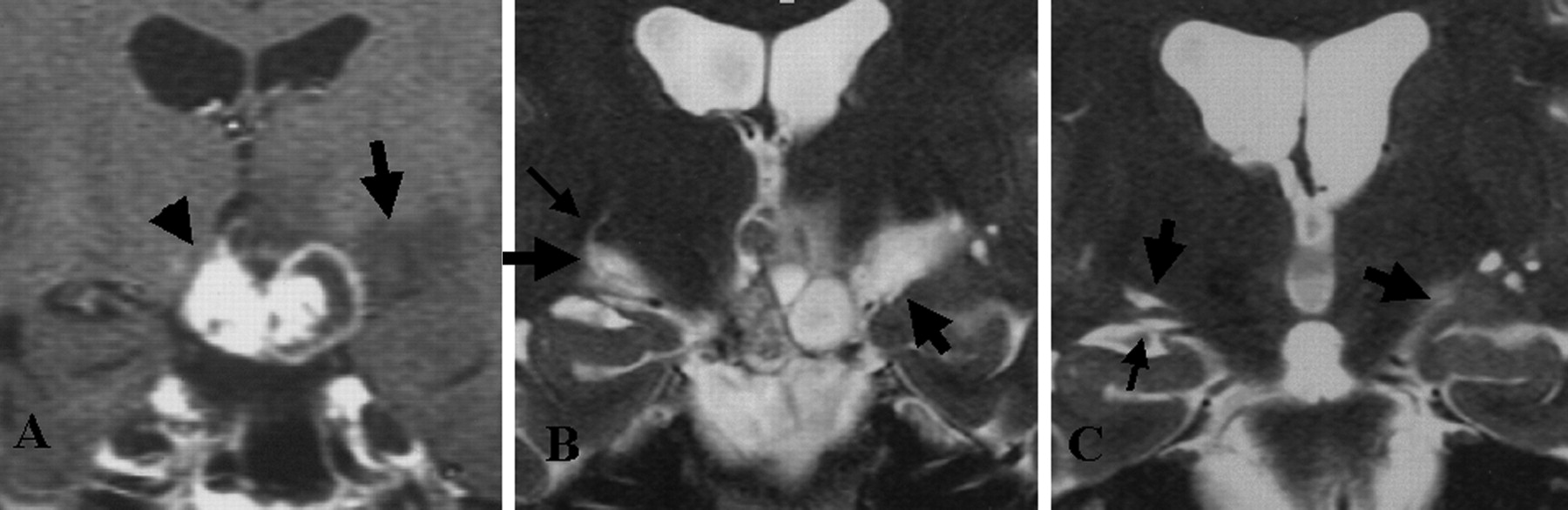
- Fig 1.
Representative MR images show an edema along the OT in a 33-year-old woman with craniopharyngioma (reproduced from reference 8 with the permission of the ©American Society of Neuroradiology).
A, T1-weighted contrast-enhanced coronal MR image before treatment. High- and low-signal-intensity mass (arrowhead) is visible at the suprasellar cistern. Low-signal-intensity edema (arrow) is noted along the left OT.
B, Heavily T2-weighted coronal MR image before treatment shows bilateral edemas (thick arrows) along the OT. The left side is more prominent. The OTs are difficult to differentiate from the edemas. On the right side, a curvilinear area of high signal intensity (thin arrow) originates from the edema.
C, On this heavily T2-weighted coronal MR image obtained 3 months after surgery (similar level as in B), the edema disappears on the right side. A large PV space (left thick arrow), present in normal conditions, is visible along the right OT (thin arrow). Edema (right thick arrow) remains on the left side.
- Fig 2.
Schematic representation of the anatomy in an 85-year-old man shows the basal view of the OT and surrounding brain tissue in the left hemisphere. The whole length of the OT is exposed by removing the left temporal lobe. The tract is divided into three equal-sized portions: anterior, middle, and posterior. Numerous perforation points are present medial to (thick arrows) and through (thin arrows) the OT. These are mainly located in the middle and posterior portions of the OT.
- Fig 3.
Histologic samples obtained in a 79-year-old woman.
A, Coronal section of the middle portion of the OT shows the cerebral peduncle (CP) (hematoxylin-eosin stain, original magnification ×20) Along the OT, a large 1.5-mm-high space has a pial layer (thin arrow) that lines its inner surface. Vessels (thick arrows) are present in the space, and no necrotic or ischemic changes are visible in the surrounding brain tissue. The histologic features are compatible with those of PV spaces around the anterior commissure shown in B. Small spaces (arrowheads) are present adjacent to the OT.
B, Coronal section at the anterior commissure (AC) and the lower basal ganglia shows multiple PV spaces (hematoxylin-eosin stain, original magnification × 40). Adjacent to the anterior commissure are multiple PV spaces with vessels lined by a pial layer (arrows). No necrotic or ischemic changes are visible in the surrounding brain tissue.
- Fig 4.
Histologic samples obtained in an 86-year-old woman.
A, Section shows lines corresponding to sections in B–D.
B–D, Continuous thin sections show a penetrating vessel (arrow) from the perforation point at the subarachnoid space (B) to the largest PV space adjacent to the OT (C and D). Its maximum height is 0.5 mm. Sections show the perforation point medial to the OT, which corresponds to the large arrows in Figure 2. Small PV spaces (arrowheads) are present (myelin staining, original magnification ×25). CP indicates cerebral peduncle.
- Fig 5.
Histologic samples obtained in an 81-year-old man. CP indicates cerebral peduncle.
A, Section shows lines corresponding to sections in B–F.
B–F, Continuous thin sections show a penetrating vessel from the perforation point in the subarachnoid space (B) to the largest PV space within the upper part of the OT (F). Its maximum height is 0.7 mm. Sections show the perforation point through the OT, which corresponds to the small arrows in Figure 2. A few small PV spaces (arrowheads) are visible along the OT (myelin staining, original magnification ×25). Bracket in E = 1 mm.
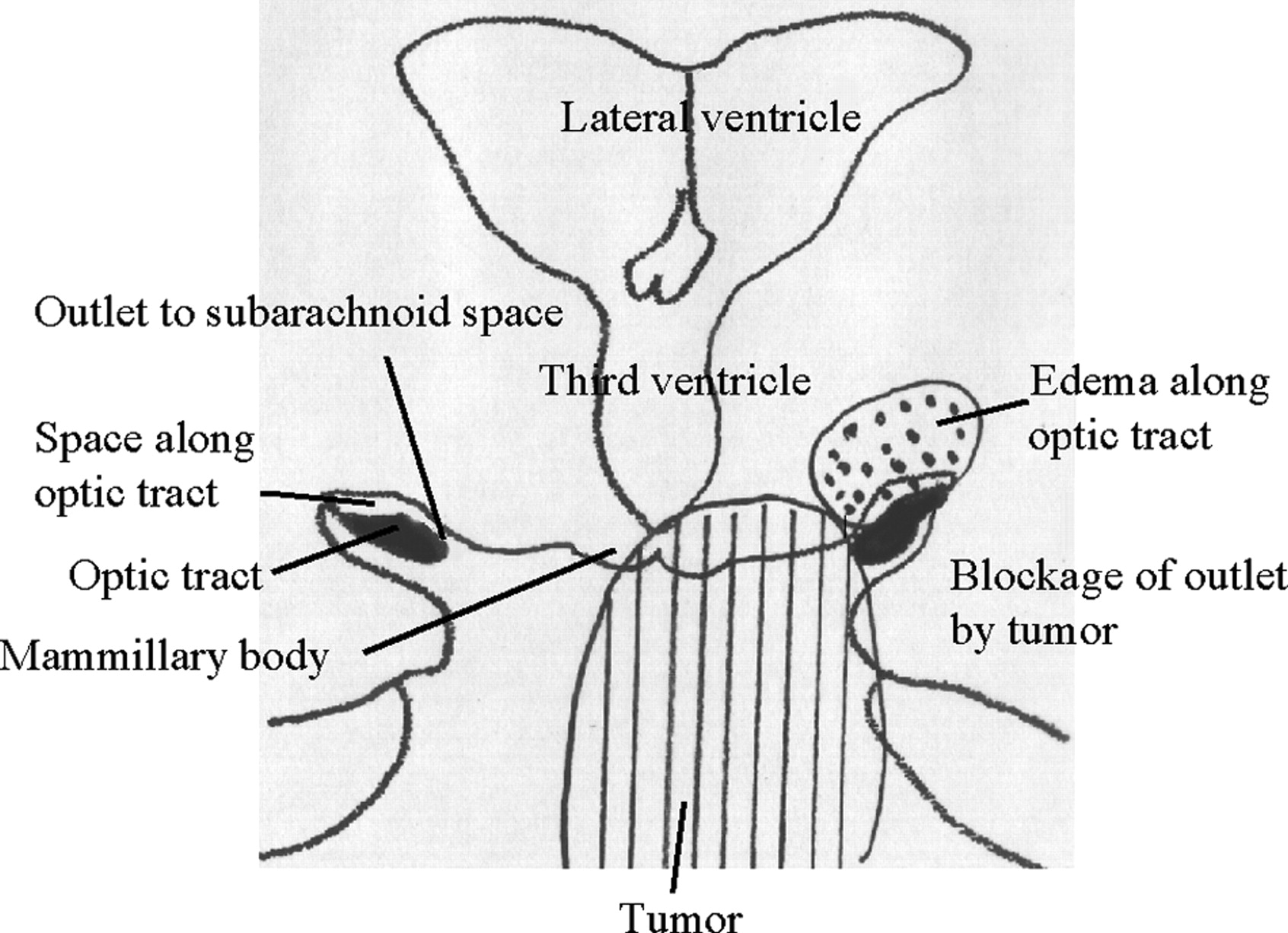
- Fig 6.
Schematic representation shows a possible new mechanism for edema along the OT in pituitary-region tumors. Right, In the normal anatomy, the PV space along the OT communicates with the adjacent subarachnoid space through a thin channel medial to the OT. Left, Tumor can block this channel with mechanical, inflammatory, or adhesive processes. The PV space retains interstitial fluid and distends along the OT (dotted area).


