Advertisement

Main menu

  • Home
  • Content
    • Current Issue
    • Accepted Manuscripts
    • Article Preview
    • Past Issue Archive
    • Video Articles
    • AJNR Case Collection
    • Case of the Week Archive
    • Case of the Month Archive
    • Classic Case Archive
  • Special Collections
    • AJNR Awards
    • Low-Field MRI
    • Alzheimer Disease
    • ASNR Foundation Special Collection
    • Photon-Counting CT
    • View All
  • Multimedia
    • AJNR Podcasts
    • AJNR SCANtastic
    • Trainee Corner
    • MRI Safety Corner
    • Imaging Protocols
  • For Authors
    • Submit a Manuscript
    • Submit a Video Article
    • Submit an eLetter to the Editor/Response
    • Manuscript Submission Guidelines
    • Statistical Tips
    • Fast Publishing of Accepted Manuscripts
    • Graphical Abstract Preparation
    • Imaging Protocol Submission
    • Author Policies
  • About Us
    • About AJNR
    • Editorial Board
    • Editorial Board Alumni
  • More
    • Become a Reviewer/Academy of Reviewers
    • Subscribers
    • Permissions
    • Alerts
    • Feedback
    • Advertisers
    • ASNR Home

User menu

  • Alerts
  • Log in

Search

  • Advanced search
American Journal of Neuroradiology

American Journal of Neuroradiology

ASHNR American Society of Functional Neuroradiology ASHNR American Society of Pediatric Neuroradiology ASSR
  • Alerts
  • Log in

Advanced Search

  • Home
  • Content
    • Current Issue
    • Accepted Manuscripts
    • Article Preview
    • Past Issue Archive
    • Video Articles
    • AJNR Case Collection
    • Case of the Week Archive
    • Case of the Month Archive
    • Classic Case Archive
  • Special Collections
    • AJNR Awards
    • Low-Field MRI
    • Alzheimer Disease
    • ASNR Foundation Special Collection
    • Photon-Counting CT
    • View All
  • Multimedia
    • AJNR Podcasts
    • AJNR SCANtastic
    • Trainee Corner
    • MRI Safety Corner
    • Imaging Protocols
  • For Authors
    • Submit a Manuscript
    • Submit a Video Article
    • Submit an eLetter to the Editor/Response
    • Manuscript Submission Guidelines
    • Statistical Tips
    • Fast Publishing of Accepted Manuscripts
    • Graphical Abstract Preparation
    • Imaging Protocol Submission
    • Author Policies
  • About Us
    • About AJNR
    • Editorial Board
    • Editorial Board Alumni
  • More
    • Become a Reviewer/Academy of Reviewers
    • Subscribers
    • Permissions
    • Alerts
    • Feedback
    • Advertisers
    • ASNR Home
  • Follow AJNR on Twitter
  • Visit AJNR on Facebook
  • Follow AJNR on Instagram
  • Join AJNR on LinkedIn
  • RSS Feeds

AJNR Awards, New Junior Editors, and more. Read the latest AJNR updates

OtherBRAIN

Leptomeningeal Metastasis from Malignant Pleural Mesothelioma

Boris D. Petrovic, Dusko B. Kozic, Robert R. Semnic, Mladen Prvulovic, Dragana Djilas-Ivanovic, R. Nuri Sener and Istvan Klem
American Journal of Neuroradiology August 2004, 25 (7) 1223-1224;
Boris D. Petrovic
  • Find this author on Google Scholar
  • Find this author on PubMed
  • Search for this author on this site
Dusko B. Kozic
  • Find this author on Google Scholar
  • Find this author on PubMed
  • Search for this author on this site
Robert R. Semnic
  • Find this author on Google Scholar
  • Find this author on PubMed
  • Search for this author on this site
Mladen Prvulovic
  • Find this author on Google Scholar
  • Find this author on PubMed
  • Search for this author on this site
Dragana Djilas-Ivanovic
  • Find this author on Google Scholar
  • Find this author on PubMed
  • Search for this author on this site
R. Nuri Sener
  • Find this author on Google Scholar
  • Find this author on PubMed
  • Search for this author on this site
Istvan Klem
  • Find this author on Google Scholar
  • Find this author on PubMed
  • Search for this author on this site
  • Article
  • Figures & Data
  • Info & Metrics
  • Responses
  • References
  • PDF
Loading

Abstract

Summary: Leptomeningeal metastasis from malignant mesothelioma is very rare; to our knowledge, only one imaging report exists in the literature. We present the case of widespread leptomeningeal lesions secondary to a malignant mesothelioma in a 61-year-old woman.

CNS involvement due to metastatic mesothelioma is very rare. The metastatic intracranial lesion may present as a solitary mass or as leptomeningeal infiltration (1–7). Leptomeningeal metastasis is extremely rare, and to our knowledge, only one report of the MR imaging findings of this condition exists in the literature (1). Herein, we believe we report the imaging findings in the second such reported case of this condition.

Case Report

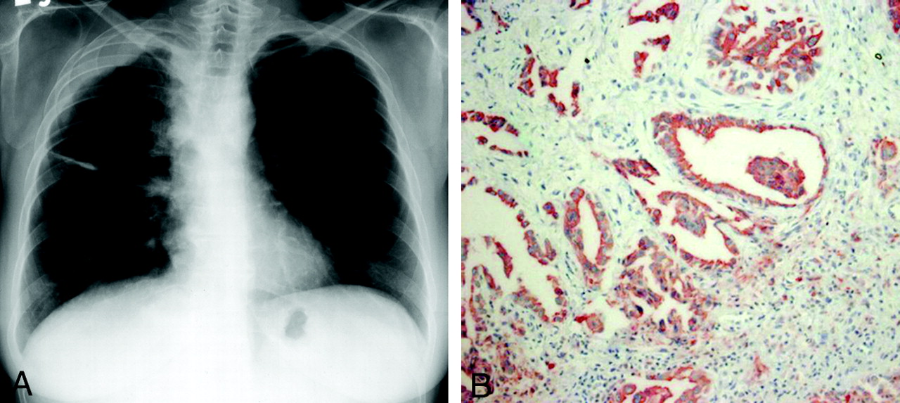
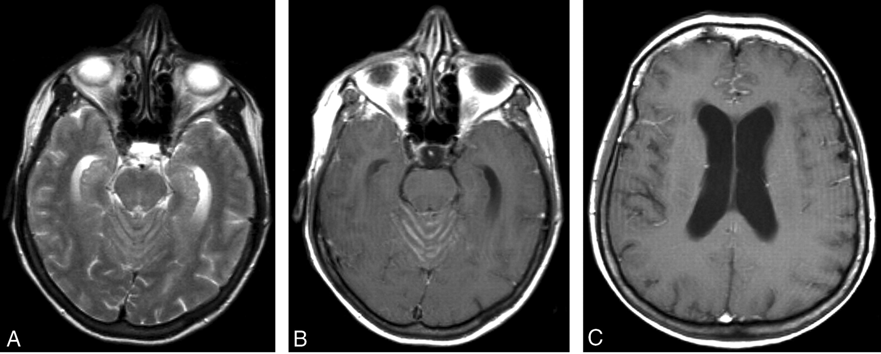
Malignant mesothelioma of the right pleura in a 61-year-old woman was confirmed by pleurolysis and biopsy (Fig 1A and B). Although she responded well to chemotherapy, 1 year later she was admitted to the hospital because of vomiting, for which she received symptomatic therapy. A year thereafter, she was admitted again with confusion and ataxia, at which time T2-weighted images revealed an impression of widened cerebellar sulci (Fig 2A). On postcontrast T1-weighted images, widespread leptomeningeal infiltrations were evident in the cerebellar and cerebral sulci. The lesions were more prominent in the cerebellar sulci compared with in the cerebral sulci. The third and lateral ventricles were dilated, which suggested development of acute hydrocephalus (Fig 2B and C). At that stage, findings of a chest radiograph and an abdominal sonogram were noted to be normal. Lumbar puncture revealed findings consistent with those of neoplastic infiltration.

Fig 1.
  • Download figure
  • Open in new tab
  • Download powerpoint
Fig 1.

Mesothelioma.

A, Conventional radiograph of the chest, revealing the mesothelioma in the right pleura.

B, Positive Calretinin reaction of the mesothelioma (tumor biopsy obtained by pleuroscopy; ×250 magnification).

Fig 2.
  • Download figure
  • Open in new tab
  • Download powerpoint
Fig 2.

Leptomeningeal metastasis.

A, T2-weighted image, revealing widening of vermian sulci.

B, Postcontrast T1-weighted image, revealing enhancement of the vermian sulci corresponding to leptomeningeal infiltration. Note dilatation of temporal horns.

C, Postcontrast T1-weighted image, revealing leptomeningeal enhancement in the frontal sulci. Lateral ventricular dilatation is noted.

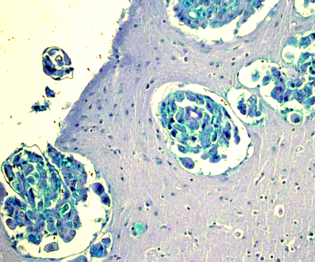
The patient died 30 days after the MR imaging examination. At autopsy, mesothelioma with infiltration of the lower lobe of the right lung was evident. Metastases to the mediastenial lymph nodes and micrometastases to the liver were noted. Leptomeningeal infiltration was confirmed by histologic analysis revealing presence of sarcomatous cells (Fig 3).

Fig 3.
  • Download figure
  • Open in new tab
  • Download powerpoint
Fig 3.

Leptomeningeal metastasis of the mesothelioma (Alcian blue staining; ×250 magnification).

Discussion

It has been reported that hematogenous metastases of malignant mesothelioma usually involves the liver, adrenal glands, kidneys, and the contralateral lung. Metastatic spread to the CNS is rare. Most of these had been encountered as masses in the brain parenchyma (2–6), and one case was noted with orbital metastasis (7). Falconieri et al (2) reported three autopsy cases of malignant pleural mesothelioma with brain metastases and provided a review of 15 similar previously published reports. They noted that in two patients, the brain metastases were discovered incidentally at autopsy. In one patient, the brain metastasis was discovered ante mortem, when a CT scan suggested a primary tumor of the brain. With respect to the histopathologic findings, the tumors had spindle-shaped malignant cells, pseudopalisading, necrosis, and vascular buds suggestive of glioblastoma multiforme (2). Other authors also had the impression that the metastatic tumors of malignant mesothelioma in the brain mimicked the pattern of glioblastoma multiforme both histologically and radiologically (2–6). On the other hand, leptomeningeal involvement is very rare. The only imaging report is by Oksuzoglu et al (1), who used MR imaging. Their patient was a 44-year-old woman who presented clinically with convulsions, and the diagnosis of malignant mesothelioma was confirmed by biopsies of lymph nodes (1).

It is known that the causes of leptomeningeal metastasis include adenocarcinomas originating from the lung, stomach, breast, ovary, malignant melanoma, leukemia, lymphoma, and primary CNS malignancies. Clinically, these usually present as a low-grade meningitis syndrome. Our patient also presented with such a syndrome in that she had confusion and ataxia, which was an indication for a cranial MR imaging examination. This revealed leptomeningeal metastases with contrast enhancement, more prominent in the cerebellar sulci, and changes consistent with acute hydrocephalus, which likely accounted for ataxia and confusion.

Conclusion

This patient reveals that, although very rare, leptomeningeal metastases can be associated with malignant mesothelioma of the pleura. In the event that these patients present clinically with a low-grade meningitis syndrome, brain MR imaging should be performed with administration of intravenous paramagnetic contrast medium to uncover lesions in the leptomeninges.

References

  1. ↵
    Oksuzoglu B, Yalcin S, Erman M, Dagdelen S. Leptomeningeal infiltration of malignant mesothelioma. Med Oncol 2002;19:167–169
    CrossRefPubMed
  2. ↵
    Falconieri G, Grandi G, DiBonito L, et al. Intracranial metastases from malignant pleural mesothelioma: report of three autopsy cases and review of the literature. Arch Pathol Lab Med 1991;115:591–595
    PubMed
  3. Kitai R, Kabuto M, Kawano H, et al. Brain metastasis from malignant mesothelioma: case report. Neurol Med Chir (Tokyo) 1995;35:172–174
    PubMed
  4. Kawai A, Nagasaka Y, Muraki M, et al. Brain metastasis in malignant pleural mesothelioma. Intern Med 1997;36:591–594
    PubMed
  5. Schwechheimer K, Butzengeiger M. Brain metastases in malignant fibrous mesothelioma: case report and review of the literature. Acta Neuropathol (Berl) 1983;60:301–304
    PubMed
  6. ↵
    Kobayashi S, Ida M, Matsui O, et al. Lipomatous change in a brain metastasis from malignant pleural mesothelioma. Neuroradiology 2001;43:159–161
    PubMed
  7. ↵
    Kubota K, Furuse K, Kawahara M, et al. A case of malignant pleural mesothelioma with metastasis to the orbit. Jpn J Clin Oncol 1996;26:469–471
    Abstract/FREE Full Text
  • Received November 6, 2003.
  • Accepted after revision November 20, 2003.
  • Copyright © American Society of Neuroradiology
View Abstract
PreviousNext
Back to top

In this issue

American Journal of Neuroradiology: 25 (7)
American Journal of Neuroradiology
Vol. 25, Issue 7
1 Aug 2004
  • Table of Contents
  • Index by author
Advertisement
Print
Download PDF
Email Article

Thank you for your interest in spreading the word on American Journal of Neuroradiology.

NOTE: We only request your email address so that the person you are recommending the page to knows that you wanted them to see it, and that it is not junk mail. We do not capture any email address.

Enter multiple addresses on separate lines or separate them with commas.
Leptomeningeal Metastasis from Malignant Pleural Mesothelioma
(Your Name) has sent you a message from American Journal of Neuroradiology
(Your Name) thought you would like to see the American Journal of Neuroradiology web site.
CAPTCHA
This question is for testing whether or not you are a human visitor and to prevent automated spam submissions.
Cite this article
Boris D. Petrovic, Dusko B. Kozic, Robert R. Semnic, Mladen Prvulovic, Dragana Djilas-Ivanovic, R. Nuri Sener, Istvan Klem
Leptomeningeal Metastasis from Malignant Pleural Mesothelioma
American Journal of Neuroradiology Aug 2004, 25 (7) 1223-1224;

Citation Manager Formats

  • BibTeX
  • Bookends
  • EasyBib
  • EndNote (tagged)
  • EndNote 8 (xml)
  • Medlars
  • Mendeley
  • Papers
  • RefWorks Tagged
  • Ref Manager
  • RIS
  • Zotero
0 Responses
Respond to this article
Share
Bookmark this article
Leptomeningeal Metastasis from Malignant Pleural Mesothelioma
Boris D. Petrovic, Dusko B. Kozic, Robert R. Semnic, Mladen Prvulovic, Dragana Djilas-Ivanovic, R. Nuri Sener, Istvan Klem
American Journal of Neuroradiology Aug 2004, 25 (7) 1223-1224;
del.icio.us logo Twitter logo Facebook logo Mendeley logo
  • Tweet Widget
  • Facebook Like
  • Google Plus One

Jump to section

  • Article
    • Abstract
    • Case Report
    • Discussion
    • Conclusion
    • References
  • Figures & Data
  • Info & Metrics
  • Responses
  • References
  • PDF

Related Articles

  • No related articles found.
  • PubMed
  • Google Scholar

Cited By...

  • Meningeal and brainstem infiltration by a malignant mesothelioma
  • Crossref
  • Google Scholar

This article has not yet been cited by articles in journals that are participating in Crossref Cited-by Linking.

More in this TOC Section

  • Progression of Microstructural Damage in Spinocerebellar Ataxia Type 2: A Longitudinal DTI Study
  • SWI or T2*: Which MRI Sequence to Use in the Detection of Cerebral Microbleeds? The Karolinska Imaging Dementia Study
  • Statin Therapy Does Not Affect the Radiographic and Clinical Profile of Patients with TIA and Minor Stroke
Show more BRAIN

Similar Articles

Advertisement

Indexed Content

  • Current Issue
  • Accepted Manuscripts
  • Article Preview
  • Past Issues
  • Editorials
  • Editor's Choice
  • Fellows' Journal Club
  • Letters to the Editor
  • Video Articles

Cases

  • Case Collection
  • Archive - Case of the Week
  • Archive - Case of the Month
  • Archive - Classic Case

More from AJNR

  • Trainee Corner
  • Imaging Protocols
  • MRI Safety Corner

Multimedia

  • AJNR Podcasts
  • AJNR Scantastics

Resources

  • Turnaround Time
  • Submit a Manuscript
  • Submit a Video Article
  • Submit an eLetter to the Editor/Response
  • Manuscript Submission Guidelines
  • Statistical Tips
  • Fast Publishing of Accepted Manuscripts
  • Graphical Abstract Preparation
  • Imaging Protocol Submission
  • Evidence-Based Medicine Level Guide
  • Publishing Checklists
  • Author Policies
  • Become a Reviewer/Academy of Reviewers
  • News and Updates

About Us

  • About AJNR
  • Editorial Board
  • Editorial Board Alumni
  • Alerts
  • Permissions
  • Not an AJNR Subscriber? Join Now
  • Advertise with Us
  • Librarian Resources
  • Feedback
  • Terms and Conditions
  • AJNR Editorial Board Alumni

American Society of Neuroradiology

  • Not an ASNR Member? Join Now

© 2025 by the American Society of Neuroradiology All rights, including for text and data mining, AI training, and similar technologies, are reserved.
Print ISSN: 0195-6108 Online ISSN: 1936-959X

Powered by HighWire